Dispatches from the GUILD Conference, Series #74

Fertility and Use of Assisted Reproductive Technology among Women with InflammatoryBowel Disease: A Practical Guide

Read Article


Women with Inflammatory Bowel Disease (IBD) increasingly utilize assisted reproductive technologies (ART), such as in vitro fertilization (IVF) and oocyte cryopreservation. Women with active IBD or a history of pelvic surgery may have higher rates of infertility compared to the general population. Currently, data regarding ART and induction of IBD flare are limited. However, recent evidence demonstrates that ART is safe in women with well-controlled IBD, with the rate of disease flare during ART procedures remaining low. This article reviews the current evidence on fertility in IBD, when to refer patients for evaluation, safety and efficacy of ART, and cost considerations. Physicians should counsel patients that achieving disease remission prior to conception is strongly recommended and reassure patients that most IBD medications do not appear to impair the efficacy of ART. With a multidisciplinary approach, we can improve fertility rates and pregnancy outcomes among women with IBD. 

Introduction

The incidence of IBD, comprising Crohn’s disease (CD) and Ulcerative Colitis (UC), peaks between the ages of 15–30 years across Western Europe and North America, with a second smaller peak occurring between the ages of 60–79 years. As 25% of all IBD patients are diagnosed before the age of twenty, the disease burden is often concentrated during a woman’s reproductive years.1

Caring for a young woman with IBD often involves addressing concerns regarding medication safety and risk of flares during pregnancy. Women with IBD face higher rates of voluntary childlessness compared to the general population, a phenomenon caused by misconceptions rather than physical inability.2,4

This article addresses practical questions providers will face behind closed doors. Could I ever have children? Should I see a fertility specialist? Is IVF safe for me? Will medications hurt my baby? Will I flare during IVF treatment? By using up-to-date literature, we offer a roadmap for optimizing reproductive health in this population. 

Understanding Fertility in IBD

It is a common misconception that a diagnosis of IBD means infertility. Up to 18% of women with CD and 14% women with UC may experience infertility.12 However, this increased risk of infertility is not generalizable across all women with IBD. In fact, multiple systematic reviews and national cohort studies suggest there are similar fertility rates in women with UC and slightly decreased fertility rates in women with CD compared to the general population.2 The primary drivers of reduced fertility are active disease at the time of conception, prior pelvic surgery (particularly ileal pouch-anal anastomosis), and active peri-anal disease.1,2 Decreased fertility is most often seen after ileal-pouch-anal anastomosis (J-pouch) surgery from pelvic adhesions and tubal obstruction while active disease can lead to a dysfunctional placenta and reduced ovarian reserve, reflected by decreased anti-Mullerian hormone levels. Fortunately, systematic reviews have reported that women with IBD in remission have similar ovarian reserves to healthy controls.2,10 This data supports the key clinical target of maintaining 3-6 months of remission prior to conception. 


Table 1. Myths and Facts About IBD And Pregnancy

MYTHSFACTS
If I have IBD, my child will get itWhile genetics can play a role, the absolute risk to a child is lower than expected.  If one parent has IBD, the risk of a child developing the disease is around 6-9%
(compared to ~1% in the general population).  If both parents have IBD, the risk increases to 30%.1,2 
There is nothing I can do to lower the risk for my child getting IBDEnvironmental exposures during pregnancy may play a role in IBD pathogenesis, and many are modifiable.  Smoking: Maternal smoking is associated with a 1.5-fold increased risk of IBD in the offspring. Smoking cessation is the single most effective action a patient can take.2,3 Antibiotics: Prenatal exposure to antibiotics, especially multiple courses or use during the third trimester, is associated with increased risk of IBD in the offspring, likely due to maternal microbiome disruption. While antibiotics should be used for necessary indications, antibiotic stewardship is encouraged.1,2 Diet and Food Additives: The MOMMY-IBD study, a prospective birth cohort from China, found that mothers with IBD consumed higher amounts of food additives compared to non-IBD mothers. Higher intake of food additives was associated with depletion of beneficial gut Bacteroides species and proliferation of Streptococcus species. Fecal calprotectin levels were significantly higher in infants born to mothers with higher food additive intake, regardless of parental IBD status. Common dietary emulsifiers have been shown to directly increase pro-inflammatory potential and promote dysbiosis. Encouraging a diet rich in natural, whole foods while limiting ultra-processed foods containing emulsifiers and additives may support the development of a healthy infant microbiome and potentially reduce IBD risk in offspring.17
I need to stop my IBD medications before getting pregnantDiscontinuing effective therapy is rarely necessary and often dangerous. The risk of disease flare poses a far greater risk of poor outcomes to both the pregnancy and the fetus than the medications themselves. Maintenance therapies, including 5-ASAs and biologics, should be continued throughout gestation. Thiopurines are also considered low risk, as current data show no increased risk of congenital malformations.1,2,4  The only exception is methotrexate (stop 1-3 months prior to conception). JAK inhibitors (tofacitinib, upadacitinib, filgotinib), and S1P modulators should be discontinued 4 weeks prior to attempting conception unless there is no other viable option for the mother.1,2,13
IBD medications will cause birth defectsMultiple systematic reviews, including the PIANO registry covering > 1,400 live births, found no increased risk of congenital malformations associated with anti-TNFs, thiopurines, or combination therapy.  Biologics consist of large monoclonal antibodies that cannot cross the placenta passively during first trimester during key fetal organogenesis (weeks 2-8), making teratogenicity unlikely. Most monoclonal antibody transfers actively in the third trimester (~80%). However, small molecules (JAK inhibitors, S1P receptor modulators) can cross the placenta throughout pregnancy, raising concerns about potential teratogenicity.2  Overall, the rate of congenital anomalies in exposed infants is similar to the general population.  Conversely, while corticosteroids are not teratogenic, they increase the risk of preterm birth and gestational diabetes; therefore, providers should strive to get patients on steroid-sparing agents and into remission.1,2,3,5
I cannot breastfeed while on IBD medicationsBreastfeeding is safe and encouraged for able mothers with IBD.  Monoclonal antibodies (biologics) have very poor transfer into breast milk, with less than 1% of maternal serum levels.2  Any amount ingested is largely broken down by the infant’s digestive system before it can be absorbed.1,2  Clinical data from the PIANO registry confirms that breastfed infants exposed to biologics meet developmental milestones at the same rates as unexposed or non-breastfed infants and do not have an increased risk of infection or other complications.1,2 While global guidelines advocate for breastfeeding for at least 6-12 months, a clear distinction must be made for small molecule therapies. Women treated with methotrexate, cyclosporine, allopurinol, JAK inhibitors, and S1P modulators should avoid breastfeeding, as these smaller compounds can readily cross into breast milk.2
All women with IBD need a C-sectionAn IBD diagnosis does not always require a C-section. For many patients, vaginal delivery is safe and appropriate.2,5 C-sections are recommended in women with active perianal Crohn’s disease (including complex perianal fistulas, rectovaginal fistulas, active rectal inflammation, rectal abscesses) and a history of ileal pouch-anal anastomosis (IPAA).1,2,5 
My baby cannot receive vaccines if I was on biologics during pregnancyInfants exposed to biologics in utero can and should receive most vaccinations on schedule. The Bacillus Calmette-Guérin (BCG) vaccine and live oral polio (U.S. uses inactive polio vaccine) should be avoided for the first 6 months of life among infants exposed to biologics in utero.2  New data indicate that the live rotavirus vaccination is safe in this population, with studies showing no serious adverse events in infants with in utero biologic exposure.2 Regarding breastfeeding, there should be minimal to no transfer of biologic medications into the breastmilk, and thus, do not cause systemic immunosuppression in the infant. Live vaccines should therefore be given on the standard schedule regardless of breastfeeding.1,2 
I cannot take aspirin during pregnancy because it will cause an IBD flareWhile IBD patients are typically counseled to avoid NSAIDs to prevent disease flare, low-dose aspirin is an exception. There is no evidence of increased IBD disease activity while on low-dose aspirin (150–162 mg). Recent studies have confirmed that flare rates were similar between women taking low-dose aspirin compared to those who were not.1,2,5 Low-dose aspirin can reduce the risk of preterm preeclampsia by over 60%. Because women with IBD are at increased risk for preeclampsia, it is recommended to initiate low-dose aspirin (~150-162 mg) between 12 and 16 weeks of gestation and continue throughout pregnancy.2 

For gastroenterologists, it is also important to screen for non-biological causes of infertility. Decreased libido and dyspareunia resulting from active perianal or pelvic disease are frequently overlooked. Additionally, misinformation regarding medication teratogenicity, heritability, and poor pregnancy outcomes also contributes to voluntary infertility.2 A review of sexual function and common misconceptions is the best first step in identifying patients who need gastroenterology or fertility support. 

Combating Misinformation

Patients often rely on unverified online sources for health information. Gastroenterologists can use the evidence-based talking points in Table 1 to address some common myths. Additionally, they can refer them to the PIANOstudy.org website for a patient education video in seven languages.

When to Refer for Fertility Evaluation

Gastroenterologists should have a lower threshold for referral to a reproductive endocrinologist in patients with IBD compared to the general population.  While standard guidelines recommend referral after 12 months of unsuccessful conception, women with IBD, particularly those with CD or prior pelvic surgery, should be referred after 6 months of trying. For patients older than 40 years or those with extensive pelvic surgeries (such as J-pouch), referral after 4 months of unsuccessful conception is appropriate.3,4

Early referral to a fertility expert is important for three reasons:

Anatomical complications from prior surgeries may create a challenging pathway to natural conception.3 

If a patient has active disease, the path to remission may require medication changes and imaging/endoscopic evaluation, a process that can take several months.2,3

Natural age-related fertility decline compounds the existing fertility challenges in those with chronic inflammatory conditions such as IBD.2,3,5

Preconception/Pre-ART Optimization

All women with IBD of childbearing age should be offered pre-conception counseling. The goal should be made clear: endoscopic and steroid-free clinical remission for 3-6 months prior to pursuing ART or natural conception.2,5

The rationale for this recommendation is that disease activity within six months of conception is associated with a 5-fold increased risk of disease activity during pregnancy. Active IBD increases the risk of adverse outcomes, including pre-term birth, low fetal birth weight, pre-eclampsia, and C-section delivery.2,4,5

Confirmation of remission is more than the lack of clinical symptoms. Gastroenterologists should assess for:

Fecal calprotectin <150 μg/g

Normal C-Reactive Protein (CRP)

Mucosal healing on colonoscopy or flexible sigmoidoscopy or transmural healing on intestinal ultrasound

Adequate drug levels for thiopurines and anti-TNFs

If disease activity is present, the patient should ideally optimize therapy and delay conception until optimal control is achieved.2 For those pursuing ART, remission is also recommended to optimize tolerance of hormonal therapies and maximize the success of implantation.

Pre-conception/Pre-ART Management 

The safety of IBD medication during pregnancy planning is a frequent source of confusion. The current data suggest that most IBD medications have no negative effects on egg harvesting, ART efficacy, or pregnancy rates. 

1. Safe to continue: Anti-TNFs, 5-ASA (mesalamine), thiopurines (azathioprine), corticosteroids, integrin blockers (vedolizumab), and IL-12/23 and IL-23 inhibitors (ustekinumab, risankizumab, mirikizumab, guselkumab).2,13

2. Must discontinue:
a. Methotrexate: A known teratogen and abortifacient, this should be discontinued at least 1 month prior to attempting conception.1,2,13
b. Small Molecules: JAK inhibitors (e.g., upadacitinib, tofacitinib) and S1P receptor modulators (e.g., ozanimod) should be discontinued at least 4 weeks prior to conception due to limited safety data unless there is no other viable option for the mother.1,13 While there is growing, but small, data on use in pregnancy, there is no data on use during cryopreservation. However, there is currently no evidence or theoretical reason to think a risk may exist. A risk-to-benefit discussion should be had with the patient on these medications. In many cases, given disease severity, the drug is continued. 

Safety and Efficacy of ART in IBD

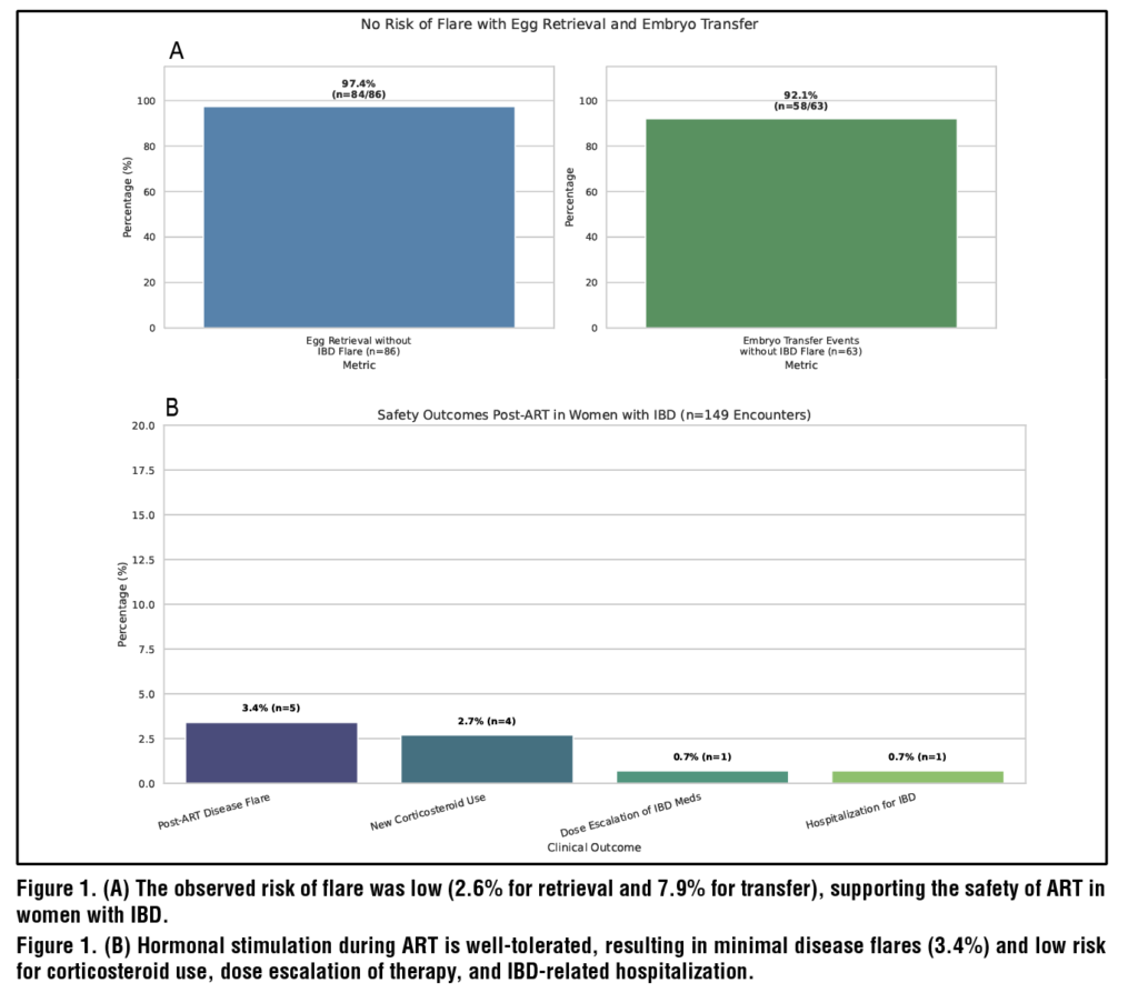
Flares occurred in 3.4% of post-ART encounters with minimal IBD-related hospitalization (0.7%), steroid use post-ART (2.7%), and medication escalation (1.3%). (Figure 1B)

Efficacy was also high, with egg retrieval rates exceeding 97% and an embryo transfer rate of approximately 92% occurring without an IBD flare. (Figure 1A)

 Multiple cohort studies confirm these findings, showing that women with medically managed IBD achieve live birth rates comparable to healthy controls.8 These findings suggest that ART is low risk for flare among women with IBD and is effective.8,11,14 

However, those with prior surgeries have different outcomes. Women with a J-pouch have a 64% lower live birth rate after IVF compared to those who have their UC medically managed. Similarly, women with CD who have had prior pelvic surgeries have a 49%-71% lower live birth rate after ART compared to those with medically managed CD.3 

Options and Financial Considerations

Women with IBD should be aware of the many ways to have a family – natural conception, ART, surrogacy, and adoption. For those with significant prior or current disease burden or with medication concerns, these options should be discussed. For women undergoing colectomy for UC, many centers offer the option of a subtotal proctocolectomy with ileostomy and rectal stump during childbearing to avoid scarring in the pelvis.  

The financial burden of surrogacy and ART represents a significant barrier for many patients struggling to conceive naturally. The average cost of one cycle of IVF ranges from $15,000-$30,000 when medications are used.6,16 Since many patients require multiple cycles to achieve pregnancy, the costs can quickly escalate. A financing industry survey noted that 70% of women who underwent IVF went into debt, and 34% of respondents reported cessation of treatment due to high treatment costs.15

Insurance coverage for ART is not standardized. As of 2025, 25 states have some form of fertility insurance by law, but coverage often includes spending caps or cycle limits. Patients should understand their insurance benefits and plan accordingly. Financial stress can add to the emotional toll of fertility treatment.15,16

Summary

  • Fertility among women with IBD in remission is similar to the general population. Infertility is usually driven by active disease or prior pelvic surgery. 
  • Misinformation often drives voluntary childlessness. Reassure patients that the risk of passing IBD to a child is low and that most medications are safe to use.  
  • Stop methotrexate during conception, pregnancy, and breastfeeding. 
  • JAK inhibitors and S1P modulators should be stopped unless there is no viable option for maternal health
  • Continue biologics and thiopurines. 
  • Referral to a fertility expert should be made early – after 6 months of concerted attempts or 4 months if the patient is older than 40 and/or has had prior pelvic surgery. 
  • Pre-conception optimization requires 3-6 months of steroid-free remission. Active disease at conception increases the risk of flares by 5-fold and increases risk for pre-term birth, low birth weight, C-section deliveries, and NICU admission. 
  • ART is safe in IBD patients, with low rates of flares (~3%), steroid use (2.7%), and hospitalizations (<1%). 
  • Discuss the various options to have a family and prepare patients for the high cost of IVF ($15,000-$30,000 per cycle) with variable insurance coverage.
  • With proper counseling and multidisciplinary support, we can even the scales, giving women with IBD a safe path to a successful pregnancy and a healthy family. 

References

1. Musso G, Cassader M, Gambino R. Endoscopic duodenal mucosa
ablation: the future of diabetes treatment? Trends Mol Med. 2024
Jul;30(7):612-616. doi: 10.1016/j.molmed.2024.03.003. Epub
2024 Mar 29. PMID: 38553333.
2. Parikh A, Thevenin C. Physiology, Gastrointestinal Hormonal
Control. [Updated 2023 May 1]. In: StatPearls [Internet]. Treasure
Island (FL): StatPearls Publishing; 2025 Jan-. Available from:
https://www.ncbi.nlm.nih.gov/books/NBK537284/#
3. Rønnestad I, Akiba Y, Kaji I, Kaunitz JD. Duodenal luminal
nutrient sensing. Curr Opin Pharmacol. 2014 Dec;19:67-75. doi:
10.1016/j.coph.2014.07.010. Epub 2014 Aug 9. PMID: 25113991;
PMCID: PMC4845631.
4. Breen DM, Rasmussen BA, Côté CD, Jackson VM, Lam TK.
Nutrient-sensing mechanisms in the gut as therapeutic targets for
diabetes. Diabetes. 2013 Sep;62(9):3005-13. doi: 10.2337/db13-
0523. PMID: 23970519; PMCID: PMC3749331.
5. Holesh JE, Aslam S, Martin A. Physiology, Carbohydrates.
[Updated 2023 May 12]. In: StatPearls [Internet]. Treasure Island
(FL): StatPearls Publishing; 2025 Jan-. Available from: https://
www.ncbi.nlm.nih.gov/books/NBK459280/#
6. Nakrani MN, Wineland RH, Anjum F. Physiology, Glucose
Metabolism. [Updated 2023 Jul 17]. In: StatPearls [Internet].
Treasure Island (FL): StatPearls Publishing; 2025 Jan-. Available
from: https://www.ncbi.nlm.nih.gov/books/NBK560599/
7. Habegger KM, Al-Massadi O, Heppner KM, Myronovych A,
Holland J, Berger J, Yi CX, Gao Y, Lehti M, Ottaway N, Amburgy
S, Raver C, Müller TD, Pfluger PT, Kohli R, Perez-Tilve D,
Seeley RJ, Tschöp MH. Duodenal nutrient exclusion improves
metabolic syndrome and stimulates villus hyperplasia. Gut. 2014
Aug;63(8):1238-46. doi: 10.1136/gutjnl-2013-304583. Epub 2013
Oct 9. PMID: 24107591; PMCID: PMC3981953.
8. Haidry RJ, van Baar AC, Galvao Neto MP, Rajagopalan H, Caplan
J, Levin PS, Bergman JJ, Rodriguez L, Deviere J, Thompson
CC. Duodenal mucosal resurfacing: proof-of-concept, procedural
development, and initial implementation in the clinical setting.
Gastrointest Endosc. 2019 Oct;90(4):673-681.e2. doi: 10.1016/j.
gie.2019.03.024. Epub 2019 Mar 29. PMID: 30935932
9. van Baar ACG, Haidry R, Rodriguez Grunert L, Galvao MPN,
Bisschops R, Hayee BH, Costamagna G, Deviere J, Bergman
JJGHM. Duodenal mucosal resurfacing: Multicenter experience
implementing a minimally invasive endoscopic procedure for
treatment of type 2 diabetes mellitus. Endosc Int Open. 2020
Nov;8(11):E1683-E1689. doi: 10.1055/a-1244-2283. Epub 2020
Oct 22. PMID: 33140025; PMCID: PMC7581486.
10. van Baar ACG, Holleman F, Crenier L, Haidry R, Magee C,
Hopkins D, Rodriguez Grunert L, Galvao Neto M, Vignolo
P, Hayee B, Mertens A, Bisschops R, Tijssen J, Nieuwdorp
M, Guidone C, Costamagna G, Devière J, Bergman JJGHM.
Endoscopic duodenal mucosal resurfacing for the treatment of
type 2 diabetes mellitus: one year results from the first international,
open-label, prospective, multicentre study. Gut. 2020
Feb;69(2):295-303. doi: 10.1136/gutjnl-2019-318349. Epub 2019
Jul 22. PMID: 31331994; PMCID: PMC6984054
11. Mingrone G, van Baar AC, Devière J, Hopkins D, Moura
E, Cercato C, Rajagopalan H, Lopez-Talavera JC, White K,
Bhambhani V, Costamagna G, Haidry R, Grecco E, Galvao Neto
M, Aithal G, Repici A, Hayee B, Haji A, Morris AJ, Bisschops
R, Chouhan MD, Sakai NS, Bhatt DL, Sanyal AJ, Bergman
JJGHM; Investigators of the REVITA-2 Study. Safety and efficacy
of hydrothermal duodenal mucosal resurfacing in patients with
type 2 diabetes: the randomised, double-blind, sham-controlled,
multicentre REVITA-2 feasibility trial. Gut. 2022 Feb;71(2):254-
264. doi: 10.1136/gutjnl-2020-323608. Epub 2021 Feb 17. PMID:
33597157; PMCID: PMC8761999.
12. Kovarova V, Lankova I, Machytka E, Knotkova K, Kratochvílová
H, Beneš M, Spicak J, Vasura A, Goldin E, Munter G, Zima
T, Mraz M, Dagan H, Levy B, Haluzik M, Kral J. Duodenal
Laser Ablation for Treatment of Type 2 Diabetes: Results of
First in Human Study. United European Gastroenterol J. 2025
Jun;13(5):750-758. doi: 10.1002/ueg2.12762. Epub 2025 Feb 20.
PMID: 39976927; PMCID: PMC12188360.
13. Busch CBE, Meiring S, van Baar ACG, Holleman F, Nieuwdorp
M, Bergman JJGHM. Recellularization via electroporation therapy
of the duodenum combined with glucagon-like peptide-1 receptor
agonist to replace insulin therapy in patients with type 2 diabetes:
12-month results of a first-in-human study. Gastrointest Endosc.
2024 Nov;100(5):896-904. doi: 10.1016/j.gie.2024.04.2904. Epub
2024 Apr 29. PMID: 38692517.
14. Meiring S, Aydin Ö, van Baar ACG, van der Vossen EWJ,
Rampanelli E, van Grieken NCT, Holleman F, Nieuwdorp
M, Bergman JJGHM. From Endoscopic Inspection to Gene-
Expression: A Thorough Assessment of the Duodenal Mucosa
After Resurfacing-A Prospective Study. Dig Dis Sci. 2025
Mar;70(3):1052-1063. doi: 10.1007/s10620-024-08710-4. Epub
2025 Jan 8. PMID: 39779586; PMCID: PMC11920325.
15. Chuang TJ, Ko CW, Shiu SI. The metabolic influence of duodenal
mucosal resurfacing for nonalcoholic fatty liver disease.
Medicine (Baltimore). 2023 Oct 6;102(40):e35147. doi:
10.1097/MD.0000000000035147. PMID: 37800801; PMCID:
PMC10553053.
16. Meiring S, Busch CBE, van Baar ACG, Hemke R, Holleman
F, Nieuwdorp M, Bergman JJGHM. Eliminating exogenous
insulin therapy in patients with type 2 diabetes by duodenal
ablation and GLP-1RA decreases risk scores for cardiovascular
events. Cardiovasc Diabetol. 2022 Sep 22;21(1):191. doi: 10.1186/
s12933-022-01628-z. PMID: 36138441; PMCID: PMC9503196.
17. van Baar ACG, Meiring S, Smeele P, Vriend T, Holleman F,
Barlag M, Mostafavi N, Tijssen JGP, Soeters MR, Nieuwdorp M,
Bergman JJGHM. Duodenal mucosal resurfacing combined with
glucagon-like peptide-1 receptor agonism to discontinue insulin
in type 2 diabetes: a feasibility study. Gastrointest Endosc. 2021
Jul;94(1):111-120.e3. doi: 10.1016/j.gie.2020.12.021. Epub 2020
Dec 24. PMID: 33359437.
18. van Baar ACG, Devière J, Hopkins D, Crenier L, Holleman F,
Galvão Neto MP, Becerra P, Vignolo P, Rodriguez Grunert L,
Mingrone G, Costamagna G, Nieuwdorp M, Guidone C, Haidry RJ,
Hayee B, Magee C, Carlos Lopez-Talavera J, White K, Bhambhani
V, Cozzi E, Rajagopalan H, J G H M Bergman J. Durable metabolic
improvements 2 years after duodenal mucosal resurfacing
(DMR) in patients with type 2 diabetes (REVITA-1 Study).
Diabetes Res Clin Pract. 2022 Feb;184:109194. doi: 10.1016/j.
diabres.2022.109194. Epub 2022 Jan 13. PMID: 35032562.
19. Wallenius V, Dirinck E, Fändriks L, Maleckas A, le Roux CW,
Thorell A. Glycemic Control after Sleeve Gastrectomy and Roux-
En-Y Gastric Bypass in Obese Subjects with Type 2 Diabetes
Mellitus. Obes Surg. 2018 Jun;28(6):1461-1472. doi: 10.1007/
s11695-017-3061-3. PMID: 29264780; PMCID: PMC5973990.
20. Umeda LM, Silva EA, Carneiro G, Arasaki CH, Geloneze B,
Zanella MT. Early improvement in glycemic control after bariatric
surgery and its relationships with insulin, GLP-1, and glucagon
secretion in type 2 diabetic patients. Obes Surg 2011;21:896–901
21. Mitchell BG, Collier SA, Gupta N. Roux-en-Y Gastric Bypass.
[Updated 2024 Nov 9]. In: StatPearls [Internet]. Treasure Island
(FL): StatPearls Publishing; 2025 Jan-. Available from: https://
www.ncbi.nlm.nih.gov/books/NBK553157/
22. Seeras K, Sankararaman S, Lopez PP. Sleeve Gastrectomy.
[Updated 2023 Jan 23]. In: StatPearls [Internet]. Treasure Island
(FL): StatPearls Publishing; 2025 Jan-. Available from: https://
www.ncbi.nlm.nih.gov/books/NBK519035/
23. Pories WJ, Swanson MS, MacDonald KG, Long SB, Morris PG,
Brown BM, Barakat HA, deRamon RA, Israel G, Dolezal JM, et
al. Who would have thought it? An operation proves to be the most
effective therapy for adult-onset diabetes mellitus. Ann Surg. 1995
Sep;222(3):339-50; discussion 350-2. doi: 10.1097/00000658-
199509000-00011. PMID: 7677463; PMCID: PMC1234815.
24. Seeras K, Acho RJ, Lopez PP. Roux-en-Y Gastric Bypass Chronic
Complications. [Updated 2023 Jun 5]. In: StatPearls [Internet].
Treasure Island (FL): StatPearls Publishing; 2025 Jan-. Available
from: https://www.ncbi.nlm.nih.gov/books/NBK519489/#
25. Nauck MA, Quast DR, Wefers J, Meier JJ. GLP-1 receptor agonists
in the treatment of type 2 diabetes – state-of-the-art. Mol
Metab. 2021 Apr;46:101102. doi: 10.1016/j.molmet.2020.101102.
Epub 2020 Oct 14. PMID: 33068776; PMCID: PMC8085572.
26. Rajagopalan H, Cherrington AD, Thompson CC, Kaplan LM,
Rubino F, Mingrone G, Becerra P, Rodriguez P, Vignolo P, Caplan
J, Rodriguez L, Galvao Neto MP. Endoscopic Duodenal Mucosal
Resurfacing for the Treatment of Type 2 Diabetes: 6-Month
Interim Analysis From the First-in-Human Proof-of-Concept
Study. Diabetes Care. 2016 Dec;39(12):2254-2261. doi: 10.2337/
dc16-0383. Epub 2016 Aug 12. PMID: 27519448.

Download Tables, Images & References

Nutrition Reviews in Gastroenterology, SERIES #31

Disorders of Gut-Brain Interaction: Behavioral Therapies and Integration of the GI-Psychologist and GI-Registered Dietitian Care

Read Article


Effective management of Disorders of Gut-Brain Interaction (DGBIs) often requires an interdisciplinary approach that extends beyond the gastrointestinal (GI) provider. DGBIs are characterized by dysregulation of the gut–brain axis, and growing evidence supports behavioral and dietary therapies that target these bidirectional pathways. Behavioral health providers and registered dietitians (RD) with specialized gastroenterology training — hereafter referred to as GI-psychologists and GI-RDs — deliver evidence-based interventions that have potential to improve patient symptoms and quality of life (QoL), with GI-psychologists using Brain-Gut Behavioral Therapies (BGBT), such as cognitive-behavioral therapy, to address psychosocial contributors, and GI-RDs applying diet strategies, including the low FODMAP diet, to optimize gastrointestinal function. Collaboration between these providers enables comprehensive evaluation of symptoms and DGBI subtype, supporting appropriate referrals. This review is to summarize current evidence for therapy approaches and delineates the distinct and complementary contributions of GI-psychologists and GI-RDs in DGBI, with the aim of guiding interdisciplinary referral practices.

The Role of the Brain-Gut Axis in Disorders of Gut-Brain Interactions

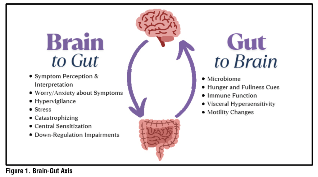
Symptoms and sensations in the digestive tract can be related to structural organ malfunction or from sensory malfunction via the Gut-Brain Axis (GBA). The central nervous and enteric nervous systems are connected to one another via complex nervous, endocrine and immune bidirectional pathways.1–3. When the GBA is dysregulated, benign sensations in the GI organs such as those of normal digestive processes may be perceived as threatening and more severe due to cognitive, behavioral and affective processes which can function to amplify those signals.4 For example, relevant processes often include symptom or illness specific anxiety and catastrophizing, avoidance of feared activities and foods, hypervigilance to bodily sensations, and activation of the stress response.5,6 These factors interact with central processes which contribute to symptoms including visceral hypersensitivity of the peripheral nerves in the digestive tract and to enhanced pain perception through central sensitization.7 (See Figure 1) Disorders derived from disruption of the GBA formerly called Functional Gastrointestinal (GI) Disorders and are now termed Disorders of Gut-Brain Interaction (DGBI) (e.g., irritable bowel syndrome (IBS), functional dyspepsia).8

A challenge for many patients with DGBI driven symptoms is a lack of a clear mechanism for their concern, as typically objective testing does not provide an explanation. An interdisciplinary GI team can engage in a collaborative discussion starting from a clear explanation of the GBA, what DGBI diagnosis they meet criteria for, and what evidence-based treatments they may qualify for to manage and treat their symptoms with the various specialists available to them.9 This can offer much needed assurance that the symptoms are real and expected in DGBI, can help reduce stigma associated with these disorders and can help patients develop a clear treatment plan.10

Brain-Gut Behavioral Therapies 

When accessible and appropriate for the patient, Brain-Gut Behavioral Therapies (BGBTs) can offer a pathway to symptom relief for patients with DGBIs. To date, BGBTs with a robust evidence base include Cognitive Behavioral Therapy (CBT), gut-directed hypnotherapy (GDH)11 and mindfulness therapies such as Acceptance and Commitment Therapy (ACT).12 (See Table 1) The aim of these therapies is to target the functioning of the brain and gut through modifying underlying interpretations about symptoms and illness. 

Cognitive Behavioral Therapy

CBT is a skills-based therapy which emphasizes the interconnectedness of thoughts, emotions, behaviors, and physical sensations. While CBT was initially developed and studied in mental health, it has been successfully applied in medical settings including DGBI. Components of the treatment vary based on patient presentation but often include relaxation strategies (including instruction in diaphragmatic breathing), cognitive awareness and reframing, exposure exercises to avoided foods or situations and stress management with problem solving skills. Specifically in IBS, CBT has demonstrated benefit to symptom experience and severity,quality of life and impact on the brain’s interpretation of symptoms.13–17 For non-cardiac chest pain and functional dyspepsia, smaller studies have also shown benefit in symptom perception.18,19 CBT has also been recommended as an intervention target for functional heartburn, though robust trials to test efficacy are still needed.20 Beyond traditional face to face delivery, evidence suggests that CBT for IBS can be successfully administered in group format and online/telephone.21 

Table 1. Evidence-based Treatments: Cognitive Behavioral Therapy, Gut-Directed Hypnotherapy, and Acceptance and Commitment Therapy 

Evidence Based Treatment for DGBIMain ComponentsTypical CoursePatient Characteristic Considerations
Cognitive Behavioral Therapy (CBT)15,21,46 An intervention which includes psychoeducation about the condition and gut-brain axis, skills training to target and change maladaptive cognitions, skills training to target physiological arousal and promote relaxation, and strategies to help broaden behavioral responses to symptoms and interoceptive and behavioral exposure.~3-12 sessionsPatient endorses symptoms specific anxiety and behavioral avoidance.   Patient is psychologically minded, meaning they are interested and able to see themselves and their own thoughts and behavior from a 3rd person perspective, analyzing these and apply skills to make change. 
Gut-Directed Hypnotherapy23,27,47  (GDH)A facilitated state of deep relaxation, known as trance, in which patients have increased receptiveness to suggestion. Suggestions are tailored to patient’s specific symptoms and quality of life concerns.~4-12 sessions Patient endorses pain, tension/tightness, or visceral hypersensitivity as a primary symptom/experience.  Patients do NOT have active symptoms of post-traumatic stress disorder (PTSD), or PTSD symptoms are stable at present.
Acceptance and Commitment Therapy (ACT)30,48Identification of valued life domains and explicit formulation of committed actions patient can take to live life in greater accordance with their values, mindfulness practice, reduction of emotional avoidance, identification of “fusion” with maladaptive thoughts and skills taught to help “de-fuse” from these thoughts.~5-12 sessionsPatient endorses difficulty putting space between themselves and negative thoughts and emotions related to GI symptoms or condition or is feeling “stuck.” Patient is noting that their GI condition or symptoms are preventing them from living life in accordance with the values that are important to them. 

Gut-Directed Hypnotherapy

GDH is a form of therapy that, over a series of sessions, guides patients into a deep relaxation and further into a hypnotic or trance state prior to the delivery of tailored suggestions to modify their visceral sensations and pain experience. Therapeutic suggestions also often include those to increase patient’s engagement in valued life activities over attending to bodily sensations. Frequent practice, often with audio, is a common component of the therapy. GDH has shown to have a substantial impact on IBS symptoms and abdominal pain.22,23 Smaller studies examining GDH in the esophagus, such as in functional heartburn and globus, have also shown promise for improving symptoms but warrant replication in larger trials.24–26 Like CBT, GDH can be effectively delivered in a variety of modalities including via video and in groups.23,27,28


Table 2. Nutrition Care Plan: The GI-RDs Contribution to the Interdisciplinary Team41-43

Nutrition Assessment ComponentsRD Considerations Nutrition Interventions/GI-RD “toolbox”Nutrition Monitoring
and Evaluation
Nutritional Status Weight BMI Weight History Malnutrition  Appropriate labs
(e.g., prealbumin, electrolytes, zinc, iron studies) Nutrition Focused Physical Exam (NFPE)
Weight restoration with slow caloric increase; may use oral nutrition supplement as first line treatment Address micronutrient concerns with supplementation  Nutrition education focused on role of weight restoration on GI symptom management Weight gain Dietary components (e.g.,
% of estimated energy needs in dietary intake, fiber intake, micronutrient intake) Labs Improvement of muscle wasting & fat loss on NFPE
Nutrition/Food HistoryPrevious diet therapy attempts (Gluten-Free Diet (GFD), Low FODMAP Diet, National Institute for Health and Care Excellence (NICE), etc.) Herbal supplement usage History of eating disorder (ED) for shape/weight concerns Active ED Current diet restrictions are significant enough to cause concern for diet quality Prescence and severity of sitophobia Assess for avoidant restrictive food intake disorder (ARFID). Consider using ARFID screener, though not currently validated in the GI patient population specifically (e.g., PARDI-AR-Q,49 Nine Item ARFID Screen43)Pausing other supplements; trial of peppermint oil,50 melatonin,51 psyllium husk52 Personalized therapeutic dietary interventions that could include changes in eating behaviors and meal timing, consistent fiber intake, FODMAP restrictions among others  For patients with a history of ED, consider gentle nutrition, modified FODMAP diet, as well as mindful eating  Triage level of care to eating disorder treatment Referral to GI-psychologist for additional food exposure support for those with sitophobia Develop food hierarchy and exposures trials in tandem with GI-psychologist in mild sitophobia or ARFID Refer to an ARFID treatment center for patients with moderate-severe ARFID, AFTER evaluation by GI-psychologist Nutrition counseling: Optimize nutritional intake and health, improve or stabilize GI symptoms as part of DGBI while providing validationGI symptom frequency GI symptom severity Diet quality Development of maladaptive dietary behaviors e.g., over restriction, skipping meals, reducing intake to <75% of estimated energy needs
Psychosocial History  Mental health Resources, including financial, time, logistical Motivation  Social factors, environment  Cultural factors Health literacy Collaboration of care meeting with current therapist, and/or refer to general mental health community provider Motivational interviewing  Nutrition education materials appropriate for knowledge level Meal delivery services, brands, recipes developed appropriate for diet application if kitchen skills are limitedReadiness to change Knowledge recall

Acceptance and Commitment Therapy

Mindfulness based therapies, such as ACT, aim to assist patients in finding grounding in the present moment in order to experience the transient nature of thoughts, emotions and urges, a process known as psychological flexibility.29 In DGBI, this may look like a patient acknowledging that their symptoms are present today, accepting that it is frustrating and ultimately choosing to attend an important social gathering and being open to finding joy in that experience. Another key component of ACT is assisting patients in identifying their personal values and finding opportunities to commit to behaviors and actions that are consistent with those values. In general, the literature on ACT as a BGBT for DGBI is more nascent than that of CBT or GDH and deserves further study. However, those studies implementing a full ACT protocol for patients with DGBI, namely, IBS, have found ACT may help reduce symptom severity and acceptance of their diagnosis.30 

Though all the therapies are described separately above, it is not uncommon for a skilled provider to use one or multiple BGBT skills during the intervention course with a patient.

Diet Therapy in DGBI

When patients are asked about their preference of medical, dietary or behavioral intervention, one study found that patients with DGBI prefer diet-focused interventions as first line therapy.31 The American Gastroenterological Association 2022 Clinical Practice Update on the Role of Diet in Irritable Bowel Syndrome highlighted several best practice advice statements that focus on diet and the role of a dietitian in IBS care.32 These include nutrition assessment and screening for eating disorders prior to dietary restriction, providing nutrition education about the role of food and meal-related symptoms, and personalization of meal choices. Instructing patients to keep a 3-day food and symptom log prior to the first GI-RD visit may help illuminate these patterns.32

Therapeutic diets, such as low FODMAP diet that have shown efficacy in reducing symptoms of IBS, should be used for a finite period and may not be an appropriate starting point for patients who are consuming low culprit foods, have active eating, or psychiatric disorders or are food insecure.32 Additionally, alterations of the microbes of the gut, food chemistry and GI infections have been identified as mechanisms which have potential to increase intestinal permeability and hypersensitivity in DGBI.3,33,34 Recent understanding of gut microbiome and its role in the GBA suggest that therapeutic diets may have several impacts by directly modulating both the microbiota and its metabolome which play a role in the GBA.35


Table 3. DGBI Counseling Services Referral Considerations

DGBI Patient PresentationPsychology ReferralDietitian Referral
Observed association between higher levels of stress and worsening GI symptoms,
regardless of diet consumption
X
Presence of or anticipation of GI symptoms creates worry or anxietyX
Significantly changed behavior in an effort to control symptomsXX
Avoidance of eating foods in general or broad general classes of foodsXX
Discordance between objective, diagnostic testing and patients report of symptomsX
Meal-related GI symptoms
X
Specific concern for dietary Intolerance, such as carbohydrate malabsorption
or gluten intolerance 

X
Unintentional weight loss and/or a concern for malnutritionXX
History of disordered eating in newly diagnosed DGBIXX
Nutritionally pertinent medical diagnosis, such as diabetes, CKD, CVD
X
Interested in holistic lifestyle-based therapies vs. medication, or in addition to medical therapyXX

Role of GI-Psychologist: Assessment and Treatment Options 

GI-psychologists, with clinical skills and specialized training in the functioning of the GI tract, are well positioned to apply BGBTs. The process begins with an evaluation to ascertain components of the biopsychosocial model.36–38 (See Figure 2) Patients are typically asked to provide a timeline of their GI symptom onset, as well as potential gut-brain dysregulation triggers, such as stressful life events, exposure to physical (e.g., abdominal surgery) or psychological (e.g., sexual abuse) trauma, or the experience of infections. The GI-psychologist seeks to understand progression of symptoms over time, including whether symptoms worsen with higher levels of stress, and how GI symptoms or their management impact QoL, functioning, and relationships, with attention to symptom-specific anxiety, hypervigilance, and visceral hypersensitivity.25 

During the evaluation, the GI-psychologist gathers information on life domains that may affect  GI symptoms. Patients are typically asked about any current and past psychiatric symptoms and history, including frank and subclinical eating disorders, psychotherapy, and psychotropic medications.39 Accordingly, patients are also asked to describe their global levels of perceived stress and the coping strategies they may use to handle stress to help determine an approach versus avoidance style. Physical health behaviors, including use of substances like alcohol, cannabis and nicotine, quality and duration of sleep, typical diet, food triggers, and engagement of physical activity are reviewed for potential to worsen symptoms. Patients assigned female at birth may also be asked questions in the evaluation regarding reproductive health, such as whether GI symptoms coincide or worsen with menstrual cycles. Typically, the assessment will include conceptualization, treatment planning, and potential referrals to outside providers. 

The GI-psychologist may refer patients to external providers and services needed to augment their care. Among others, these commonly include community resources, pelvic-floor physical therapy (PFPT), specialists or programs for eating disorders (e.g., laxative abuse, severe restriction with or without body and shape concerns), or specialists in psychiatric concerns. Depending on the symptom severity of a patient’s mental health presentation, the GI-psychologist will collaborate with the patient to determine the sequence of treatment, i.e., whether BGBT can occur in parallel to or after a patient’s primary mental health or eating disorder treatment.  

Similarly, the GI-psychologist may make a referral to a GI-RD to help elucidate various components of diet, nutritional status and if a therapeutic diet trial is warranted.40 

Role of GI-RD: Assessment and Treatment Options 

The first step with a GI-RD is the initial visit, during which the dietitian conducts a nutrition assessment to obtain detailed information regarding a patient’s medical history, nutrition history, diet recall, lifestyle habits, cultural considerations and knowledge, beliefs, and attitudes regarding food. Specifically, a GI-RD closely evaluates presentation of a patient’s symptoms, appropriateness of current diet, past or current eating behaviors that may indicate a disordered relationship with food, known food allergies, intolerances, or sensitivities, and the extent of dietary restriction. 

Based on these findings, the GI-RD collaborates on a treatment plan and provides tailored recommendations for nutrition interventions to treat the DGBI. Follow-up visits with a GI-RD include assessments of the outcome of the initial nutrition interventions, adjusts as needed, and provide ongoing education and counseling to optimize progress. If the GI-RD identifies factors that inform the need for a referral to a GI-psychologist, the dietitian may make a direct referral to support integrative care.41–43 (See Table 2)

Intersection and Overlap of GI-Psychologist and GI-RD

In an interdisciplinary care model, GI-RDs’ and GI-psychologists’ roles often overlap. It can be challenging to define the boundaries of nutrition interventions centered on diet as providers of both disciplines have applicable skillsets. A collaborative treatment plan and active communication regarding a patient’s goals and intervention barriers and facilitators can be exceptionally helpful in these situations to enhance progress. Importantly, medical therapy is less than 50% effective at treating global GI symptoms, suggesting the necessity of several modalities to best address both GI and extra-intestinal symptoms.44 

Food and eating play a critical role in the experience of GI symptoms; beliefs about food, eating and digestion can trigger maladaptive processes such as symptom-specific anxiety, avoidance of food(s), and hypervigilance to the body after eating, reinforcing the DGBI. Approximately 80% of patients with DGBI implicate food as a catalyst for symptom onsetand develop adaptive eating behaviors to manage symptoms.40,45 Inappropriate avoidance of foods can lead to overly restricted diets and malnutrition. For example, patients with sitophobia often have highly restricted diets and struggle to reintroduce a wider variety of foods, even after food intolerances and allergies have been ruled out. A GI-psychologist and GI-RD team can collaborate to create a food avoidance hierarchy and assist the patient in utilizing psychological and behavioral skills to incorporate foods systematically. Other considerations for referrals are outlined in Table 3.

Case Scenario and Treatment Course 

To further illustrate interdisciplinary care and the components of treatment, please see a case example: 

Case: 

Patient is a 45-year-old white, married straight cis-woman with IBS-C, gastritis with possible GERD (awaiting objective testing). She is 20 years post-cholecystectomy.  

The patient is currently experiencing days of constipation followed by a period of diarrhea, post-prandial abdominal discomfort, nausea, heartburn and bloating. She is taking plecanatide (3mg) and omeprazole (20mg). Prior therapeutics include linaclotide, lubiprostone, prucalopride, cholestyramine, and nortriptyline. 


Table 4. Case Scenario. Between GI-Psych Evaluation and intervention session #1, the patient had consultation with a motility specialist, underwent anorectal manometry (ARM), was referred and started pelvic floor physical therapy (PFPT) and scheduled consultation with GI-RD.

Patient was referred to GI-psychologist by her general gastroenterologist for treatment of IBS-C and other GI symptoms. Condensed and pertinent data from that evaluation is below:  

Patient reported that lower GI symptoms began over 20 years ago, around the time she was in an abusive relationship. In the last several years, her upper GI and abdominal symptoms have worsened. In addition to symptoms, she reports a concern about obtaining an accurate diagnosis that captures the range of problems with her GI system. She described several factors that are likely contributing to current symptoms and associated distress including long history of anxiety, OCD and experience of trauma which may contribute to GBA disruption. 

The patient described vigilance to her body sensations, her body weight/shape and to her diet as her symptoms occur reliably after eating. This is further distressing to her as she has noticed weight gain in recent years. The patient described a tendency to avoid leaving home for fear of not knowing where bathrooms will be. She described a strong relationship between stress and GI symptoms and noted an overall high level of stress which she attributed to her own anxiety and tendency toward perfectionism and people-pleasing. She reported no concerns regarding social influences on health.

Patient reported treatment goals are to: 

  • Understand etiology of symptoms
  • Improve consistency of her bowel movements 
  • Understand dietary triggers 
  • Decrease worries and anxiety about her symptoms and increase comfort leaving home 
  • Reduce weight and bloating as this is a body image concern

Table 4. Case Scenario (Continued  ). Following her engagement with a multi-disciplinary treatment team including motility specialist, PFPT, GI-psychologist and GI-RD, patient reported a significant improvement in symptoms of constipation and only occasional post-prandial abdominal discomfort, nausea, heartburn and bloating. She expressed satisfaction with response to treatment and the integration of her care team. At this point, GI-psychologist and GI-RD terminated care as treatment goals had been met.

GI-psychologist next steps: 

  • Request referral to motility specialist to evaluate for pelvic floor disorder given patient’s experience of alternating constipation and diarrhea 
  • Referral to GI-RD as patient is experiencing post-prandial symptoms 
  • Referral to a trauma specialist as a patient is experiencing current post-traumatic stress symptoms
  • Plan for treatment including CBT and ACT. Plan to defer gut-directed hypnotherapy as patient is experiencing current trauma symptoms

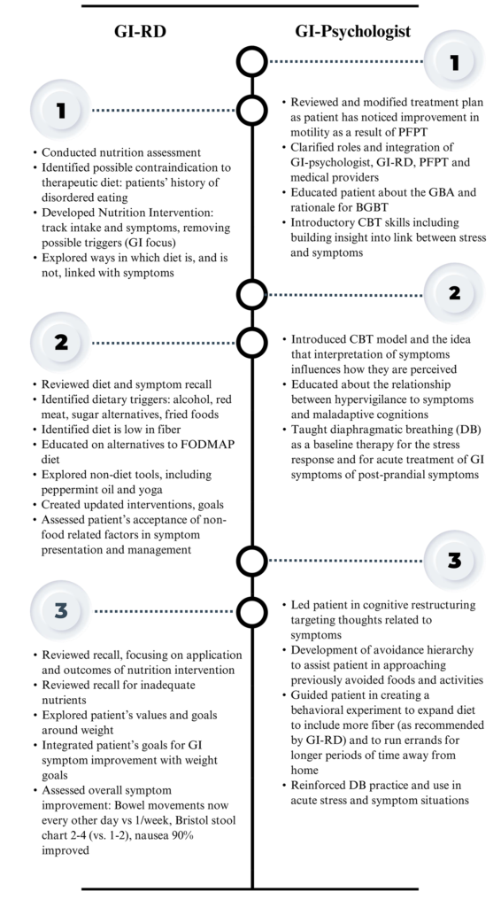
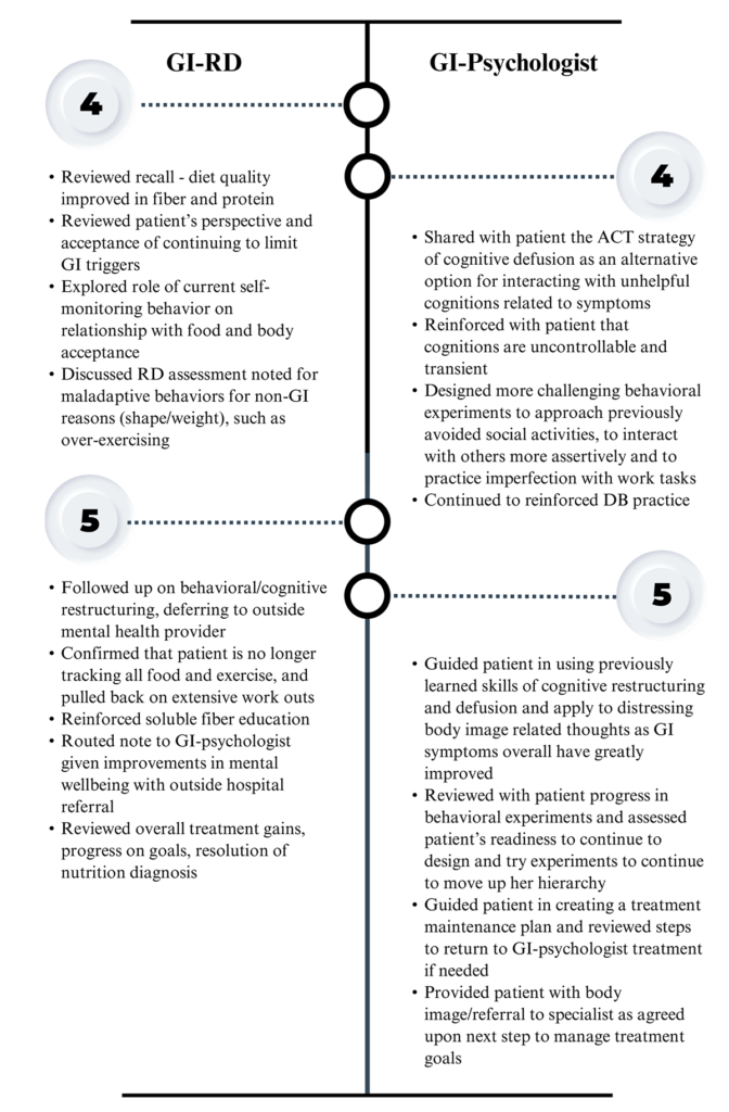
See Table 4 for the patient’s treatment course with GI-psychologist and GI-RD working as a team in integrated care. 

References

1. Agirman G, Yu KB, Hsiao EY. Signaling inflammation across the gut-brain axis. Science. 2021;374(6571):1087-1092. 

2. Palsson OS, Ballou S. Hypnosis and cognitive behavioral therapies for the management of gastrointestinal disorders. Curr Gastroenterol Rep. 2020;22(7):1-9. 

3. Mayer EA, Nance K, Chen S. The gut-brain axis. Annu Rev Med. 2022;73:439-453. 

4. Labanski A, Langhorst J, Engler H, Elsenbruch S. Stress and the brain-gut axis in functional and chronic-inflammatory gastrointestinal diseases: A transdisciplinary challenge. Psychoneuroendocrinology. 2020;111:104501. 

5. Labus JS, Bolus R, Chang L, et al. The Visceral Sensitivity Index: development and validation of a gastrointestinal symptom-specific anxiety scale. Aliment Pharmacol Ther. 2004;20(1):89-97. 

6. Ballou S, Vasant DH, Guadagnoli L, et al. A primer for the gastroenterology provider on psychosocial assessment of patients with disorders of gut-brain interaction. Neurogastroenterol Motil. 2024;36(12):e14894. 

7. Keefer L, Ko CW, Ford AC. AGA clinical practice update on management of chronic gastrointestinal pain in disorders of gut-brain interaction: Expert review. Clin Gastroenterol Hepatol. 2021;19(12):2481-2488.e1. 

8. Schmulson MJ, Drossman DA. What is new in Rome IV. Journal of Neurogastroenterology and Motility. 2017;23(2):151-163. 

9. Keszthelyi D, Keefer L. “A rose by any other name”? — Beyond diagnostic nomenclature in neurogastroenterology. Gastroenterology. 2025;169(1):19-21. doi:10.1053/j.gastro.2025.01.229

10. Feingold JH, Drossman DA. Deconstructing stigma as a barrier to treating DGBI: Lessons for clinicians. Neurogastroenterol Motil. 2021;33(2):e14080. 

11. Black CJ, Thakur ER, Houghton LA, Quigley EMM, Moayyedi P, Ford AC. Efficacy of psychological therapies for irritable bowel syndrome: systematic review and network meta-analysis. Gut. 2020;69(8):1441-1451. 

12. Keefer L, Ballou SK, Drossman DA, Ringstrom G, Elsenbruch S, Ljótsson B. A Rome working team report on brain-gut behavior therapies for disorders of gut-brain interaction. Gastroenterology. 2022;162(1):300-315. 

13. Goodoory VC, Khasawneh M, Thakur ER, et al. Effect of brain-gut behavioral treatments on abdominal pain in irritable bowel syndrome: Systematic review and network meta-analysis. Gastroenterology. 2024;167(5):934-943.e5. 

14. Bonnert M, Olén O, Lalouni M, et al. Internet-delivered cognitive behavior therapy for adolescents with irritable bowel syndrome: A randomized controlled trial. Official journal of the American College of Gastroenterology | ACG. 2017;112(1):152. 

15. Owusu JT, Sibelli A, Moss-Morris R, van Tilburg MAL, Levy RL, Oser M. A pilot feasibility study of an unguided, internet-delivered cognitive behavioral therapy program for irritable bowel syndrome. Neurogastroenterol Motil. 2021;33(11):e14108. 

16. Hunt M, Miguez S, Dukas B, Onwude O, White S. Efficacy of Zemedy, a mobile digital therapeutic for the self-management of irritable bowel syndrome: crossover randomized controlled trial. JMIR Mhealth Uhealth. 2021;9(5):e26152. 

17. Jacobs JP, Gupta A, Bhatt RR, et al. Cognitive behavioral therapy for irritable bowel syndrome induces bidirectional alterations in the brain-gut-microbiome axis associated with gastrointestinal symptom improvement. Microbiome. 2021;9(1):236. 

18. Jonsbu E, Martinsen EW, Morken G, Moum T, Dammen T. Change and impact of illness perceptions among patients with non-cardiac chest pain or benign palpitations following three sessions of CBT. Behavioural and Cognitive Psychotherapy. 2013;41(4):398-407. 

19. Keefer L, Ballou SK, Drossman DA, Ringstrom G, Elsenbruch S, Ljótsson B. A Rome working team report on brain-gut behavior therapies for disorders of gut-brain interaction. Gastroenterology. 2022;162(1):300-315. 

20. Guadagnoli L, Yadlapati R, Pandolfino J, et al. Behavioral therapy for functional heartburn: Recommendation statements. Clin Gastroenterol Hepatol. 2024;22(8):1709-1718.e3. 

21. Chen LJ, Kamp K, Fang A, Heitkemper MM. Delivery methods of cognitive behavior therapy for patients with irritable bowel syndrome. Gastroenterol Nurs. 2022;45(3):149-158. 

22. Lövdahl J, Törnblom H, Ringström G, Palsson OS, Simrén M. Randomised clinical trial: individual versus group hypnotherapy for irritable bowel syndrome. Aliment Pharmacol Ther. 2022;55(12):1501-1511. 

23. Adler EC, Levine EH, Ibarra AN, et al. Gut-directed hypnotherapy for irritable bowel syndrome: A systematic review and meta-analysis. Neurogastroenterology & Motility. 2025;37(7):e70037. 

24. Kiebles JL, Kwiatek MA, Pandolfino JE, Kahrilas PJ, Keefer L. Do patients with globus sensation respond to hypnotically assisted relaxation therapy? A case series report. Accessed January 30, 2026. https://dx.doi.org/10.1111/j.1442-2050.2010.01064.x

25. Riehl ME, Kinsinger S, Kahrilas P, Pandolfino J, Keefer L. The role of a health psychologist in the management of functional esophageal complaints. Dis Esophagus. 2015;28(5):428-436. 

26. Riehl ME, Pandolfino JE, Palsson OS, Keefer L. The feasibility and acceptability of esophageal-directed hypnotherapy for functional heartburn. Dis Esophagus. 2016;29(5):490-496. 

27. Anderson EJ, Peters SL, Gibson PR, Halmos EP. Comparison of digitally delivered gut-directed hypnotherapy program with an active control for irritable bowel syndrome. Official journal of the American College of Gastroenterology | ACG. 2025;120(2):440. 

28. Peters SL, Gibson PR, Halmos EP. Smartphone app-delivered gut-directed hypnotherapy improves symptoms of self-reported irritable bowel syndrome: A retrospective evaluation. Neurogastroenterol Motil. 2023;35(4):e14533. 

29. Hayes SC, Levin ME, Plumb-Vilardaga J, Villatte JL, Pistorello J. Acceptance and commitment therapy and contextual behavioral science: Examining the progress of a distinctive model of behavioral and cognitive therapy. Behav Ther. 2013;44(2):180-198. 

30. Marchese SH, Naftaly JP, Pandolfino J. Acceptance and commitment therapy for the treatment of irritable bowel syndrome and inflammatory bowel disease: a narrative review. Transl Gastroenterol Hepatol. 2024;9:43. 

31. Sturkenboom R, Keszthelyi D, Masclee AAM, Essers BAB. Discrete choice experiment reveals strong preference for dietary treatment among patients with irritable bowel syndrome. Clinical Gastroenterology and Hepatology. 2022;20(11):2628-2637. 

32. Chey WD, Hashash JG, Manning L, Chang L. AGA Clinical practice update on the role of diet in irritable bowel syndrome: Expert review. Gastroenterology. 2022;162(6):1737-1745.e5. 

33. Barbara G, Aziz I, Ballou S, et al. Rome Foundation working team report on overlap in disorders of gut-brain interaction. Nat Rev Gastroenterol Hepatol. 2025;22(4):228-251.

34. Lacy BE, Pimentel M, Brenner DM, et al. ACG clinical guideline: Management of irritable bowel syndrome. Official journal of the American College of Gastroenterology | ACG. 2021;116(1):17. 

35. Schneider E, O’Riordan KJ, Clarke G, Cryan JF. Feeding gut microbes to nourish the brain: unravelling the diet–microbiota–gut–brain axis. Nat Metab. 2024;6(8):1454-1478. 

36. Engel GL. The need for a new medical model: A challenge for biomedicine. Science. 1977;196(4286):129-136.

37. Bolton D. A revitalized biopsychosocial model: core theory, research paradigms, and clinical implications. Psychological Medicine. 2023;53(16):7504-7511. 

38. Van Oudenhove L, Levy RL, Crowell MD, et al. Biopsychosocial aspects of functional gastrointestinal disorders: How central and environmental processes contribute to the development and expression of functional gastrointestinal disorders. Gastroenterology. 2016;150(6):1355-1367.e2. 

39. Kinsinger SW. Practical Approaches to Working with a gastrointestinal psychologist. Gastroenterol Clin North Am. 2022;51(4):711-721.

40. Böhn L, Störsrud S, Törnblom H, Bengtsson U, Simrén M. Self-reported food-related gastrointestinal symptoms in IBS are common and associated with more severe symptoms and reduced quality of life. Am J Gastroenterol. 2013;108(5):634-641. 

41. Scarlata K, Catsos P, Smith J. From a dietitian’s perspective, diets for irritable bowel syndrome are not one size fits all. Clinical Gastroenterology and Hepatology. 2020;18(3):543-545. 

42. Guadagnoli L, Mutlu EA, Doerfler B, Ibrahim A, Brenner D, Taft TH. Food-related quality of life in patients with inflammatory bowel disease and irritable bowel syndrome. Qual Life Res. 2019;28(8):2195-2205. 

43. Murray HB, Dreier MJ, Zickgraf HF, et al. Validation of the Nine Item ARFID Screen (NIAS) subscales for distinguishing ARFID presentations and screening for ARFID. Int J Eat Disord. 2021;54(10):1782-1792. 

44. Chey WD, Keefer L, Whelan K, Gibson PR. Behavioral and diet therapies in integrated care for patients with irritable bowel syndrome. Gastroenterology. 2021;160(1):47-62. 

45. Scarlata K, Zickgraf HF, Satherley RM, et al. A call to action: Unraveling the nuance of adapted eating behaviors in individuals with gastrointestinal conditions. Clinical Gastroenterology and Hepatology. 2025;23(6):893-901.e2. 

46. Sugaya N, Shirotsuki K, Nakao M. Cognitive behavioral treatment for irritable bowel syndrome: a recent literature review. BioPsychoSocial Med. 2021;15(1):1. 

47. Sasegbon A, Hasan SS, Whorwell PJ, Vasant DH. Experience and clinical efficacy of gut-directed hypnotherapy in an Asian population with refractory irritable bowel syndrome. JGH Open. 2022;6(7):447-453. 

48. Taghvaeinia A, Karami M, Azizi A. Comparison of the effect of dialectical behavior therapy, acceptance and commitment therapy mindfulness-based stress reduction on irritable bowel syndrome symptoms, quality of life, anxiety and depression: A pilot randomized controlled trial. Psychiatr Q. 2024;95(1):53-68. 

49. Bryant-Waugh R, Stern CM, Dreier MJ, et al. Preliminary validation of the pica, ARFID and rumination disorder interview ARFID questionnaire (PARDI-AR-Q). J Eat Disord. 2022;10:179. 

50. Alammar N, Wang L, Saberi B, et al. The impact of peppermint oil on the irritable bowel syndrome: a meta-analysis of the pooled clinical data. BMC Complement Altern Med. 2019;19:21. 

51. Siah KTH, Wong RKM, Ho KY. Melatonin for the treatment of irritable bowel syndrome. World J Gastroenterol. 2014;20(10):2492-2498. 

52. Garg P, Garg PK, Bhattacharya K. Psyllium husk positively alters gut microbiota, decreases inflammation, and has bowel-regulatory action, paving the way for physiologic management of irritable bowel syndrome. Gastroenterology. 2024;166(3):545-546.

Download Tables, Images & References

Frontiers in Endoscopy, Series #105

Endoscopic Duodenal Mucosal Resurfacing

Read Article

With prevalence of type 2 diabetes and metabolic dysfunction-associated steatotic liver disease (MASLD) rising in parallel, there is an increased need for alternatives to surgical and pharmacological treatments for these conditions. Pharmacologic therapy can achieve good results in many patients, but for those with side effects, rising costs, or difficulty with adherence, the main alternative is bariatric surgery (which comes with significant cost and risk).17 Duodenal mucosal resurfacing (DMR) offers a less invasive alternative to bariatric surgery without the side effects and inconvenience of medication for patients with type 2 diabetes and MASLD.11 

The duodenum is targeted in DMR because it is a major site of nutrient sensing and signaling in the gastrointestinal tract.2,3,4 The combination of bile acids, lipases, and incretin hormones help to control glucose levels in the blood and liver. Bariatric surgeries that bypass the duodenum or regulate the flow of nutrients into the duodenum show rapid improvements in glycemic control, making the duodenum a plausible site contributing to metabolic dysfunction.17 

DMR is an endoscopic procedure that targets the superficial mucosa of the descending and inferior duodenum.8 The ablation of the mucosa is thought to reset the nutrient sensing and improve glycemic control.14 There is complete regrowth of the mucosa with minimal scarring and collagen deposition, suggesting the metabolic benefits are not structural.12 

DMR is still under investigation, but early human feasibility studies and safety data show promising evidence. There have been consistent decreases in HbA1c and improved insulin sensitivity at follow-up, though these have not been studied long term.12  As MASLD is closely linked to insulin resistance and type 2 diabetes, DMR is being investigated as a potential adjunctive treatment with GLP-1 agonists and lifestyle modifications.15 Early evidence suggests that DMR has been associated with a decrease in liver lipid measurements, but this is still investigational.15 There are also multiple different technologies for DMR that are emerging, but have not been compared head-to-head. 

Physiology and Pathophysiology of the Duodenum

Altered nutrient sensing pathways in the duodenum have been implicated in the development of insulin resistance and fatty liver disease, forming the basis for duodenal mucosal resurfacing as a targeted endoscopic intervention. Following a meal, there is a rise in the concentration of nutrients in the duodenum that are subsequently absorbed in the small bowel and released into the bloodstream. 

Lipids are initially digested by lipases released by the stomach and pancreas, with a small contribution from salivary lipases. The presence of fatty acids and monoglycerides in the duodenum triggers the release of CCK from I-cells. CCK stimulates contraction of the gallbladder and release of bile acids into the duodenum. Bile acids help to emulsify the lipids, providing more surface area for lipases, and they also trigger the release of glucagon-like peptide-1 (GLP-1) from L-cells. GLP-1 helps to stimulate the release of insulin from the pancreas while also delaying gastric emptying and contributing to satiety, acting as a negative feedback mechanism. 

Digestion of carbohydrates starts with salivary lipase, but this enzyme is quickly deactivated in stomach acid. The majority of carbohydrate digestion occurs in the small bowel via a combination of pancreatic amylase and brush border enzymes. Production and release of pancreatic amylase is stimulated by CCK. Starches and sugars are broken down into monosaccharides and are absorbed across enterocytes. 

The main regulation of carbohydrate absorption is through nutrient sensing that adjusts the number of glucose transporters (SGLT-1 and GLUT-2) on the surface of enterocytes. The main nutrient sensors are T1R2 and T1R3 which are sweet taste receptors located on enterocytes and enteroendocrine cells. Activation of T1R2 and T1R3 increases GLUT-2 insertion into the apical membrane and SLGT-1 expression. In addition, it activates GLP-1 and GLP-2 levels to decrease the rate of gastric emptying. SLGT-1 also acts as a carbohydrate sensor that increases GLUT-2 and stimulates GLP-1 release.2 

Systemic glucose levels are regulated through insulin and glucagon. Insulin is released following meals and it stimulates the uptake and storage of glucose. Glucagon opposes the action of insulin and is released during fasting to raise blood glucose levels. 

In patients with type 2 diabetes and metabolic dysfunction-associated steatotic liver disease (MASLD) there is an exaggerated nutrient sensing response caused by mucosal hypertrophy, altered chemosensory receptor activity, and increased nutrient transporter expression. This impairs the regulation of glucose output from the liver and decreases sensitivity to insulin. These signaling changes are thought to contribute to the development and progression of type 2 diabetes and MASLD and form the basis for DMR. DMR aims to normalize these signaling pathways in the duodenum to augment the disease progression. DMR is thought to improve the signaling in the duodenum through ablating the tissue with dysfunctional signaling to act as a “reset” for the mucosa. 

DMR Techniques, Devices, and Procedural Details

Hydrothermal DMR

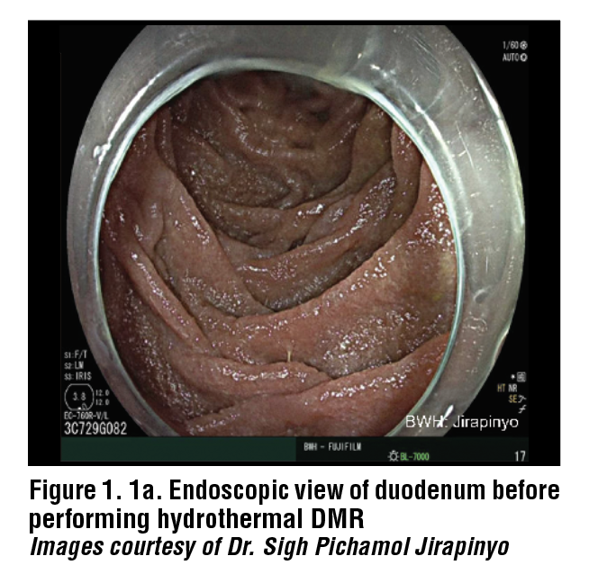
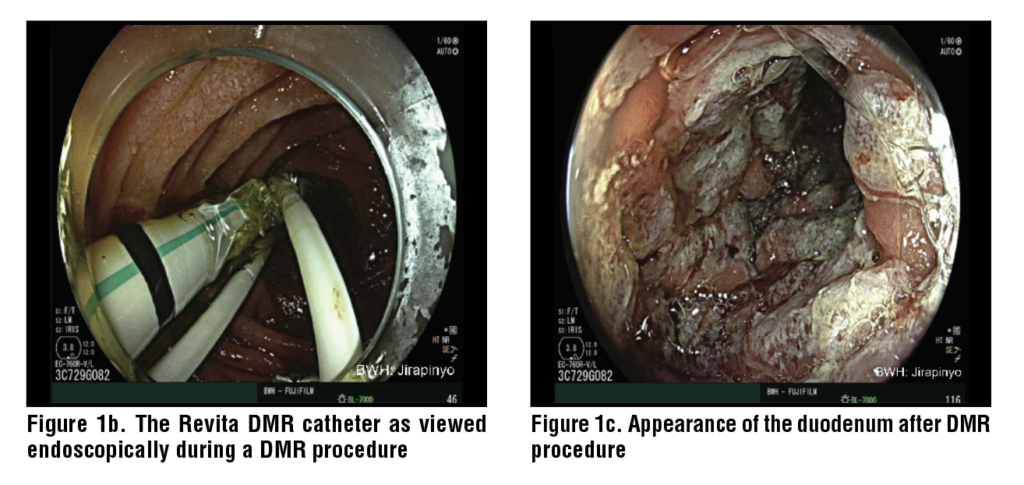
Hydrothermal DMR starts with the injection of a saline solution into the duodenal submucosa to lift the submucosa. This provides a barrier to prevent damage to the underlying muscularis propria and creates an even surface to ablate. Hydrothermal DMR uses a Revita catheter (Fractyl Health, Inc, Burlington, MA, USA) that has three submucosal injectors and a 2 cm balloon on the end. The balloon uses heated water to ablate the surface of the mucosa. The water inside the balloon reaches 80°C and ablates the surface of the mucosa for 10 seconds. The mucosa only requires one ablation before being advanced to the next segment of the duodenum until 10 cm of the duodenum has been treated. The ablation induces coagulative necrosis in the mucosal cells which form the most superficial 0.6mm of the duodenal wall. The muscularis propria lies at 1.0mm depth, and the ablation is not strong enough to disrupt that layer. The treatment is performed distal to the major duodenal papilla to ensure its safety. (Figure 1)

Prior to performing DMR, an upper endoscopy is required to evaluate the patient’s anatomy and exclude diseases of the mucosa such as duodenitis with ulcers, celiac disease, etc. Other conditions that may be contraindications to DMR include strictures, varices, and gastroduodenal ulcers, in addition to those previously mentioned. It is recommended that the wall of the duodenum be marked contralaterally to the major duodenal papilla to provide a proximal margin for treatment. A 0.035” guidewire should be passed to the proximal jejunum to act as a guide for the DMR catheter. Fluoroscopy can be utilized to ensure that the placement of the guidewire is correct. The Revita catheter can be placed over the guidewire and positioned just distal to the major duodenal papilla. For direct visualization during the procedure, an endoscope can be positioned in the duodenum just proximal to the balloon on the catheter. The gross appearance of the duodenum should be evaluated following the procedure to ensure the entire segment has been ablated. There are five ablation cycles to complete one treatment of 10cm of the duodenum.  

Following the procedure, patients are generally discharged the same day or after an overnight hospitalization. Patients are prescribed a diet after the procedure that slowly advances from clear liquids to solid foods over two weeks during healing of the mucosa. After the two-week diet progression, patients should be counseled on important dietary changes that can help manage glucose control.

The ideal candidate for DMR is between age 28 and 75 with type 2 diabetes and a HbA1c between 7.5% and 10%, a body mass index (BMI) between 24-40 kg/m2 and preserved intrinsic insulin production. There are several contraindications for DMR including type 1 diabetes, low endogenous insulin production, previous gastrointestinal surgery that prohibits access to the duodenum, history of acute and chronic pancreatitis, history of duodenal inflammatory disease such as Crohn’s or Celiac disease, or anticoagulant therapy that cannot be discontinued. It is important that endogenous insulin production is maintained because the goal of DMR therapy is to improve insulin sensitivity, and those without endogenous insulin production may not benefit from the procedure.

Laser Duodenal Ablation

An emerging alternative to Hydrothermal DMR is Laser Duodenal Ablation using the Digma System (Digma Medical Ltd., Givat Shmuel, Israel). The Digma system consists of a control console and a single use catheter that is compatible with the working channel greater than 3.7 mm. The catheter uses a polyethylene terephthalate (PET) balloon that inflates to distend the duodenum. A continuous wave 5-15 W laser then delivers a focused beam to the duodenum, ablating 6 cm of the mucosa circumferentially per ablation cycle. The treatment starts at the duodenojejunal flexure and moves proximal, with up to seven ablations in three to four positions, ablating a total of 24 cm of the duodenal mucosa.

Irreversible Electroporation (IRE) Mucosal Ablation

Irreversible Electroporation (IRE) is an alternative to thermal ablation and uses pulsed electrical fields to create pores in cell membranes and induce apoptosis. The pulsed electrical fields of IRE also induce the renewal of the duodenal mucosa. The combination therapy is called recellularization via electroporation therapy (ReCET), performed using the Endogenex system (Endogenex Inc, Plymouth, Minnesota, USA). To perform ReCET, a guidewire is placed into the jejunum endoscopically. The position is confirmed using fluoroscopy and then the Endogenex catheter is introduced over the guidewire to the 2nd section of the duodenum. The ablation is performed proximally to distally, starting just distal to the major duodenal papilla. The catheter ablates 2 cm and two-thirds of the circumference of the duodenum at a time. The total segment of the duodenum that is treated is 10 cm.

Mechanism of Action

DMR ablates the mucosa and is thought to provide a reset for the signaling of enterocytes back to a healthier phenotype. This procedure allows for regrowth of the mucosa with minimal scarring and has shown significant decreases in HbA1c and blood glucose at follow-up.12 The mechanism of resulting decrease in HbA1c and blood glucose is uncertain, but is thought to be related to changes in enteroendocrine signaling. In follow-up histological evaluation, there was no change in the density of L and K cells after DMR, which are a key source of GLP-1 and GIP.13 This could mean that the mechanism of DMR alters hormonal signaling or cellular function rather than enteroendocrine cell number. There do not appear to be any structural changes to the mucosa, suggesting that the metabolic benefits of DMR are not mediated by structural damage or by inflammation.13

Western blot molecular analysis showed increased expression of PDZK1 and GATA6, which are involved in epithelial differentiation and nutrient signaling.13 This supports the hypothesis that DMR provides a reset for the duodenal mucosa which improves glucose homeostasis and downstream metabolic signaling. 

Following DMR, there are significant decreases in insulin, glucagon and C-peptide levels in the blood. There are also significant decreases in postprandial glucose and glucagon concentrations.12 

DMR is being investigated as a potential adjunctive therapy for non-alcoholic steatohepatitis (NASH) as there are limited treatment options beyond lifestyle modifications and pharmacologic therapy with agents such as GLP-1 agonists. Bariatric surgery is an option for some patients with NASH, but there are higher levels of surgical complications and morbidity in patients with cirrhosis, most of whom would be denied surgical therapy on these grounds. Current studies indicate there is improved glycemic control following DMR, but further studies are needed to show if there is a benefit in NASH.14

Combination Therapy with GLP-1 Receptor Agonists

With DMR acting locally to increase insulin levels in the blood and GLP-1 receptor agonists acting systemically to increase insulin secretion, the combination has potential to reduce insulin dependance and enhance glycemic control in patients with type 2 diabetes.2, 12  Early clinical studies have combined hydrothermal duodenal ablation with liraglutide or irreversible electroporation with semaglutide. All three studies included patients between ages 28 and 75 with type 2 diabetes on basal insulin with an HbA1c less than 8.0%.13,, The primary endpoint of all studies was the number of patients following DMR that were able to stay off of basal insulin with adequate glycemic control defined as an HbA1c of less than 7.5%. Of the 16 patients that underwent hydrothermal DMR with liraglutide, 11 of them (69%) had adequate control at 6 month follow up.16,17 At 18 month follow up, 8 patients (53%) remained off insulin therapy.16 A total of 14 patients underwent IRE with a range of electroporation doses from one 600V treatment to two 750V treatments.13 At 12 month follow up, 12 patients (86%) did not require insulin therapy, with the two patients needing basal insulin having only one 600V dose of electroporation.13 

Safety and Adverse Events

Overall Safety Profile in Trials

DMR is generally well tolerated, with studies reporting mostly mild adverse events including nausea and abdominal pain following the procedure.7,10 In animal studies, there were no adverse events following the procedure, with only a mild inflammatory response noted in the duodenal mucosa. Histologically, the mucosal healing process was completed within six weeks. Some animals displayed increased collagen deposition in the submucosa of the treated region, but they displayed no functional limitations.7

In the first in human study of DMR with 29 patients, there were no adverse events reported during the procedure. Postprocedural adverse events were limited, with three patients out of the 29 experiencing duodenal stenosis following the ablation, that were treated using endoscopic dilation. The duodenal stenosis was determined to be caused by inadequate submucosal lift.7 There was complete regrowth of the mucosa observed at three months following the procedure in all 29 patients. There were 11 patients out of 29 that had evidence of low-to-intermediate fibrosis.

In follow up clinic visits, there were transient and mild adverse events immediately following the procedure, most commonly abdominal pain, diarrhea, hyperglycemia, hypoglycemia, nasopharyngitis, and headache.10 On follow-up endoscopy 30 days after the procedure, the duodenum exhibited complete healing. After 30 days of healing, the most common adverse events were hypoglycemia and abdominal pain.10  There were no reported duodenal strictures and one instance of jejunal perforation that was caused by manipulation of the endoscope.10  There were also no clinical signs of pancreatitis, malabsorption, anemia, or infection.10 

Long-Term Safety

Long-term safety profiles are still being developed, as DMR is an emerging/experimental procedure. After two years following DMR, trials indicate that the procedure was well tolerated with no device or procedure related serious adverse effects. Of the 46 patients that were followed, there were two reported adverse events that could have been linked to the procedure including one patient with constipation and one patient with general malaise and vitamin B12 deficiency.15  A larger sample size and a longer follow-up will be necessary, as data is very limited. 

Comparative and Alternative Therapies

Bariatric Operations and Foregut Surgeries

Roux-en-Y gastric bypass and sleeve gastrectomy are bariatric surgical procedures that improve glycemic control and decrease weight in patients with type 2 diabetes and obesity. A Roux-en-Y gastric bypass is a restrictive and malabsorptive bariatric surgical procedure than involves bypassing the majority of the stomach and the whole duodenum to reduce food intake to promote weight loss., A sleeve gastrectomy involves removing 80% of the stomach along the greater curvature to promote early satiety and decrease the release of the hormone ghrelin that stimulates hunger signals.

Bariatric surgeries such as the Roux-en-Y gastric bypass and sleeve gastrectomy are indicated for people with a BMI of 35 kg/m2 or greater or a BMI of 30 kg/m2 with type 2 diabetes.18  Gastric bypass has been shown to provide long-term control of type 2 diabetes with decreased antidiabetic medicine use and higher rates of type 2 diabetes remission. There is a possibility of severe adverse events following gastric bypass surgery including bleeding, infection, and bowel leaks following the procedure. There can also be long-term issues afterwards such as dumping syndrome, nutritional deficiencies, gallstones, ulcers, and strictures. 

While DMR aims to control blood glucose, Roux-en-Y gastric bypass and sleeve gastrectomy provide glycemic control while also controlling weight. Roux-en-Y gastric bypass causes greater weight loss than sleeve gastrectomy, but they provide similar improvements in glycemic control.16  Roux-en-Y gastric bypass can also help control symptoms of acid reflux. Both procedures encourage caloric restriction which aids in glycemic control, but they also alter nutrient flow, absorption, and incretin hormone release.16 Altered nutrient flow and incretin hormone release are also thought to be the basis for DMR.13

Pharmacologic Therapies

Pharmacologic therapies to provide glycemic control in patients with type 2 diabetes are alternatives or supplements to DMR that act through a variety of metabolic pathways. GLP-1 receptor agonists such as semaglutide and GLP-1/GIP agonists such as tirzepatide mimic the naturally occurring incretin hormone GLP-1 that is released from L-cells and GIP released from K-cells. Semaglutide and tirzepatide augment insulin and glucagon secretion while also slowing gastric emptying and decreasing caloric intake. 

Slowed gastric emptying and decreased caloric intake result in decreased HbA1c and significant weight loss.22  These medications augment the same hormonal pathways that are hypothesized to decrease HbA1c levels in DMR.13 Both bariatric surgery and pharmacologic treatment cause weight loss, while weight changes following DMR have been modest and not always statistically significant.10,  There is variability between cohorts and further studies would need to be completed to quantify the weight changes following DMR. It seems likely that, if widely implemented, DMR would often be used alongside GLP-1 agents. 

Conclusion: Evidence Gaps, Limitations, and Research Priorities

As DMR is still an emerging and, to some extent, experimental procedure, clinical trial data is extremely limited. There is a need for trials in more patients to establish significant safety data for the procedure as well as examine the outcomes of a larger cohort. Trials have had varied inclusion criteria including HbA1c range, insulin use, and BMI. By standardizing the patient population, it allows for greater pooling of the data and comparison of results between trials. 

Establishing long term safety and efficacy of DMR is difficult with trials for long term follow-up limited to 2 years and cohorts of less than 50 patients. Additional long-term follow-up data need to become available, and additional studies need to be done to increase the sample size. 

The significant uncertainty about the mechanism of DMR needs to be investigated. By understanding the exact mechanism by which DMR improves metabolic outcomes, the procedure can be refined to be as least invasive as possible as well as more targeted. This can help determine the ideal candidate, the most effective treatment length as well as treatment frequency. Long term adverse events and metabolic changes need to be further elucidated.

Head-to-head comparisons between hydrothermal ablation and IRE have not been performed at this time but having trials of this type would help to clarify safety profiles of each modality. This could help direct further device development and provide future guidelines for treatment. 

References

1. Musso G, Cassader M, Gambino R. Endoscopic duodenal mucosa
ablation: the future of diabetes treatment? Trends Mol Med. 2024
Jul;30(7):612-616. doi: 10.1016/j.molmed.2024.03.003. Epub
2024 Mar 29. PMID: 38553333.
2. Parikh A, Thevenin C. Physiology, Gastrointestinal Hormonal
Control. [Updated 2023 May 1]. In: StatPearls [Internet]. Treasure
Island (FL): StatPearls Publishing; 2025 Jan-. Available from:
https://www.ncbi.nlm.nih.gov/books/NBK537284/#
3. Rønnestad I, Akiba Y, Kaji I, Kaunitz JD. Duodenal luminal
nutrient sensing. Curr Opin Pharmacol. 2014 Dec;19:67-75. doi:
10.1016/j.coph.2014.07.010. Epub 2014 Aug 9. PMID: 25113991;
PMCID: PMC4845631.
4. Breen DM, Rasmussen BA, Côté CD, Jackson VM, Lam TK.
Nutrient-sensing mechanisms in the gut as therapeutic targets for
diabetes. Diabetes. 2013 Sep;62(9):3005-13. doi: 10.2337/db13-
0523. PMID: 23970519; PMCID: PMC3749331.
5. Holesh JE, Aslam S, Martin A. Physiology, Carbohydrates.
[Updated 2023 May 12]. In: StatPearls [Internet]. Treasure Island
(FL): StatPearls Publishing; 2025 Jan-. Available from: https://
www.ncbi.nlm.nih.gov/books/NBK459280/#
6. Nakrani MN, Wineland RH, Anjum F. Physiology, Glucose
Metabolism. [Updated 2023 Jul 17]. In: StatPearls [Internet].
Treasure Island (FL): StatPearls Publishing; 2025 Jan-. Available
from: https://www.ncbi.nlm.nih.gov/books/NBK560599/
7. Habegger KM, Al-Massadi O, Heppner KM, Myronovych A,
Holland J, Berger J, Yi CX, Gao Y, Lehti M, Ottaway N, Amburgy
S, Raver C, Müller TD, Pfluger PT, Kohli R, Perez-Tilve D,
Seeley RJ, Tschöp MH. Duodenal nutrient exclusion improves
metabolic syndrome and stimulates villus hyperplasia. Gut. 2014
Aug;63(8):1238-46. doi: 10.1136/gutjnl-2013-304583. Epub 2013
Oct 9. PMID: 24107591; PMCID: PMC3981953.
8. Haidry RJ, van Baar AC, Galvao Neto MP, Rajagopalan H, Caplan
J, Levin PS, Bergman JJ, Rodriguez L, Deviere J, Thompson
CC. Duodenal mucosal resurfacing: proof-of-concept, procedural
development, and initial implementation in the clinical setting.
Gastrointest Endosc. 2019 Oct;90(4):673-681.e2. doi: 10.1016/j.
gie.2019.03.024. Epub 2019 Mar 29. PMID: 30935932
9. van Baar ACG, Haidry R, Rodriguez Grunert L, Galvao MPN,
Bisschops R, Hayee BH, Costamagna G, Deviere J, Bergman
JJGHM. Duodenal mucosal resurfacing: Multicenter experience
implementing a minimally invasive endoscopic procedure for
treatment of type 2 diabetes mellitus. Endosc Int Open. 2020
Nov;8(11):E1683-E1689. doi: 10.1055/a-1244-2283. Epub 2020
Oct 22. PMID: 33140025; PMCID: PMC7581486.
10. van Baar ACG, Holleman F, Crenier L, Haidry R, Magee C,
Hopkins D, Rodriguez Grunert L, Galvao Neto M, Vignolo
P, Hayee B, Mertens A, Bisschops R, Tijssen J, Nieuwdorp
M, Guidone C, Costamagna G, Devière J, Bergman JJGHM.
Endoscopic duodenal mucosal resurfacing for the treatment of
type 2 diabetes mellitus: one year results from the first international,
open-label, prospective, multicentre study. Gut. 2020
Feb;69(2):295-303. doi: 10.1136/gutjnl-2019-318349. Epub 2019
Jul 22. PMID: 31331994; PMCID: PMC6984054
11. Mingrone G, van Baar AC, Devière J, Hopkins D, Moura
E, Cercato C, Rajagopalan H, Lopez-Talavera JC, White K,
Bhambhani V, Costamagna G, Haidry R, Grecco E, Galvao Neto
M, Aithal G, Repici A, Hayee B, Haji A, Morris AJ, Bisschops
R, Chouhan MD, Sakai NS, Bhatt DL, Sanyal AJ, Bergman
JJGHM; Investigators of the REVITA-2 Study. Safety and efficacy
of hydrothermal duodenal mucosal resurfacing in patients with
type 2 diabetes: the randomised, double-blind, sham-controlled,
multicentre REVITA-2 feasibility trial. Gut. 2022 Feb;71(2):254-
264. doi: 10.1136/gutjnl-2020-323608. Epub 2021 Feb 17. PMID:
33597157; PMCID: PMC8761999.
12. Kovarova V, Lankova I, Machytka E, Knotkova K, Kratochvílová
H, Beneš M, Spicak J, Vasura A, Goldin E, Munter G, Zima
T, Mraz M, Dagan H, Levy B, Haluzik M, Kral J. Duodenal
Laser Ablation for Treatment of Type 2 Diabetes: Results of
First in Human Study. United European Gastroenterol J. 2025
Jun;13(5):750-758. doi: 10.1002/ueg2.12762. Epub 2025 Feb 20.
PMID: 39976927; PMCID: PMC12188360.
13. Busch CBE, Meiring S, van Baar ACG, Holleman F, Nieuwdorp
M, Bergman JJGHM. Recellularization via electroporation therapy
of the duodenum combined with glucagon-like peptide-1 receptor
agonist to replace insulin therapy in patients with type 2 diabetes:
12-month results of a first-in-human study. Gastrointest Endosc.
2024 Nov;100(5):896-904. doi: 10.1016/j.gie.2024.04.2904. Epub
2024 Apr 29. PMID: 38692517.
14. Meiring S, Aydin Ö, van Baar ACG, van der Vossen EWJ,
Rampanelli E, van Grieken NCT, Holleman F, Nieuwdorp
M, Bergman JJGHM. From Endoscopic Inspection to Gene-
Expression: A Thorough Assessment of the Duodenal Mucosa
After Resurfacing-A Prospective Study. Dig Dis Sci. 2025
Mar;70(3):1052-1063. doi: 10.1007/s10620-024-08710-4. Epub
2025 Jan 8. PMID: 39779586; PMCID: PMC11920325.
15. Chuang TJ, Ko CW, Shiu SI. The metabolic influence of duodenal
mucosal resurfacing for nonalcoholic fatty liver disease.
Medicine (Baltimore). 2023 Oct 6;102(40):e35147. doi:
10.1097/MD.0000000000035147. PMID: 37800801; PMCID:
PMC10553053.
16. Meiring S, Busch CBE, van Baar ACG, Hemke R, Holleman
F, Nieuwdorp M, Bergman JJGHM. Eliminating exogenous
insulin therapy in patients with type 2 diabetes by duodenal
ablation and GLP-1RA decreases risk scores for cardiovascular
events. Cardiovasc Diabetol. 2022 Sep 22;21(1):191. doi: 10.1186/
s12933-022-01628-z. PMID: 36138441; PMCID: PMC9503196.
17. van Baar ACG, Meiring S, Smeele P, Vriend T, Holleman F,
Barlag M, Mostafavi N, Tijssen JGP, Soeters MR, Nieuwdorp M,
Bergman JJGHM. Duodenal mucosal resurfacing combined with
glucagon-like peptide-1 receptor agonism to discontinue insulin
in type 2 diabetes: a feasibility study. Gastrointest Endosc. 2021
Jul;94(1):111-120.e3. doi: 10.1016/j.gie.2020.12.021. Epub 2020
Dec 24. PMID: 33359437.
18. van Baar ACG, Devière J, Hopkins D, Crenier L, Holleman F,
Galvão Neto MP, Becerra P, Vignolo P, Rodriguez Grunert L,
Mingrone G, Costamagna G, Nieuwdorp M, Guidone C, Haidry RJ,
Hayee B, Magee C, Carlos Lopez-Talavera J, White K, Bhambhani
V, Cozzi E, Rajagopalan H, J G H M Bergman J. Durable metabolic
improvements 2 years after duodenal mucosal resurfacing
(DMR) in patients with type 2 diabetes (REVITA-1 Study).
Diabetes Res Clin Pract. 2022 Feb;184:109194. doi: 10.1016/j.
diabres.2022.109194. Epub 2022 Jan 13. PMID: 35032562.
19. Wallenius V, Dirinck E, Fändriks L, Maleckas A, le Roux CW,
Thorell A. Glycemic Control after Sleeve Gastrectomy and Roux-
En-Y Gastric Bypass in Obese Subjects with Type 2 Diabetes
Mellitus. Obes Surg. 2018 Jun;28(6):1461-1472. doi: 10.1007/
s11695-017-3061-3. PMID: 29264780; PMCID: PMC5973990.
20. Umeda LM, Silva EA, Carneiro G, Arasaki CH, Geloneze B,
Zanella MT. Early improvement in glycemic control after bariatric
surgery and its relationships with insulin, GLP-1, and glucagon
secretion in type 2 diabetic patients. Obes Surg 2011;21:896–901
21. Mitchell BG, Collier SA, Gupta N. Roux-en-Y Gastric Bypass.
[Updated 2024 Nov 9]. In: StatPearls [Internet]. Treasure Island
(FL): StatPearls Publishing; 2025 Jan-. Available from: https://
www.ncbi.nlm.nih.gov/books/NBK553157/
22. Seeras K, Sankararaman S, Lopez PP. Sleeve Gastrectomy.
[Updated 2023 Jan 23]. In: StatPearls [Internet]. Treasure Island
(FL): StatPearls Publishing; 2025 Jan-. Available from: https://
www.ncbi.nlm.nih.gov/books/NBK519035/
23. Pories WJ, Swanson MS, MacDonald KG, Long SB, Morris PG,
Brown BM, Barakat HA, deRamon RA, Israel G, Dolezal JM, et
al. Who would have thought it? An operation proves to be the most
effective therapy for adult-onset diabetes mellitus. Ann Surg. 1995
Sep;222(3):339-50; discussion 350-2. doi: 10.1097/00000658-
199509000-00011. PMID: 7677463; PMCID: PMC1234815.
24. Seeras K, Acho RJ, Lopez PP. Roux-en-Y Gastric Bypass Chronic
Complications. [Updated 2023 Jun 5]. In: StatPearls [Internet].
Treasure Island (FL): StatPearls Publishing; 2025 Jan-. Available
from: https://www.ncbi.nlm.nih.gov/books/NBK519489/#
25. Nauck MA, Quast DR, Wefers J, Meier JJ. GLP-1 receptor agonists
in the treatment of type 2 diabetes – state-of-the-art. Mol
Metab. 2021 Apr;46:101102. doi: 10.1016/j.molmet.2020.101102.
Epub 2020 Oct 14. PMID: 33068776; PMCID: PMC8085572.
26. Rajagopalan H, Cherrington AD, Thompson CC, Kaplan LM,
Rubino F, Mingrone G, Becerra P, Rodriguez P, Vignolo P, Caplan
J, Rodriguez L, Galvao Neto MP. Endoscopic Duodenal Mucosal
Resurfacing for the Treatment of Type 2 Diabetes: 6-Month
Interim Analysis From the First-in-Human Proof-of-Concept
Study. Diabetes Care. 2016 Dec;39(12):2254-2261. doi: 10.2337/
dc16-0383. Epub 2016 Aug 12. PMID: 27519448.

Download Tables, Images & References

from the pediatric literature

Capsule Endoscopy Yield in Pediatric Patients

Read Article

Video capsule endoscopy (VCE) uses a small, wireless, capsule camera that is swallowed or placed endoscopically to evaluate the entirety of the gastrointestinal (GI) tract often beyond the reach of standard endoscopy. This study evaluated the efficacy of VCE in making a diagnosis as well as assisting in potentially changing therapy in children with GI conditions.

Data from this retrospective study consisted of all VCE studies performed at one tertiary children’s hospital in the United States from 2004 to 2022. The VCE equipment utilized consisted of the Rapid™ PillCam Reader (Medtronic).  VCE studies were considered incomplete if the small bowel could not be visualized.  VCE studies performed in patients greater than 18 years of age were excluded. Patient data associated with each study was compiled including past medical history, VCE indication, type of VCE deployment, and study results. VCE studies were split into study indications which included inflammatory bowel disease (IBD), GI bleeding, anemia, polyposis syndromes, protein-losing enteropathy, abdominal pain, and “other” (diarrhea, nausea, emesis, weight loss, constipation). VCE results were classified as positive or negative based on findings although normal findings were not considered positive.

In total, 478 VCE studies were completed successfully in 427 patients. The mean age of the patient study group was 12 years (range 10 months to 18 years) with 58% of the study patients being male.  256 patients (54%) swallowed the capsule while 222 patients (46%) had endoscopic placement of the capsule. Positive findings were present in 245 studies (51.3%), and 169 studies (35.4%) led to changes in therapy or diagnostic planning.

When VCE was performed for IBD (153 studies), 81 studies (52.9%) had positive findings with 62 studies (40.5%) leading to a change in therapy or diagnostic planning. When VCE was performed for GI bleeding (114 studies), 54 studies (46.9%) had positive findings with 43 studies (36.8%) leading to a change in therapy or diagnostic planning. When VCE was performed for anemia (84 studies), 51 studies (62.2%) had positive findings with 32 studies (36.8%) leading to a change in therapy or diagnostic planning.  When VCE was performed for polyposis syndromes (48 studies), 29 studies (60.4%) had positive findings with 13 studies (27.1%) leading to a change in therapy or diagnostic planning. When VCE was performed for abdominal pain (41 studies), 10 studies (24.4%) had positive findings with 6 studies (14.6%) leading to a change in therapy or diagnostic planning. When VCE was performed for protein-losing enteropathy (14 studies), 8 studies (57.1%) had positive findings with 7 studies (50%) leading to a change in therapy or diagnostic planning. When VCE was performed for other reasons (24 studies), 12 studies (50%) had positive findings with 6 studies (25%) leading to a change in therapy or diagnostic planning. It should be noted that 61 studies (12.8%) found possible disease states outside of the small intestine, including findings present in the esophagus, stomach, and colon.

Statistical analysis demonstrated that all indications for VCE were significantly associated with positive findings or subsequent changs in therapy and diagnostic planning except for the indication of abdominal pain without other features. All the indications for VCE except for abdominal pain had no significant difference between them regarding their rate of positive findings or the subsequent changes in therapy and diagnostic planning.

This study suggests that VCE is an excellent diagnostic tool for finding small bowel disease possibly leading to therapy changes in children except for the indication of abdominal pain without other symptoms. This study should persuade clinicians to not perform VCE in most cases of abdominal pain children who do not have other symptoms or diseases such as GI bleeding or inflammatory bowel disease.

Kaihlanen K, Zhang S, Phen C, Rojas I.  Clinical impact and diagnostic yield of small bowel capsule endoscopy in children. Journal of Pediatric Gastroenterology and Nutrition 2026; 82: 389-397.

Comparing pH-Impedance Monitoring with Barium Esophagram Results for Children with Reflux

Gastroesophageal reflux (GER) is often visualized when children undergo a barium esophagram or upper gastrointestinal (UGI) barium study. GER in this setting is typically considered a false positive finding, and the authors of this study evaluated the association of true reflux with such findings on barium studies.

All pediatric patients who underwent pH-impedance monitoring over a 4-year retrospective period at a tertiary children’s hospital in the United States were included in the study if they had undergone a barium study of the esophagus which included barium esophagrams or UGI barium studies. The barium studies and pH-impedance study had to occur within 1 year of each other.  Information including patient demographics, medical history, medications, potential biopsies obtained during upper endoscopy, and potential findings on esophageal manometry were collected. The Lyon Consensus 2.0 criteria (see https://gut.bmj.com/content/gutjnl/73/2/361.full.pdf) were used to diagnose GER or gastroesophageal reflux disease (GERD) by pH-impedance monitoring for patients off acid suppression medication.

A total of 90 children (median age 10 years, 56.7% female) with potential GER qualified for the study. The most common indications for testing were emesis / regurgitation (75.6%), chronic cough (37.8%), and heartburn (35.6%). No difference in upper endoscopy and esophageal manometry findings were present in patients with or without reflux noted by barium or by pH-impedance monitoring.

Patients with pH-impedance studies completed both on and off acid suppression medication and who also had diagnostic criteria positive for GERD based on acid exposure had no significant correlation with the presence or absence of reflux noted on barium studies. No significant correlation was seen between reflux determined by esophageal pH from pH-impedance monitoring or by reflux seen by barium studies in patients both on and off acid suppression medication regardless of age, median body mass index, clinical symptoms, and time off acid suppression medication for patients. The overall sensitivity and specificity for diagnosing GERD by barium study compared to pH-impedance monitoring was 33.3% and 44.9%, respectively. When considering only those patients off acid suppression medication, the sensitivity and specificity were 31.3% and 54.3%, respectively. The positive predictive value and the negative predictive value of diagnosing GERD by barium in the setting of a positive GERD seen by pH-impedance monitoring was 23.8% and 63.3%, respectively. Ten of the study patients had hiatal hernias, and no significant correlation was found between the presence and absence of reflux noted on barium studies and esophageal acid exposure identified by pH-impedance monitoring in this specific patient group.

This study confirms again UGI barium studies and barium esophagrams should not be used to diagnose true GERD. Barium studies are very helpful in diagnosing UGI anatomic abnormalities, but the diagnosis of GERD in children should be based on pH-impedance monitoring.

Davis T, Rogers B, Bhardwaj R, Gyawali C. Diagnostic value of barium oesophagram compared to pH-impedance monitoring in the detection of paediatric gastro-oesophageal reflux.  Archives of Diseases in Childhood. 2026; 111: 334-338.

Download Tables, Images & References

Dispatches from the GUILD Conference, Series #73

Prevention and Management of Postoperative Crohn’s Disease

Read Article

Postoperative recurrent Crohn’s disease is common and often clinically silent at onset, requiring objective assessments for diagnosis and surveillance. Patients with a history of multiple bowel resections, penetrating disease, or who smoke cigarettes after surgery are at highest risk for disease recurrence. Antibiotics, aminosalicylates, and immunomodulators modestly reduce the risk of clinical disease recurrence. In contrast, monoclonal antibodies, specifically anti-tumor necrosis factor (TNF) medications as well as the anti-integrin agent vedolizumab, are effective at suppressing disease recurrence and may have the potential to alter the natural course of disease after surgery. In this manuscript, the management of postoperative Crohn’s disease is summarized, and a simplified approach to prevention, monitoring, and treatment is provided.

Risk and Diagnosis of Postoperative Crohn’s Disease 

Despite significant medical therapeutic advances, as many as 20-30% Crohn’s disease (CD) patients require bowel surgery, mostly commonly for stricturing or penetrating complications, e.g., fistula, intraabdominal abscess. Surgery is not curative of CD, and postoperative recurrence (POR) of CD occurs in the majority of patients. In the pre-biologic era (circa 2000), natural history studies found that 70–90% of CD patients developed endoscopic evidence of disease activity within 1 year of their surgery, and that 30–60% of postoperative CD patients became symptomatic from recurrent disease within 3–5 years of their surgery. Consequently, up to 50% of postoperative CD patients in the pre-biologic era required repeat surgery within 5 years of their first surgery. 

Postoperative CD recurrence is often clinically silent with only 1/3 of patients reporting symptoms of CD despite endoscopic disease activity in the majority postoperatively. Thus, relying on symptoms postoperatively significantly underestimates mucosal disease activity and may miss an opportunity for disease interception. Fortunately, with increased utilization of postoperative management strategies, more modern estimates suggest the risk of recurrence is diminishing.

The degree of endoscopic disease activity correlates with subsequent progression to symptomatic recurrence. Thus, ileocolonoscopy is the current gold standard for POR assessment and is recommended to be performed within 6-12 months postoperatively. More delayed endoscopic assessments are associated with worse clinical outcomes. Disease activity is traditionally estimated using the Rutgeerts score, which defines activity on a 0 (normal) to 4 (severe) scale based on the extent of aphthous ulcerations in the neoterminal ileum. The more severe the endoscopic recurrence, e.g. i3 or i4, the more likely the development of clinical symptoms, i.e. clinical recurrence, and requirement for future surgery, i.e. surgical recurrence.  

Table 1. Summary of Gastroenterological Society Definitions of High-Risk Features for Postoperative Crohn’s Disease Recurrence.

American College of Gastroenterology (ACG)American Gastroenterological Association (AGA) European Crohn’s and Colitis Organization (ECCO) British Society of Gastroenterology (BSG) 
Classified as high risk with ≥ 1:
• Active tobacco use (especially in women and heavy smokers)
• ≥2 prior surgeries
• presence of penetrating disease*
Classified as high risk with ≥ 1:
• Active tobacco use
• ≥2 prior surgeries for penetrating disease, with or without perianal disease
• Age <30 years
Classified as high risk with ≥ 1:
• Smoking
• ≥ 1 prior intestinal surgery
• Penetrating disease at index surgery
• Perianal location
• Granulomas or myenteric plexitis in
• resection specimen
• Extensive small bowel resection (>50 cm)

2023 ECCO workshop includes:
• Immune-mediated inflammatory diseases
• Extra-intestinal manifestations
• Prior colon involvement
Classified as high risk if actively smoking or with ≥ 2:
• Multiple resections
• Preoperative penetrating disease behavior Perianal disease
• Granulomas in resection specimen
• Myenteric plexitis in the proximal resection margin
• Extensive bowel disease (>50 cm)
*Penetrating disease classified as fistulas, abscesses, intestinal perforations


Though ileocolonoscopy is sensitive at detecting POR, the invasive nature of the test is associated with patient discomfort, high cost, and procedural risk. Thus, noninvasive assessments are of particular interest. Fecal calprotectin (FCP) levels, produced by gastrointestinal leukocytes and epithelial cells at sites of mucosal injury and inflammation, correlate with endoscopic recurrence while normal levels have a strong negative predictive value. Based on available data, FCP cutoffs between 100-150 ug/g have been proposed, identifying endoscopic recurrence with 70-89% sensitivity, 58-69% specificity, and negative predictive values > 90%. Consequently, the American Gastroenterological Association (AGA) now advocates for fecal calprotectin-guided surveillance strategies as a cost-effective alternative to universal endoscopic evaluation in lower-risk patients within the first year postoperatively.1 AGA guidelines recommend assessing FCP between 6 and 12 months postoperatively. Earlier assessments are being explored.  

Imaging modalities including computed tomography (CT) and magnetic resonance (MR) have both shown acceptable correlation to endoscopic disease activity, though they have disadvantages of cost, poorly tolerated oral contrast, radiation (CT), and limited availability (MR). Intestinal ultrasound (IUS) has emerged as a promising radiation-free, cost-effective point-of-care imaging modality capable of detecting POR with comparable accuracy to ileocolonoscopy in expert hands.2 Combining FCP assessments with IUS can further improve test characteristics. Consequently, postoperative monitoring algorithms are increasingly incorporating these non-invasive tools when available.  

Risk Factors for Postoperative Recurrence

Factors associated with POR include clinical, disease, surgical, histologic, microbiotic, and molecular characteristics. Active smoking after surgery doubles the risk of endoscopic, clinical, and surgical recurrence and smoking cessation can reduce recurrence rates. Younger age at disease onset and rapid progression (<10 years) to surgical resection may increase recurrence risk. A history of prior surgical resections for Crohn’s may impart the strongest risk for future POR.3,4 Penetrating disease behavior (fistula, abscess) at the time of surgery is associated with increased clinical and surgical recurrence. 

Surgical approach and anastomosis technique may influence POR. These include more extensive surgical resections of the mesentery as well as novel anastomotic orientations such as the Kono-S configuration. Such approaches are being explored in randomized trials. Furthermore, histologic findings in the resection specimen including presence of granulomas, myenteric and submucosal plexitis, and positive surgical margins may identify individuals at increased risk for POR.5,6

Table 2. Efficacy of various therapies and knowledge gaps for the prevention of postoperative Crohn’s disease recurrence.

Therapy/InterventionPostoperative Recurrence Prevention Relative Efficacy
Curcumin
Vitamin D 
Enteral Nutrition+
Probiotics
Nitroimidazole/Antibiotics+
Mesalamine
Budesonide
Thiopurines+
Anti-TNF+++
Vedolizumab++
Ustekinumab+
Anti-IL-23s?
Janus kinase inhibitors?

Microbiome, serologic, genetic, and other “-omics” signatures have been described in individuals who progress to POR, but data remains inconclusive for routine clinical care at the current time.

Risk stratification has been adopted in various gastroenterological societal guidelines (Table 1). Generally, patients at high risk for recurrence include those who are actively smoking, multiple prior surgical resections, and penetrating disease behavior, with or without perianal disease. Such risk stratification may help identify patients warranting more aggressive treatment and monitoring after surgery.

Medical Prophylaxis for Preventing Postoperative Crohn’s Disease

Conventional medical therapies including antibiotics (e.g. metronidazole), aminosalicylates, and immunomodulators modestly reduce the risk of clinical disease recurrence and may be considered particularly in resource-limited settings; however, comparative analyses suggest that more novel targeted therapies are more efficacious for this purpose (Table 2).7 

Anti-tumor necrosis factor (TNF) therapy postoperatively prevents endoscopic POR and may have the potential to change the natural course of Crohn’s disease after surgery. The seminal PREVENT trial demonstrated that infliximab when used prophylactically (within 2-4 weeks postoperatively) in individuals at high risk for recurrence significantly reduced endoscopic recurrence at week 76 compared to placebo (22.4% vs. 51.3%, P < 0.001), although not clinical recurrence (12.9% vs 20.0%, P=0.097).8 This protective effect appears to extend to other anti-TNFs as adalimumab has also been found to prevent POR in several studies.9-11

In addition to anti-TNFs, the anti-integrin molecule vedolizumab (VDZ) has been recently assessed for postoperative prevention. The REPREVIO randomized trial demonstrated that CD patients with at least one POR risk factor assigned to receive VDZ within 4 weeks of ICR had a 77.8% probability of having a significantly lower modified Rutgeerts score and lower proportion with severe endoscopic recurrence (mRS ≥i2b) than the placebo group.12 Rates of severe endoscopic recurrence were 23.3% and 62.2% for the VDZ and placebo groups respectively. Notably, 62.8% and 62.2% of the VDZ group and placebo group respectively were anti-TNF experienced. These findings suggest that VDZ can be an efficacious preventative strategy either in those who are anti-TNF experienced or as an initial postoperative therapy. No prospective studies have evaluated comparative efficacy between anti-TNFs and VDZ.  

Data is emerging on the effectiveness and comparative efficacy of newer biologics or oral small molecules. These include the anti-interleukin 12/23 agent ustekinumab, anti-interleukin 23 molecules, and Janus kinase inhibitors. While these molecules are often utilized clinically for the purpose of preventing recurrence, data supporting efficacy in the postoperative setting is currently lacking.

Watch and Wait: Endoscopically-Guided Postoperative Management

Given that most, but not all, patients will develop postoperative recurrent disease suggests that universal prophylactic biologic therapy in all postoperative Crohn’s disease patients would mean overtreating a subset with consequent risks and costs. An alternative strategy would be to objectively monitor individuals postoperatively and if disease activity is identified and confirmed endoscopically, then to initiative or adjust treatment at that time. Such endoscopically-guided detection and treatment of POR was assessed in the pivotal POCER study.13 The authors demonstrated that colonoscopy at 6 months after surgery with treatment escalation for identified recurrence improved endoscopic rates at 18 months compared to routine care without a 6 month colonoscopy (49% vs. 67%, P = 0.03). Furthermore, a multicenter, retrospective cohort study comparing medical prophylaxis vs. endoscopically-guided strategies found that while there was a significant reduction in 12-month  endoscopic POR in high-risk patients on medical prophylaxis when compared to the endoscopy-driven group (24.3% vs 44.5%, p=0.03), there was no significant difference in time to clinical POR for both low- and high-risk groups over the 3 year study follow up period.14 However, other retrospective studies have suggested that medical prophylaxis was associated with a significantly lower risk of endoscopic POR.15 

Strategies for Postoperative Crohn’s Disease Management

Consequently, key questions that remain in the practical management of postoperative Crohn’s disease are: (1) which patients should receive immediate postoperative therapy as prophylaxis against POR, and (2) which patients would it be reasonable to wait to treat endoscopic recurrence? Until evidence more clearly determines the optimal strategy for individual patients, the current prevailing approach for postoperative Crohn’s disease management is to stratify postoperative treatment based on risk and treat those patients at high risk for recurrence with prophylactic medical therapy (Figure 1). High risk factors include active smoking, multiple prior Crohn’s-related surgeries, penetrating disease behavior (e.g., intraabdominal fistula or abscess). The authors also consider those with residual disease (gross or positive margins) after surgery to be at high risk for POR. High-risk individuals should be considered for initiation of prophylactic biologic therapy within 4 weeks of surgery. Such risk-stratified utilization of medical prophylaxis has been shown to reduce rates; however, it should be noted that when utilized in low-risk patients, prophylaxis was similarly effective.14,16

For individuals at high risk on medical prophylaxis, or with surgical or histopathologic factors for recurrence, e.g., myenteric plexitis, transmural lesions, granulomas all requiring validation studies, one can consider incorporating early biomarker monitoring with FCP as well as IUS at 3 months postop. If FCP is elevated > 150 ug/ml or IUS features of disease activity, earlier colonoscopy (prior to month 6) to assess disease activity and adjust treatment regimen is reasonable. If these early assessments are normal or minimal, a gold standard colonoscopy between 6-12 months should be used to guide therapy and follow up decisions.  Concurrent FCP assessment (measured prior to colonoscopy preparation) is helpful if future biomarker monitoring is desired to align FCP levels to contemporaneous endoscopy findings.  

In high-risk patients who are receiving preoperative biologic therapy and plan to utilize biologic therapy postoperatively, it is important to distinguish preoperative therapeutic failure (e.g., active disease progression despite adequate drug exposure) from “failure” due to preexisting damage (e.g., fibrostenotic stricture) or complication (e.g., penetrating disease). With verified therapeutic failure, the biologic mechanism of action should be changed postoperatively. It is the authors opinion that with a preexisting stricture or complication, the preoperative biologic exposure does not necessarily represent a true therapeutic failure but was rather instituted too late in the disease course to reverse the existing tissue damage. Consequently, the agent or therapeutic class may be continued postoperatively for prophylaxis, particularly for anti-TNFs (+/- immunomodulator). Despite historical concerns about risk of perioperative complications with biologics, more recent large prospective studies controlling for confounding factors (e.g., malnutrition, steroids) have not seen a detrimental effect of perioperative biologic exposure.17 Thus, in this situation, the authors also frequently continue the biologic dosing throughout the perioperative period after discussing with the surgical team.

Low-risk patients are identified by those without prior surgical history, nonsmokers, and lacking other high-risk factors. Individuals identified as low risk for POR could refrain from prophylactic biologic therapy and instead be monitored with FCP and IUS at 3 months with subsequent colonoscopy within 12 months of surgery with treatment decisions guided by endoscopic findings.

Importantly, early endoscopic remission does not guarantee subsequent remission. Up to 50% of individuals with endoscopic remission on their index postoperative colonoscopy can experience disease progression regardless of medical therapy utilization with median time to progression of 18 months.18 Thus, those without endoscopic recurrence could be monitored with serial FCP every 3-6 months, periodic IUS, and ongoing colonoscopy surveillance in 1-2 years with subsequent intervals determined by findings. 

Figure 1. Proposed simplified algorithm for the management of postoperative Crohn’s disease stratified by postoperative recurrence risk

Symptoms that mimic active Crohn’s disease can occur following an ileocecal resection and it is important for providers to understand possible etiologies and diagnostic plans. Postsurgical abdominal pain or discomfort is common in the days to weeks following the event, but typically steadily dissipates with time. Non-Crohn’s potential pain etiologies to be considered include postoperative complications (anastomotic leak, abscess, hematoma), impaired gastrointestinal motility (e.g. ileus, opioid-induced constipation or gastroparesis), adhesive disease, cholelithiasis, cholecystitis, nephrolithiasis, or urinary tract infections. Increased frequency and loose consistency of bowel movements can be normal gastrointestinal consequences of a resection surgery and intestinal adaption can occur in the months following. Fiber supplementation can often improve this clinical situation. Other postoperative diarrheal states should also be considered including Clostridium difficile infection, bile acid diarrhea secondary to ileal resection, and small intestinal bacterial overgrowth. Stool pathogen panels, glucose or lactulose breath testing, and a trial of bile acid sequestrant (e.g., cholestyramine, colestipol) can be considered. Finally, individuals with extensive or multiple prior resections may be at risk of short gut syndrome with consequent malabsorptive diarrhea, dehydration, weight loss, and electrolyte disturbances.  

Postoperative health maintenance should include periodic assessments of nutritional status including Vitamin B12 and Vitamin D, immunization considerations for those on advanced therapy, monitoring weight and dietary intake, smoking cessation when applicable, and ensuring execution of the postoperative Crohn’s disease management and monitoring plan.

Conclusions

Despite medical and management advances, a significant portion of CD patients require resective surgery. Postoperative recurrence of CD is common, often silent, and requires appropriate therapeutic and monitoring strategies to prevent disease progression. Preoperative risk stratification can help identify patients who may benefit most from prophylactic medical therapy postoperatively. To date, anti-TNFs remain the most effective and studied therapy for prevention of Crohn’s disease in high-risk patients, however, vedolizumab is emerging as an option. Ongoing surveillance with fecal calprotectin and IUS, when available, and colonoscopy at 6-12 months postoperatively allows for early recurrence identification and treatment. There remain many key knowledge gaps in risk factors, biomarkers, and management algorithms for postoperative Crohn’s disease. 

Financial Disclosures

Dr. Click – Consulting for AbbVie 

Dr. Regueiro – Advisory Boards and Consultant (both) for Abbvie, Johnson and Johnson, UCB, Takeda, Pfizer, BMS, Organon, Amgen, Genentech, Gilead, Salix, Prometheus, Lilly, Celgene, Boehringer Ingelheim Pharmaceuticals Inc. (BIPI), Celltrion, Roche, Merck, Sanofi, Biocon, Abavax


References

References

1. Ananthakrishnan AN, Adler J, Chachu KA, et al. AGA Clinical Practice Guideline on the Role of Biomarkers for the Management of Crohn’s Disease. Gastroenterology. Dec 2023;165(6):1367–1399. doi:10.1053/j.gastro.2023.09.029

2. Bohra A, Van Langenberg DR, Vasudevan A. Intestinal Ultrasound in the Assessment of Luminal Crohn’s Disease. Gastrointestinal Disorders. 2022;4(4):249–262. 

3. De Cruz P, Kamm MA, Prideaux L, Allen PB, Desmond PV. Postoperative recurrent luminal Crohn’s disease: a systematic review. Inflamm Bowel Dis. Apr 2012;18(4):758-77. doi:10.1002/ibd.21825

4. Shah RS, Nakamura T, Bachour S, et al. S0825 rior Surgical History Is the Strongest Risk Factor for Postoperative Crohn’s Disease Recurrence: A Guideline-Based Risk-Stratified Analysis. Official journal of the American College of Gastroenterology | ACG. 2020;115:S424. doi:10.14309/01.ajg.0000705348.73867.ec

5. Simillis C, Jacovides M, Reese GE, Yamamoto T, Tekkis PP. Meta-analysis of the role of granulomas in the recurrence of Crohn disease. Dis Colon Rectum. Feb 2010;53(2):177-85. doi:10.1007/DCR.0b013e3181b7bfb0

6. Li Y, Stocchi L, Rui Y, Remzi FH, Shen B. Comparable outcomes of the consistent use versus switched use of anti- tumor necrosis factor agents in postoperative recurrent Crohn’s disease following ileocolonic resection. Int J Colorectal Dis. Nov 2016;31(11):1751-1758. doi:10.1007/s00384-016-2632-4

7. Singh S, Garg SK, Pardi DS, Wang Z, Murad MH, Loftus EV, Jr. Comparative efficacy of pharmacologic interventions in preventing relapse of Crohn’s disease after surgery: a systematic review and network meta-analysis. Gastroenterology. Jan 2015;148(1):64–76 e2; quiz e14. doi:10.1053/j.gastro.2014.09.031

8. Regueiro M, Feagan BG, Zou B, et al. Infliximab Reduces Endoscopic, but Not Clinical, Recurrence of Crohn’s Disease After Ileocolonic Resection. Gastroenterology. Jun 2016;150(7):1568-1578. doi:10.1053/j.gastro.2016.02.072

9. Papamichael K, Archavlis E, Lariou C, Mantzaris GJ. Adalimumab for the prevention and/or treatment of post-operative recurrence of Crohn’s disease: a prospective, two-year, single center, pilot study. J Crohns Colitis. Oct 2012;6(9):924-31. doi:10.1016/j.crohns.2012.02.012

10. Savarino E, Bodini G, Dulbecco P, et al. Adalimumab is more effective than azathioprine and mesalamine at preventing postoperative recurrence of Crohn’s disease: a randomized controlled trial. Am J Gastroenterol. Nov 2013;108(11):1731-42. doi:10.1038/ajg.2013.287

11. Savarino E, Dulbecco P, Bodini G, Assandri L, Savarino V. Prevention of postoperative recurrence of Crohn’s disease by Adalimumab: a case series. Eur J Gastroenterol Hepatol. Apr 2012;24(4):468-70. doi:10.1097/MEG.0b013e3283500849

12. D’Haens G, Taxonera C, Lopez-Sanroman A, et al. Vedolizumab to prevent postoperative recurrence of Crohn’s disease (REPREVIO): a multicentre, double-blind, randomised, placebo-controlled trial. Lancet Gastroenterol Hepatol. Jan 2025;10(1):26-33. doi:10.1016/s2468-1253(24)00317-0

13. De Cruz P, Kamm MA, Hamilton AL, et al. Crohn’s disease management after intestinal resection: a randomised trial. Lancet. Apr 11 2015;385(9976):1406-17. doi:10.1016/s0140-6736(14)61908-5

14. Joustra V, van Sabben J, van der Does de Willebois E, et al. Benefit of Risk-stratified Prophylactic Treatment on Clinical Outcome in Postoperative Crohn’s Disease. J Crohns Colitis. Apr 3 2023;17(3):318–328. doi:10.1093/ecco-jcc/jjac139

15. Ten Bokkel Huinink S, Bak MTJ, Beelen EMJ, et al. The Impact of Postoperative Prophylactic Medication on Long-Term Surgical, Severe Endoscopic and Endoscopic or Radiologic Recurrence Following Primary Ileocecal Resection in Patients With Crohn’s Disease. Aliment Pharmacol Ther. Mar 2025;61(6):1019–1031. doi:10.1111/apt.18496

16. Arkenbosch JHC, Beelen EMJ, Dijkstra G, et al. Prophylactic Medication for the Prevention of Endoscopic Recurrence in Crohn’s Disease: a Prospective Study Based on Clinical Risk Stratification. J Crohns Colitis. Mar 18 2023;17(2):221–230. doi:10.1093/ecco-jcc/jjac128

17. Cohen BL, Fleshner P, Kane SV, et al. Anti-Tumor Necrosis Factor Therapy is Not Associated with Post-Operative Infection: Results from Prospective Cohort of Ulcerative Colitis and Crohn’s Disease Patients Undergoing Surgery to Identify Risk Factors for Postoperative Infection I (Puccini). Gastroenterology. 2019;156(6):S-80. doi:10.1016/S0016-5085(19)36987-2

18. Pouillon L, Remen T, Amicone C, et al. Risk of Late Postoperative Recurrence of Crohn’s Disease in Patients in Endoscopic Remission After Ileocecal Resection, Over 10 Years at Multiple Centers. Clin Gastroenterol Hepatol. Jun 2021;19(6):1218-1225.e4. doi:10.1016/j.cgh.2020.05.027

Download Tables, Images & References

Nutrition Reviews in Gastroenterology, SERIES #30

Racial and Ethnic Disparities in Dietary Patterns and Micronutrient Supplementation

Read Article

Dietary disparities among racial and ethnic minority populations contribute significantly to chronic disease burdens across the lifespan. This review synthesizes evidence on dietary behaviors among middle-aged and older adults, adolescents, and low-income children, highlighting how socioeconomic and structural barriers limit access to healthy foods. Non-Hispanic Black and Latino adults often fall short of fruit and vegetable intake recommendations, while adolescents from low socioeconomic backgrounds demonstrate lower nutritional knowledge and self-efficacy. Children enrolled in supplemental nutrition programs show racial/ethnic disparities in nutrient intake, with Non-Hispanic Black children particularly affected. Supplement use, a potential strategy to reduce nutrient deficiencies, is significantly lower among minority groups, especially women of color. Cultural, educational, and economic factors shape these patterns. Culturally informed nutrition education and policy interventions are needed to address the root causes of these disparities and promote dietary equity. 

Introduction

Racial and ethnic disparities in health outcomes are a persistent concern in the United States, with growing recognition of how social determinants of health, including access to nutritious food and healthcare, add to these inequities. Studies have consistently shown that disparities in dietary patterns and micronutrient intake disproportionately affect racial and ethnic minority populations, contributing to higher rates of chronic diseases, including hypertension, diabetes, obesity, colorectal cancer, and inflammatory bowel disease.1

These disparities are rooted in a complex interplay of structural racism, socioeconomic inequality, food insecurity, limited availability of culturally appropriate dietary guidance, and historical mistrust in the medical system. For example, non-Hispanic Black and Hispanic/Latino populations are more likely to reside in “food deserts,” areas with limited access to affordable, nutritious food, and are less likely to receive adequate counseling on diet and supplementation during medical visits.1 Additionally, variations in cultural norms may further influence dietary practices.

Despite growing awareness of these issues, practical guidance for gastroenterologists and other clinicians on how to address them remains limited. Understanding the causes of underlying dietary disparities is essential for providing equitable, patient-centered care. This review examines the evidence on racial disparities in diet and micronutrient supplementation and offers practical strategies for healthcare providers to reduce these gaps in clinical practice. 

Disparities in Dietary Behaviors of Middle-Aged and Older Adults

Racial and ethnic minorities, particularly non-Hispanic Black individuals, face a disproportionate burden of chronic diseases associated with modifiable lifestyle factors, including cardiovascular disease, hypertension, and diabetes, compared to their White counterparts.1 Diet quality, particularly the consumption of fruits and vegetables, is a key determinant in the prevention and management of these chronic diseases.2 A growing body of evidence from long-term prospective studies demonstrates an inverse relationship between fruit and vegetable intake and the risk of cardiovascular disease, type 2 diabetes, certain cancers, and overall mortality – independent of other health behaviors.3 

Findings on racial and ethnic differences in dietary intake remain mixed. While some studies report that non-Hispanic Black and Latino middle-aged and older adults consume fruits and vegetables at levels comparable to non-Hispanic Whites,4 other research has shown significantly lower intake among non-Hispanic Black adults.5 August et al. reported that non-Hispanic White adults were more likely to engage in a range of health-promoting behaviors compared to their racial/ethnic minority counterparts during middle adulthood, although these differences were less pronounced in later life.1 Notably, among adults aged 45-64 years, only English-proficient Latino respondents were significantly less likely to meet daily fruit and vegetables recommendations compared to Whites. Among adults aged greater than 65 years, both non-Hispanic Black and limited English-proficient Latino respondents had significantly lower odds of meeting recommended intake levels.2 These findings are consistent with previous literature documenting lower fruit and vegetable consumption among Black adults relative to Whites.6 These findings also suggest that acculturation, including English language proficiency, may impact the dietary behaviors of non-White Hispanic adults. 

Adolescent Dietary Behavior and the Role of School-Based Interventions

Obesity represents the second leading cause of preventable death and is a key contributor to chronic diseases such as cardiovascular disease and cancer in the United States.7 Over recent decades, obesity prevalence has risen sharply, particularly among youth. Rates have tripled among children age 6-11 years and doubled among adolescents aged 12-19 years.7 Overweight children are more likely to become overweight in adulthood, placing them at elevated risk for future morbidity and mortality due to chronic disease. One contributor to this trend is the increasing consumption of food prepared away from home, which has been associated with higher intake of empty calories from sugar-sweetened beverages, elevated  saturated fat consumption, and increased sodium intake.8 Concurrently, this dietary pattern is linked to reduced intake of nutrient-rich foods, including fruits, vegetables, and fiber.8 

Adolescents from low socioeconomic status (SES) backgrounds are particularly vulnerable to suboptimal dietary habits, with studies consistently showing lower consumption of fruits and vegetables and greater intake of refined sugars and fats compared to peers from higher SES backgrounds. Low SES is also associated with increased morbidity, including hypertension, osteoarthritis, and asthma, and higher mortality due to chronic diseases such as cardiovascular disease and cancer.9 

Fahlman et al. conducted one of the first studies to examine dietary behaviors, nutritional knowledge, and self-efficacy among a large cohort of non-Hispanic Black students from low SES backgrounds compared to non-Hispanic White students from higher SES backgrounds.10 The findings revealed significant disparities across all domains, showing that non-Hispanic Black Students from low SES backgrounds demonstrated markedly poorer dietary behaviors (Table 1).10 Several factors may contribute to these disparities, including limited access to healthy foods, financial constraints, and reduced exposure to nutrition education.10 Notably, lower SES families often reside in neighborhoods with a high density of fast-food restaurants and few grocery stores offering affordable, fresh produce.10

Table 1. Dietary Behaviors and Knowledge and Knowledge by SES and Race10

VariableBlack Students
(Low SES)
White Students
(High SES)
p-Value
Daily fruit intakeLowerHigher<0.001
Daily vegetable intakeLowerHigher<0.001
Empty calorie consumptionHigherLower<0.001
Knowledge of dietary guidelinesLowerHigher<0.001
Belief in ability to eat healthfullyLowerHigher<0.001
Confidence in eating healthy at fast-foodLowerHigher<0.001

Table 2. Impact of Nutrition Education Programs on Dietary Knowledge and Self-Efficacy

StudyPopulationIntervention DetailsOutcome
Fahlman et al.14Black middle school students8-session programIncrease fruit/vegetable intake, increase confidence in
healthy choices
Auld et al.15Hispanic students (K-12)16 weekly lessonsIncrease confidence to eat 5+ servings of fruits/vegetables
per day
Lytle et al.13,16Diverse student populationsYear-long school-based interventionDecrease total fat/saturated fat intake, increase nutrition knowledge 

Nutritional knowledge is a modifiable factor that strongly influences adolescent dietary behaviors. Fahlman et al. found that non-Hispanic Black students from low SES backgrounds were less knowledgeable about basic nutritional guidelines (Table 1).10,11 Self-efficacy, the belief in one’s ability to perform a specific behavior, is another key determinant of health behavior. It was reported that non-Hispanic Black students of low SES were less confident in their ability to adopt healthy eating behaviors (Table 1).10 However, self-efficacy, like knowledge, can be improved through structured interventions. School-based nutrition education programs have demonstrated efficacy in increasing knowledge across diverse student populations and have shown improvement in students’ overall dietary self-efficacy (Table 2).12-15 

Table 3. Racial/Ethnic Differences in Nutrient Intake Among Children Participating in WIC (NHANES 2011–2014)17

NutrientHispanic vs Non-Hispanic WhiteNon-Hispanic Black vs
Non-Hispanic White
Notes
FiberHigher (p=0.026)Hispanic children had greater fiber intake
PotassiumHigher (p=0.038)Hispanic children had greater potassium intake
CalciumLower (p=0.009)Non-Hispanic Black children had lower intake
Vitamin DLower (p=0.012)Non-Hispanic Black children had lower intake
SodiumHigher (p=0.006)Non-Hispanic Black children had higher intake
Saturated FatLower (p=0.0016)Non-Hispanic Black children had lower intake

Although changing actual dietary behavior is more complex than improving knowledge or self-efficacy, multiple school-based programs have demonstrated meaningful behavioral change. Studies found reductions in saturated and total fat intake after a year-long intervention, with benefits observed across all racial groups (Table 2).16 Increased fruit and vegetable intake among elementary and middle school students following shorter, curriculum-based interventions have also been reported.12-14

Dietary Intake of Children in the United States Participating in WIC

The Special Supplemental Nutrition Program for Women, Infants, and Children (WIC) represents a critical opportunity to improve access to nutritious foods in low-income households and to mitigate diet-related health disparities.17 WIC food packages include provisions for grains, fruits, and vegetables, dairy and protein sources. Studies have identified nutrient inadequacies and excesses linked to adverse health outcomes in WIC populations, informing subsequent revisions to the WIC food package.18,19 However, these reviews rarely addressed racial and ethnic disparities. 

Zimmer et al. analyzed data from national databases to assess differences in nutrient and food group intake among WIC participants by race/ethnicity.17 In this nationally representative cohort of WIC-participating children, significant racial and ethnic disparities in nutrient intake were observed. Compared to non-Hispanic White children, Hispanic children had diets with lower energy density and more favorable nutrient profiles (including higher intake of fiber and potassium) (Table 3).17 In contrast, non-Hispanic Black children exhibited poorer nutrient intake profiles (except for higher intake of sodium), compared to their non-Hispanic White counterparts.17 These disparities are concerning given the well-established link between high sodium intake and hypertension, which disproportionately impacts the non-Hispanic Black population.20 Notably, non-Hispanic Black children reported lower saturated fat intake, representing one area of relative dietary advantage (Table 3).17 Despite these differences, mean nutrient intakes among WIC-participating children across all racial and ethnic groups fell short of several dietary recommendations.17

Analysis of food group intake revealed additional disparities. non-Hispanic Black children consumed significantly less dairy compared to non-Hispanic White children, a finding that may be partially explained by higher reported rates of lactose intolerance among Black Americans.17,21 Total protein consumption was higher among both non-Hispanic Black and Hispanic children compared to non-Hispanic White children.17 Across all racial and ethnic groups, mean intakes for key food groups such as seafood, total vegetables, whole grains, nuts and seeds, total dairy, and legumes were below recommended levels when converted to a daily intake basis.17, 22

Zimmer et al. emphasized the potential for WIC nutrition education efforts to address these disparities. For example, promoting the use of lactose-free alternatives, such as fortified non-dairy yogurts and cheeses, could help improve dairy intake and related nutrient deficiencies among non-Hispanic Black children.17 Future WIC interventions should integrate food package changes, nutrition education, and equity-focused strategies to reduce racial disparities and overall, to improve nutrition health.17 

Nutrient Deficiencies and the Contribution of Dietary Supplements in Racial and Ethnic Population Subgroups

Adequate intake of essential nutrients is critical for maintaining optimal health. Nevertheless, many Americans fall short of meeting the recommended nutrient intake levels. Dietary supplement use has been shown to increase overall nutrient intake and reduce the prevalence of nutrient inadequacy.23,24 The Dietary Guidelines for Americans 2015-2020 (DGA) recommend a dietary pattern rich in nutrient-dense foods, and in some cases, the use of fortified foods and dietary supplements to help achieve recommended intakes.25 The Dietary Guidelines identify potassium, dietary fiber, choline, magnesium, calcium, iron (for certain age/gender groups), and vitamins A, D, E, and C as “under consumed nutrients.” Vitamin D, calcium, potassium, and fiber are further classified as “nutrients of public health concern” due to their association with increased chronic disease risk when underconsumed.21 Dietary supplement use has risen over time in the United States, with approximately 50% of adults reporting supplement use, and two-thirds of users reporting intake of multivitamin-multimineral supplements.22,24  

Nutrient deficiencies have been associated with increased risks of adverse health outcomes, including cardiovascular disease, stroke, cognitive impairment, certain cancers, visual disorders, and poor bone health. Recent analyses of the National Health and Nutrition Examination Survey (NHANES) 2009-2012 data revealed a range of micronutrient inadequacies among U.S. racial and ethnic groups.26 Differences in key nutrient consumption may contribute to disparities in diet-related chronic diseases observed among racial/ethnic groups. 

Blumberg et al. highlighted significant racial/ethnic differences in supplement use, with non-Hispanic Whites reporting higher rates of supplement use compared to other racial/ethnic groups.26,27 These findings may contribute to higher overall nutrient intake and a lower prevalence of nutrient inadequacy among non-Hispanic Whites. The usual intakes of DGA-identified under consumed nutrients varied significantly across racial/ethnic groups, with observed mean intake differences favoring non-Hispanic Whites in all key nutrients (Table 4).26 Moreover, the proportion of individuals with intakes below the Estimated Average Requirement (EAR) and prevalence of inadequacy was significantly lower among non-Hispanic Whites in almost all key nutrients compared to other racial/ethnic groups.26 

These findings are consistent with previous studies that demonstrated significantly lower intakes of several under consumed and public health concern nutrients among non-Hispanic Black and Hispanic populations compared to non-Hispanic Whites.27 Lower intakes of these nutrients may contribute to the disparities in diet-related chronic disease observed among minority populations.28,29 

Cultural and Demographic Influence on Supplement Use Among Minority Women

Women of African American, Hispanic, Asian, Pacific Islander, Native American, and Alaskan Native descent represent approximately 29% of the female population in the United States; however, they continue to experience disproportionately greater health burdens compared with non-Hispanic White women.30 For example, regular use of multivitamin supplements has been associated with a reduced risk of congenital birth defects, coronary artery disease, colon cancer, and breast cancer, particularly among individuals who consume alcohol.30 Despite these benefits, studies identified that supplement use is most prevalent among older, well-educated, higher-income, non-Hispanic White women, particularly those residing in the western United States.30,31 This demographic trend highlights an important disparity: women who might benefit the most from supplementation are often those least likely to use these products. 

Research from iron and folic acid supplementation programs in developing countries suggests that diverse cultural practices, attitudes, and beliefs influence supplement use behaviors.30 However, in the United States, there is a paucity of literature exploring how cultural and ethnic factors shape supplement use patterns. Jasti et al. sought to address this knowledge gap by investigating supplement use behaviors among women of various ethnic backgrounds.30 Studies also examined health knowledge and attitudes among female supplement users. It was noted that a significantly higher proportion of White women reported supplement use compared to women of different racial/ethnic groups (Table 5).32 In contrast, women from minority backgrounds had poorer health knowledge and attitudes about dietary supplementation.32

Ethnographic studies on iron and folic acid supplementation programs among women of reproductive age in developing countries offer valuable insights.30 For example, in Malawi, Coca-Cola was commonly believed to “increase blood” and was often preferred over iron tablets despite its higher cost.33 Additionally, fears that iron tablets could cause “too much blood” and activate dormant illnesses were prevalent.33 In Indonesia and Honduras, women expressed concerns that iron tablets could lead to weight gain or fetal deformities.34 In India, symptoms such as fatigue, weakness, and dizziness were often normalized as part of a typical pregnancy, delaying recognition and treatment of anemia.35 These findings underscore the powerful influence of cultural beliefs on health behaviors and highlight the importance of culturally sensitive public health messaging. 

Table 4. Differences in Nutrient Inadequacy Across Racial and Ethnic Groups (NHANES 2009–2012)26

NutrientGreatest Prevalence
of Inadequacy
Lowest Prevalence
of Inadequacy
Range of Differences
in Usual Intake (%)
CalciumHispanic, NH-BlackNH-White8–20%
IronHispanicNH-White11–21%
Vitamin ANH-BlackNH-White34–55%
Vitamin CNH-BlackNH-White42–114%
Vitamin DNH-BlackNH-White82–218%
Vitamin EHispanic, NH-BlackNH-White129–236%
MagnesiumHispanic, NH-BlackNH-White14–35% (lower prevalence)
Vitamin KNH-Black, HispanicNH-WhiteNot quantified, but higher intake in NH-Whites

Table 5. Health Knowledge and Attitudes Among Female Supplement Users32

CharacteristicNon-Hispanic White WomenWomen of Other Racial/Ethnic Groups
Reported supplement useSignificantly higherLower
Chronic disease diagnosis among usersMore likelyLess likely
Belief that diet influences disease riskMore likelyLess likely
Belief that weight is modifiableMore likelyLess likely – often viewed as unmodifiable
Adoption of health-promoting behaviorsMore proactiveLess proactive

Acculturation and Dietary Patterns

In the United States, low SES is associated with poor child health outcomes—including impaired growth and development. Hispanic families, for instance, are disproportionately affected by poverty compared to non-Hispanic white families (18.5% and 5.3%, respectively).36 This discrepancy may, in part, be explained by acculturation. Acculturation is a complex, long-term process in which individuals adopt and modify cultural values, norms, and behaviors, including those related to diet and lifestyle. The degree of linguistic, social, cultural, and economic assimilation among immigrant parents shapes their children’s acculturation experience and, consequently, their health and well-being. Importantly, acculturation-related changes have been linked to increased risks of obesity and chronic disease due to altered eating patterns—such as higher caloric intake, more frequent snacking, and reduced physical activity.37

Mazur et al. demonstrated that parental language use, a proxy for acculturation, was associated with differences in dietary intake. Specifically, exclusive use of Spanish correlated with variations of energy, protein, sodium, and folate consumption, as well as percentages of energy derived from fat and saturated fat.38 Although not significant for all indicators, patterns consistently revealed that food insecurity decreased with lower acculturation (odds ratio [OR]: 0.4; 95% CI: 0.2, 0.7 for adult meal size reduction) but increased with poverty (OR: 5.9 [3.0, 11.7] and 5.4 [2.2, 13.4] for reduced child meal size).38 These findings underscore that both acculturation and economic hardship play crucial roles in shaping children’s diets and household food insecurity. 

Table 6. Acculturation and Socioeconomic Influence on Nutritional Disparities

DomainKey Findings
OverviewNutritional disparities across racial, ethnic, and socioeconomic groups stem from both cultural and structural factors.
Socioeconomic StatusLow income is linked to poorer child health; Hispanic families experience higher poverty (18.5%) than non-Hispanic White families (5.3%).
AcculturationThe process of adopting new cultural norms influences diet and lifestyle behaviors. Higher acculturation is associated with increased caloric intake, snacking, and reduced physical activity.
Parental Language and DietLanguage use reflects acculturation level; exclusive Spanish use corresponds with distinct nutrient patterns (energy, sodium, folate, fat composition).
Food SufficiencyLower acculturation is associated with less food insufficiency, while poverty substantially increases food insecurity and meal reduction.
Public Health ImplicationsInterventions should integrate culturally responsive education with strategies to improve economic stability.

Table 7. Summary of Ultra-Processed Food Consumption and Related Disparities in the United States

DomainKey Findings
DefinitionIndustrially formulated, ready-to-eat products combining food-derived ingredients and additives to enhance taste, convenience, and shelf life.
PrevalenceAbout 43% of packaged food and beverage purchases in the U.S. are ultra-processed.
Socioeconomic PatternsLower income and education groups purchase the highest proportion of UPFs; higher income and education groups purchase the least.
Dietary TrendsLower-income households consume more UPFs and fewer fruits and vegetables; higher-income households show healthier purchasing patterns.
Health EffectsHigh UPF intake is linked to increased risk of cardiovascular disease, cancer, obesity, and all-cause mortality.
Hypertension RiskGreater UPF intake (by calories or grams) increases hypertension risk. Associations differ by race — stronger among Black adults when measured by grams, suggesting higher total UPF intake.
Mediating FactorsBody mass index and dietary quality partly explain UPF–hypertension relationships, varying by racial group.
Social DeterminantsUPFs’ low cost, convenience, and energy density make them prevalent in economically constrained settings; structural inequities further reinforce these consumption patterns.

Food Insecurity in Neighborhood Food Environments

Food insecurity and limited access to affordable, nutritious foods are strongly linked to poor diet quality and elevated risks of cardiovascular disease, diabetes, and certain cancers.39 Populations with lower SES and racial or ethnic minority groups face disproportionately high rates of food insecurity and are more likely to reside in under-resourced food environments.40 Approximately 13.5 million people in the United States live in areas with limited access to supermarkets or large grocery stores, restricting their ability to purchase fresh, healthy foods.

Recent longitudinal analyses have shown that higher diet quality correlates with greater proximity to and density of food stores, neighborhood SES, and perceptions of healthy food environment. These associations are even stronger among racial and ethnic minority populations.41 Moreover, studies have found that residing in neighborhoods with limited grocery access and high fast-food density is associated with increased risks of hypertension, cardiovascular diseases, diabetes, and lower cancer survival.42 

Despite decades of research, substantial gaps remain in our understanding of the pathways linking food insecurity and neighborhood food environments to racial, ethnic, and socioeconomic disparities in health outcomes. Social determinants of health, such as food insecurity and access to affordable, nutritious food are themselves shaped by structural determinants—policies, systems, and distributions of resources that reflect structural racism and socioeconomic inequity. These structural factors interact with cultural norms, traditions, and values to shape dietary behavior.43

Ultra-Processed Foods, Socioeconomic Disparities, and Nutrition-Related Chronic Disease in the United States

American diets are increasingly dominated by ultra-processed foods (UPFs). UPFs are defined as ready-to-eat industrial products formulated from food-derived ingredients combined with additives through multiple industrial processes, designed primarily to enhance palatability, convenience, and profitability.44 Mounting evidence links UPF consumption to the global rise in diet-related chronic diseases—including cardiovascular disease, cancer, obesity, and all-cause mortality.45 

Racial, ethnic, and income disparities in obesity and nutrition-related chronic disease are well documented in the United States. Differences in dietary intake and purchasing patterns across demographic groups may contribute substantially to these inequities (Table 7).44 Dunford et al. reported that 43% of barcoded packaged foods and beverage purchases from U.S. grocery stores in 2020 were derived from UPF.44 When stratified by SES and educational status, the lowest income and education groups (high school) had the highest proportion of purchases from UPFs, whereas the highest income and education groups (college degree) had the lowest.44 These findings align with prior research showing that lower-income households consume a greater proportion of UPFs and fewer nutrient-dense foods such as fruits and vegetables, while higher-income households demonstrate healthier purchasing behaviors, including fewer processed meats and sugar-sweetened beverages.46 

Oladele et al. further demonstrated that diets with a greater proportion of calories and grams from UPFs are associated with increased risk of incident hypertension.47 Notably, race-stratified analyses revealed differences in the strength and nature of this association among Black and White adults. When UPF intake was expressed as percent kilocalories, associations were statistically significant only among White adults.47 In contrast, when UPF was expressed as percent grams, strong positive associations were observed among Black adults.47 These findings suggest that the greater hypertension risk observed in Black adults when measured by grams may reflect higher total UPF consumption, including low-calorie UPFs, compared with white adults.47 Post hoc analyses confirmed higher mean UPF intake (in grams) among Black adults. The study also identified differential mediation by body mass index (BMI) and dietary quality in the relationship between UPF intake and incident hypertension across racial groups.47  

Beyond biological mechanisms, social and structural determinants play a crucial role in shaping UPF consumption patterns. UPFs are typically inexpensive, shelf-stable, ready-to-eat, and energy-dense—characteristics that make them accessible and appealing to consumers, particularly in economically constrained settings.47 Structural inequities in housing, employment, and access to health-promoting resources further exacerbate these patterns. Limited access to affordable, high-quality foods and greater exposure to calorie-dense, ultra-processed options contribute to persistent nutritional disparities.48 

Strategies to Bridge Gaps

Practical strategies to bridge these gaps include integrating culturally sensitive nutrition education and counseling into clinical practice, supporting community-based and school-based dietary interventions, promoting policy changes to expand access to healthy foods, and encouraging appropriate use of dietary supplements where food-based strategies are insufficient. Gastroenterologists and other healthcare providers must recognize the role of structural determinants in shaping dietary behaviors and proactively engage with patients from diverse backgrounds to deliver equitable, patient-centered nutritional care (See Box 1). Future research should prioritize interventions that not only improve dietary intake across racial and ethnic groups but also address the broader systemic factors that perpetuate nutritional inequities. 

Box 1. Practical Guidelines for Clinicians — Addressing Racial and Ethnic Disparities in Diet and Nutrition

1. Conduct Culturally Informed Nutritional Assessments
– Include cultural food practices and preparation methods in dietary histories.
– Screen for common micronutrient deficiencies more prevalent in specific populations.

2. Identify Structural Barriers to Healthy Eating
Screen for food insecurity using tools like the Hunger Vital Sign™.
– Be aware of patients’ access limitations due to cost, transportation, or neighborhood food environments.
– Refer to local resources (e.g., SNAP, WIC, food pantries).

3. Deliver Tailored Nutrition Education and Counseling
– Customize guidance to reflect cultural preferences and socioeconomic realities.
– Promote healthy adaptations of traditional meals.
– Use interpreters and translated materials as needed.

4. Promote Appropriate Use of Nutritional Supplements
– Recommend supplements only when clinically indicated.
– Encourage a food-first approach, considering patients’ access to nutrient-rich foods.

5. Engage with Community and Institutional Resources
– Partner with local organizations to support community-based nutrition programs.
– Advocate for integrated dietitian services within GI practices.

6. Advocate for Policy-Level Change
– Support policies that improve food access, restrict targeted marketing of unhealthy foods, and expand coverage for nutritional counseling.

7. Build Trust Through Culturally Responsive Care
– Acknowledge systemic contributors to mistrust.
– Prioritize shared decision-making and respect for cultural values.

8. Commit to Lifelong Learning and Research
– Stay current on nutrition disparities research.
– Participate in or support research that explores nutrition-related outcomes across diverse groups.

Conclusion

Racial and ethnic disparities in diet quality, micronutrient intake, and dietary supplement use are well-documented contributors to health inequities across the lifespan. These disparities are rooted in complex socioeconomic, structural, and cultural factors that influence access to nutritious foods, dietary behaviors, and health outcomes. Middle-aged and older adults from minority groups face disproportionate burdens of diet-related chronic disease, while adolescents from low-income, minority backgrounds often experience early nutritional deficits that set the stage for lifelong health risks. Participation in nutrition assistance programs like WIC offers critical opportunities to address disparities; however, gaps in nutrient intake persist and vary by race and ethnicity. Furthermore, differences in dietary supplement use amplify existing disparities in nutrient adequacy, underscoring the need for tailored interventions.  

References

References
1. August KJ, Sorkin DH. Racial/ethnic disparities in exercise and dietary behaviors of middle-aged and older adults. J Gen Intern Med. 2011;26(3):245-250.

2. Ruigómez A, Alonso J, Antó JM. Relationship of health behaviours to five-year mortality in an elderly cohort. Age Ageing. 1995;24(2):113-119.

3. Bazzano LA, He J, Ogden LG, et al. Fruit and vegetable intake and risk of cardiovascular disease in US adults: the first National Health and Nutrition Examination Survey Epidemiologic Follow-up Study. Am J Clin Nutr. 2002;76(1):93-99.

4. Anderson NB, Bulatao RA, Cohen B, National Research Council (US) Panel on Race, Ethnicity, and Health in Later Life, eds. Critical Perspectives on Racial and Ethnic Differences in Health in Late Life. Washington (DC): National Academies Press (US); 2004.

5. Dubowitz T, Heron M, Bird CE, et al. Neighborhood socioeconomic status and fruit and vegetable intake among whites, blacks, and Mexican Americans in the United States. Am J Clin Nutr. 2008;87(6):1883-1891.

6. Bowman SA. Socioeconomic characteristics, dietary and lifestyle patterns, and health and weight status of older adults in NHANES, 1999-2002: a comparison of Caucasians and African Americans. J Nutr Elder. 2009;28(1):30-46.

7. Lutfiyya MN, Garcia R, Dankwa CM, Young T, Lipsky MS. Overweight and obese prevalence rates in African American and Hispanic children: an analysis of data from the 2003- 2004 National Survey of Children’s Health. J Am Board Fam Med. 2008;21(3):191-199.

8. St-Onge MP, Keller KL, Heymsfield SB. Changes in childhood food consumption patterns: a cause for concern in light of increasing body weights. Am J Clin Nutr. 2003;78(6):1068-1073.

9. Jaffe DH, Eisenbach Z, Neumark YD, Manor O. Individual, household and neighborhood socioeconomic status and mortality: a study of absolute and relative deprivation. Soc Sci Med. 2005;60(5):989-997.

10. Fahlman MM, McCaughtry N, Martin J, Shen B. Racial and socioeconomic disparities in nutrition behaviors: targeted interventions needed. J Nutr Educ Behav. 2010;42(1):10-16.

11. Zapata LB, Bryant CA, McDermott RJ, Hefelfinger JA. Dietary and physical activity behaviors of middle school youth: the youth physical activity and nutrition survey. J Sch Health. 2008;78(1):9-67.

12. Powers AR, Struempler BJ, Guarino A, Parmer SM. Effects of a nutrition education program on the dietary behavior
and nutrition knowledge of second-grade and third-grade students. J Sch Health. 2005;75(4):129-133.

13. Lytle L. Nutrition Education for School Aged Children: A Review of Research. Washington, DC: Office of Analysis and Evaluation; 1994.

14. Fahlman MM, Dake JA, McCaughtry N, Martin J. A pilot study to examine the effects of a nutrition intervention on nutrition knowledge, behaviors, and efficacy expectations in middle school children. J Sch Health. 2008;78(4):216-222.

15. Auld GW, Romaniello C, Heimendinger J, Hambidge C, Hambidge M. Outcomes from a school-based nutrition education program alternating special resource teachers and classroom teachers. J Sch Health. 1999;69(10):403-408.

16. Lytle LA, Stone EJ, Nichaman MZ, et al. Changes in nutrient intakes of elementary school children following a schoolbased intervention: results from the CATCH Study. Prev Med. 1996;25(4):465-477.

17. Zimmer MC, Rubio V, Kintziger KW, Barroso C. Racial/ Ethnic Disparities in Dietary Intake of U.S. Children Participating in WIC. Nutrients. 2019;11(11):2607.

18. National Academies Press. Review of WIC Food Packages; National Academies Press: Washington, DC, USA, 2017.

19. Zimmer MC, Vernarelli JA. Changes in nutrient and food group intakes among children and women participating in the Special Supplemental Nutrition Program for Women, Infants, and Children: findings from the 2005-2008 and 2011-2014 National Health and Nutrition Examination Surveys. Public Health Nutr. 2019;22(18):3309-3314.

20. Musemwa N, Gadegbeku CA. Hypertension in African Americans. Curr Cardiol Rep. 2017;19(12):129.

21. Nicklas TA, Qu H, Hughes SO, et al. Self-perceived lactose intolerance results in lower intakes of calcium and dairy foods and is associated with hypertension and diabetes in adults. Am J Clin Nutr. 2011;94(1):191-198.

22. Briefel RR, Johnson CL. Secular trends in dietary intake in the United States. Annu Rev Nutr. 2004;24:401-431.

23. Fulgoni VL 3rd, Keast DR, Bailey RL, Dwyer J. Foods, fortificants, and supplements: Where do Americans get their nutrients? J Nutr. 2011;141(10):1847-1854.

24. Bailey RL, Gahche JJ, Lentino CV, et al. Dietary supplement use in the United States, 2003-2006. J Nutr. 2011;141(2):261-266.

25. U.S. Department of Health and Human Services; U.S. Department of Agriculture. 2015–2020 Dietary Guidelines for Americans. 8th Edition. December 2015.

26. Blumberg JB, Frei B, Fulgoni VL III, Weaver CM, Zeisel SH. Contribution of Dietary Supplements to Nutritional Adequacy in Race/Ethnic Population Subgroups in the United States. Nutrients. 2017;9(12):1295.

27. Bowman SA. Socioeconomic characteristics, dietary and lifestyle patterns, and health and weight status of older adults in NHANES, 1999 2002: a comparison of Caucasians and African Americans. J Nutr Elder. 2009;28(1):30-46.

28. O’Neil CE, Nicklas TA, Keast DR, Fulgoni VL. Ethnic disparities among food sources of energy and nutrients of public health concern and nutrients to limit in adults in the United States: NHANES 2003-2006. Food Nutr Res.
2014;58:15784.

29. He FJ, MacGregor GA. Beneficial effects of potassium on human health. Physiol Plant. 2008;133(4):725-735.

30. Jasti S, Siega-Riz AM, Bentley ME. Dietary supplement use in the context of health disparities: cultural, ethnic and demographic determinants of use. J Nutr. 2003;133(6):2010S-2013S.

31. Balluz LS, Kieszak SM, Philen RM, Mulinare J. Vitamin and mineral supplement use in the United States. Results from the third National Health and Nutrition Examination Survey. Arch Fam Med. 2000;9(3):258-262.

32. Tippet KS, Cypel YS. (1997) Design and Operation: The Continuing Survey of Food Intake by Individuals and the
Diet and Health Knowledge Survey 1994–96. Nationwide Food Surveys Report 96–1. United States Department of Agriculture, Agricultural Research Service.

33. Williams L, Semu L, Behague D, Sibale C, France C. (1996) A qualitative study of constraints to reducing iron deficiency and anaemia in women of reproductive age in Thyolo district, Malawi. Arlington, VA: MotherCare, John Snow Inc.

34. PRODIM/MotherCare, John Snow Inc. (1997) Formative research on anaemia and iron supplementation during pregnancy in Honduras. Arlington, VA: MotherCare, John Snow Inc.

35. Bentley, M. E. & Parekh, A. (1998) Perceptions of anemia and health seeking behavior among women in four Indian states (Technical Working Paper #9). Arlington, VA: MotherCare, John Snow Inc.

36. US Department of Health and Human Services. Healthy people 2010. 2nd ed. Understanding and improving health and objectives for improving health. 2 vols. Washington, DC: US Government Printing Office, 2000:19 – 45.

37. Kaiser LL, Melgar-Quiñonez HR, Lamp CL, Johns MC, Harwood JO. Acculturation of Mexican-American mothers influences child feeding strategies. J Am Diet Assoc. 2001;101(5):542-547.

38. Mazur RE, Marquis GS, Jensen HH. Diet and food insufficiency among Hispanic youths: acculturation and socioeconomic factors in the third National Health and Nutrition Examination Survey. Am J Clin Nutr. 2003;78(6):1120-1127.

39. Gundersen C, Ziliak JP. Food Insecurity And Health Outcomes. Health Aff (Millwood). 2015;34(11):1830-1839.

40. Coleman-Jensen A., Rabbitt M.P., Gregory C.A., Singh A. Household food security in the United States. Economic Research Report. 2022:155.

41. Gao X, Engeda J, Moore LV, Auchincloss AH, Moore K, Mujahid MS. Longitudinal associations between objective and perceived healthy food environment and diet: The Multi-Ethnic Study of Atherosclerosis. Soc Sci Med. 2022;292:114542.

42. Kanchi R, Lopez P, Rummo PE, et al. Longitudinal analysis of neighborhood food environment and diabetes risk in the Veterans Administration diabetes risk cohort. JAMA Netw Open. 2021;4(10):e2130789.

43. Odoms-Young A, Brown AGM, Agurs-Collins T, Glanz K. Food Insecurity, Neighborhood Food Environment, and Health Disparities: State of the Science, Research Gaps and Opportunities. Am J Clin Nutr. 2024;119(3):850-861.

44. Dunford EK, Miles DR, Popkin BM. Exploring disparities in the proportion of ultra-processed foods and beverages purchased in grocery stores by US households in 2020. Public Health Nutr. 2025;28(1):e85.

45. Kim H, Hu EA, Rebholz CM. Ultra-processed food intake and mortality in the USA: results from the Third National Health and Nutrition Examination Survey (NHANES III, 1988-1994). Public Health Nutr. 2019;22(10):1777-1785.

46. Lacko AM, Maselko J, Popkin B, Ng SW. Socio-economic and racial/ethnic disparities in the nutritional quality of packaged food purchases in the USA, 2008-2018. Public Health Nutr. 2021;24(17):5730-5742.

47. Oladele CR, Khandpur N, Johnson S, et al. Ultra-processed food consumption and hypertension risk in the REGARDS cohort study. Hypertension. 2024;81(12):2520-2528.

48. Odoms-Young A, Bruce MA. Examining the Impact of Structural Racism on Food Insecurity: Implications for Addressing Racial/Ethnic Disparities. Fam Community Health. 2018;41 Suppl 2 Suppl, Food Insecurity and Obesity(Suppl 2 FOOD INSECURITY AND OBESITY):S3-S6.

Download Tables, Images & References

Frontiers in Endoscopy, Series #104

Boerhaave Syndrome: An Updated Review with an Emphasison Endoscopic Treatments

Read Article

Introduction 

Spontaneous rupture of the esophagus is also known as Boerhaave syndrome after the Dutch physician who first described it in 1724.1 Historically, Boerhaave syndrome was considered uniformly fatal.

Boerhaave syndrome is a rare entity and is typically seen in middle-aged men after heavy food or alcohol intake, occurring in about 3 per 1,000,000 patients per year.2

The sudden rise of intraesophageal and intraabdominal pressure against a closed glottis creates a severe pressure gradient across the mediastinum, where the intrathoracic pressure gradient is very low during retching. This, along with dyssynergia between the upper and lower esophageal sphincters, produces a wall stress that results in a full thickness tear – usually occurring along the left posterolateral distal esophagus at the level of the gastroesophageal junction. (Figure 1) Subsequent leakage of luminal contents into the surrounding tissue, if untreated, leads to sepsis, organ failure, and death.3,4

Classically, Boerhaave syndrome presents as Mackler’s triad: vomiting, chest pain, and subcutaneous emphysema.5 One-third of patients present atypically.5 Mortality has historically been high and often ranges from 15% to 42%, but is lower in the modern era.6,7

There has been a paradigm shift in the management of Boerhaave syndrome over the last decade. Earlier, the disorder was treated exclusively with surgery; however, it is now managed with a multidisciplinary approach and thoracic surgical interventions are rarely needed. The overall prognosis is largely determined by prompt diagnosis and treatment. Survival falls dramatically when treatment is delayed beyond 24 hours.8

The Pittsburgh Esophageal Perforation Severity Score (PSS) was originally validated for all causes of esophageal perforation and is a useful tool to stratify risk.9 The score was seen to perform particularly well in the Boerhaave syndrome subgroup (119) in a multinational multicenter retrospective cohort study (1990-2014), done on 288 adults with esophageal perforation.

The PSS assigns points to clinical variables for a maximum score of 18 and classifies patients as low risk (<2), intermediate risk (2–5), or high risk (>5) for death and major complications. Higher PSS values correlate with longer diagnostic delay, favors operative rather than conservative management, predicts increased need for ICU care, prolonged length of hospital stay, and higher in-hospital mortality.10 These findings were corroborated in a retrospective single-center cohort of 56 patients, 21.4% of whom had Boerhaave syndrome.11

Other Esophageal Conditions Predisposing to Boerhaave Syndrome 

Boerhaave syndrome may arise in an esophagus that is structurally or functionally abnormal as can be seen in patients with achalasia, eosinophilic esophagitis (EOE), benign strictures, malignancy, and esophageal varices.12 These underlying conditions affect treatment selection and prognosis. In patients with EOE, chronic inflammation leads to remodeling which weakens the esophageal wall and predisposes the patient to transmural rupture.13 On the other hand, high intraluminal pressures along with impaired emptying in patients with achalasia, predispose the esophagus to rupture spontaneously.14

Atypical and High-Risk Presentation in Boerhaave Syndrome 

Atypical and high-risk presentations often occur in older or patients with comorbidities. In a retrospective series on 18 hospitalized patients in Turkey, up to one-third of esophageal perforations were initially misdiagnosed as acute coronary syndrome, pulmonary embolism, aortic dissection, perforated peptic ulcer, pancreatitis, pneumonia, or acute hepatitis. This led to delays in definitive treatment and higher mortality.15 A multicenter study in Belarus on 103 hospitalized patients 31 % of the patients were misdiagnosed initially as perforated peptic ulcer, pneumonia, pancreatitis, or acute hepatitis. Several patients underwent unnecessary laparotomy or other procedures.16

Physical Exam/Prognosis and Diagnosis 

The majority of patients with Boerhaave syndrome present with chest pain. Other signs may include fever, dyspnea, diaphoresis, and subcutaneous or mediastinal emphysema. Up to one-fifth of cases are initially misdiagnosed.16 Various studies have determined the prognostic factors for Boerhaave syndrome. Older age, significant comorbidities, leukopenia, high CRP, larger tears, thoracic abscess or pleural contamination, overt sepsis, high ASA class and delayed treatment were consistently associated with worse outcomes.18-20 

Diagnosis

Retrospective studies support CT-based strategies as first-line imaging. It has a near-100% sensitivity and negative predictive value for ruling out perforation.21,21 CT-esophagography has been shown to be an excellent tool to rule BS in prospective series.23,24 It directly shows contrast leaks and peri-esophageal collections. Water-soluble contrast esophagography remains a useful complementary test, especially to clarify equivocal CT findings.25

Management

Historically Boerhaave syndrome has been managed almost entirely with urgent open surgery (primary repair or esophagectomy plus wide pleuro-mediastinal drainage). Early primary repair was associated with reduced leaks, but was maximally invasive and irreversible.26-28 Modern management has shifted to a multimodal, minimally invasive approach with surgery being reserved for a small minority of cases. In a retrospective cohort study on 80 patients in Germany, patients treated via endoscopic approaches achieved similar survival with less morbidity compared to those who underwent emergency surgery.26

Thoracoscopic/laparoscopic/trans-hiatal approaches are far less frequently employed today, with most patients undergoing endoscopic management as first line therapy.29,30 

Stents

Stents have been used for Boerhaave syndrome dating back to 2001.32 Fully covered and partially covered metal stents as well as plastic esophageal stents have been used (although plastic esophageal stents are now obsolete). (Figures 2 and 3) A systematic review and meta-analysis in 2024 across 18 observational studies on 171 patients with spontaneous esophageal perforations (mostly Boerhaave syndrome patients) reported that closure was achieved in 90/110 patients (82%). In the weighted pooled analysis, the closure rate was 86% (95% CI 77-93%). In the same analysis, failure with stents was reported for 27/160 (17%) with a pooled failure rate of 14% (95% CI 7-22%). Weighted mortality was 6% (95% CI 2–13%).33 

In a more recent single center study from Norway that reported on 17 consecutive patients with Boerhaave syndrome (2015-2022), overall outcomes were good. 14 self-expanding metal stents (SEMS) including 12 partially covered and 2 fully covered were utilized. Perforations sealed initially with stent alone in 10, endoscopic vacuum therapy (EVT) alone in 3, and combined stent and EVT in 4 patients. All defects were endoscopically sealed within 12 h of arrival. Mortality was reported only in one patient, however complete endoscopic healing was reported in all surviving patients. 

With regards to adverse events, early leakage was reported in 10 patients and 4 patients needed repeat stenting for persistent leaks. Stent migration was reported in 4 patients. Long term adverse events included stricture formation in three patients. Transthoracic drainage via IR was needed in 15/17 patients.34

In a single-center retrospective series from a Dutch tertiary referral center on 21 consecutive Boerhaave syndrome patients, stent-related adverse occurred in 7/19 (37%) stented patients; 3 of these 7 were migrations (two required endoscopic repositioning, one removal).35

Delayed stent migration has been reported as well with a case report of tracheoesophageal fistula forming after multiple stent placements, perforations, diversion, and gastric pull-up for Boerhaave syndrome.36 Another case of small bowel obstruction was seen in a patient with Boerhaave syndrome who was treated with a fully covered esophageal metal stent that had migrated to the jejunum.37

EVT 

Endoscopic vacuum therapy (EVT) makes use of continuous negative pressure to drain and collapse an esophageal defect so it can granulate and heal. There are two types of EVT. One is the classic open-pore polyurethane foam, commercially available as Eso-SPONGE / Endo-SPONGE (Boston Scientific, Massachusetts, U.S.A.), the other is an open-pore film sponge (OFD), commercially available as Suprasorb (Lohmann & Rauscher International GmbH & Co. KG, Rengsdorf, Germany). The Endo-SPONGEis mounted on a nasogastric or drainage tube. Then it is placed endoscopically either inside the esophageal lumen across the defect or in the peri-esophageal cavity. A vacuum pump (typically –80 to –150 mmHg) is connected to either of the tubes. It provides constant suction that evacuates pus and secretions, and stimulates granulation and reepithelization. The device needs to be exchanged every 2–4 days until the cavity has collapsed.38 OFD uses a thin open-pore drainage film wrapped around a nasogastric tube, which is often double lumen in design. Its smaller diameter can be passed through narrow strictures or be placed nasally. One lumen is connected to suction and the other can be used for feeding or decompression.39

The first use of EVT to treat patients with Boerhaave syndrome was reported in Germany in 2014. Heits et al. published a single center retrospective series on 10 patients with benign esophageal perforation including 5 Boerhaave syndrome patients who were treated with EVT as the first line therapy. They used the Endo-Sponge system with a of mean 5.4 sessions (2-12) over 19 days. Six patients started EVT within 24 h and four started after 24 h. Delayed initiation of treatment was associated with significantly longer EVT duration and hospital stay. Complete endoscopic healing was seen in 8/10 patients. Failure was reported in two patients with one requirement placement of a covered stent to speed closure. Hospital mortality was 10% though the cause was from cardiovascular collapse after successful EVT. 8/10 patients required pleural drainage either with IR or video-assisted thoracoscopic surgery (VATS). The authors strongly advocated the routine use of CT after placement to look for fluid collections that would warrant additional external drainage.38

Loske et al. used EVT to demonstrate a complete, organ preserving closure in a patient with Boerhaave syndrome. They started with the standard open-pore polyurethane foam drains; then when the perforation had shrunken to a narrow canal, they switched to OFD, allowing continued EVT through the tiny opening.40 A similar case highlighted EVT’s role as a salvage modality after failed primary closure and T-tube drainage.41

A systematic review and meta-analysis analyzing the efficacy and safety of EVT for esophageal luminal defects, including post-surgical anastomotic leaks and transmural perforations (spontaneous or iatrogenic), included 15 single center retrospective cohort studies. A total of 366 patients were included, of which 17.8% were Boerhaave syndrome patients. Clinical success was defined as complete closure of the esophageal defect and was achieved in 87.95% of all patients (95% CI 84.46–91.05%) for all indications. On subgroup analysis it was 88.89% (95% CI 83.22–93.51%) for full-thickness perforations (spontaneous and iatrogenic). The mean duration of treatment was 16.2 days (95% CI 12.6–19.9), with a mean number of 4.6 (95% CI 3.7–5.5) sponge exchanges per patient at a typical interval of 3.7 days (95% CI 3.4–4.1). The attributable mortality of EVT was 4.2% (95% CI 2.3–6.6). Any EVT-related AE was 12.6% (95% CI 10.3–14.7), stricture/dysphagia on follow-up was 5.5% (95% CI 2.1–7.8) and mean sponge migration was 2.6 episodes per patient. 8.5% of patients also received adjunctive SEMS placement in combination with EVT.42 

A 2025 retrospective study was performed to assess the efficacy of EVT in a dedicated cohort of Boerhaave syndrome. The retrospective multicenter German study of 57 patients with Boerhaave syndrome treated their patients with either EVT (25/57), stents (SEMS) (15/57) or surgery (14/57). The authors used standard intraluminal or intracavitary sponge/open pore drains with negative pressure. The median duration of treatment with EVT was 17 days (1-84) with a median cycle of 3 exchanges (0-21). Primary EVT success was achieved in 80% of patients, whereas in the non EVT group success was 43.8% and success for those treated with stents only 26.7%. On multivariable analysis, primary EVT was independently associated with treatment success. EVT failure was documented in 5/25 (20%), two patients died during EVT and three required stents or surgery. In-hospital mortality was 8% with EVT, 14% with surgery, and 33% with stenting. EVT should be concomitantly paired with pleural drainage. Forty-two percent of patients underwent percutaneous drainage or VATS, highlighting the fact that EVT often needs to be performed alongside other very invasive interventions.43

True head-to-head studies comparing the different endoscopic modalities specifically in Boerhaave syndrome are lacking, likely due to the emergent nature of the disorder. The largest comparative data come from a single center retrospective German study on 71 esophageal leaks, which included only three patients with Boerhaave syndrome. They treated their patients with either SEMS or EVT. In this study EVT was preferentially used for larger defects (> 9 mm in size) whereas the stented group had leaks of smaller size. The median duration of therapy was 23 days for the EVT group and 33 days for the stented group. EVT achieved a significantly higher leak closure rate (84.4% vs 53.8%, p < 0.05). Adverse events were higher in the stented group with 28% developing strictures and 15% experiencing stent migration. The EVT group had significantly fewer strictures (9.4%) and a low sponge dislocation rate (2.3%).44 EVT therapy, it should be noted, is very cost and labor intensive requiring multiple procedures per patient in a short period of time, which not all patients can tolerate.

VAC Stents

The main underlying challenge in the management of Boerhaave syndrome is prevention and treatment of mediastinal sepsis. Stents are often effective if placed very soon after the injury occurs and before any appreciable fluid collection has developed. Patients with fluid collections may need drains regardless of what other therapies are applied. Negative-pressure endoscopic therapies provide continuous drainage, healing and allow for endoscopic re-assessment.45 Vacuum-assisted stent systems (VACStent GmbH., Germany) combine the benefits of covered stents and EVT by maintaining luminal patency and encouraging early oral intake. VACStent consists of a fully covered self-expanding nitinol stent (similar size to a standard FCSEMS) with a black open-pore polyurethane foam in the mid-portion of the stent. A thin drainage tube exits proximally from the stent-sponge complex to connect to an external vacuum pump. Continuous negative pressure (-80 to -125 mm Hg) pulls the esophageal wall and leak cavity firmly against the sponge sleeve. The combined mechanism allows sealing of the defect with luminal diversion and continuous drainage of the periesophageal cavity. The negative pressure draws the wall snugly on to the sponge, which helps anchor the device and lowers the risk of migration compared with a standard fully covered stent.46 

A systematic review on 65 patients with 10 Boerhaave syndrome used VAC Stents for treating esophageal and GEJ transmural defects. Most patients needed a total of 1-3 VAC Stents. The mean duration of therapy was 8.8 +-8.3 days (5 to 14 days) and the average interval for changing the VAC Stent was 5.3 days (2-8). Shorter duration and fewer exchanges may improve patient comfort. Clinical success was noted in 50/65 patients (77%). 23% needed additional intervention mostly with further EVT or surgery.47

Similar outcomes were reported in another retrospective case series on the use of VAC Stents in seven patients, of which one had Boerhaave syndrome. The patient required threeVAC Stents, withsuccessful closure after 21 days.48 Although early data suggest VAC Stent is effective, with shorter treatment, fewer exchanges, and earlier oral intake, robust comparative data with standard EVT is still lacking. 

TTS (through-the-scope) Clips and Over the Scope Clips (OTSC)

Clips in the context of Boerhaave syndrome have been used for many years. (Figure 4) They have a clinical efficacy of more than 90% in small (<1–1.5 cm), sharply demarcated perforations with minimal contamination.49 The defects in patients with Boerhaave syndrome are often large with edematous, and may have necrotic ragged edges making them difficult to be approximated by OTSC.

OTSC and TTS have been reported in case series of early contained Boerhaave syndrome or as an adjunct modality.50,51

A case report described how a 2.5 cm Boerhaave syndrome tear was successfully sealed by combining an OTSC with a fully covered self-expanding metal stent. The stent was endoscopically sutured in place to prevent migration. These modalities were combined with aggressive thoracic drainage.52

In a single center series on 14 Boerhaave syndrome patients, 10 were treated primarily with surgery and two with endoscopic therapy and the others with conservative approaches. Overall, 7/14 required SEMS and 3/14 needed OTSC as an adjunct together with thoracic drainage. Primary surgery alone achieved a clinical success rate of 50% whereas endoscopic therapy had 85% clinical success. No patient needed additional surgery and only one death was reported after endoscopy.53

In a case report, a recurrent fibrotic Boerhaave syndrome tear (third transmural rupture) was successfully treated with OTSC and SEMS.54

TTS to treat Boerhaave syndrome has been described only in a handful of case reports. A case series described three patients with contained perforations and these were successfully treated with immediate clipping at presentation. However, the authors advised their use only for short linear defects.55

Conclusion

Outcomes in patients with Boerhaave syndrome are mostly driven by early diagnosis with prompt control of mediastinal sepsis. With the recent advances in therapeutic endoscopy, management has shifted more towards minimally invasive approaches via endoscopy with possible percutaneous catheter drainage, while surgery is needed in only a minority of cases. 

No single approach is ideal for every patient. Combined approaches are often required, especially in patients with complex injuries. Observational cohorts have shown that EVT is an emerging option especially for large, contaminated leaks. It has higher closure rates and fewer strictures compared to conventional stenting alone and can be combined with stents as needed. SEMS and VAC Stent systems permit early oral intake with a shorter treatment course. OTSC and TTS clips are often used in patients with early, well-localized perforations with minimal contamination. 

References

1. Derbes VJ, Mitchell RE, Jr. Hermann Boerhaave’s Atrocis, nec descripti prius, morbi historia, the first translation of the classic case report of rupture of the esophagus, with annotations. Bull Med Libr Assoc. 1955;43(2):217-40.

2. Horrocks R. Boerhaave syndrome, a rare oesophageal rupture: a case report. Br Paramed J. 2021;5(4):49-53.

3. Connelly CL, Lamb PJ, Paterson-Brown S. Outcomes following Boerhaave’s syndrome. Ann R Coll Surg Engl. 2013;95(8):557-60.

4. Sulpice L, Dileon S, Rayar M, Badic B, Boudjema K, Bail JP, et al. Conservative surgical management of Boerhaave’s syndrome: experience of two tertiary referral centers. Int J Surg. 2013;11(1):64-7.

5. Han D, Huang Z, Xiang J, Li H, Hang J. The Role of Operation in the Treatment of Boerhaave’s Syndrome. Biomed Res Int. 2018;2018:8483401.

6. Biancari F, Tauriainen T, Ylikotila T, Kokkonen M, Rintala J, Mäkäräinen-Uhlbäck E, et al. Outcome of stent grafting for esophageal perforations: single-center experience. Surg Endosc. 2017;31(9):3696-702.

7. Pan J GY, Feng T, Zheng C, Zhang X, Feng S, Sun T, Zhao F, Sha Z, Zhang H. Outcome of treatment modalities for spontaneous esophageal rupture: a meta-analysis and case series. Int J Surg. 2025 Jan 1;111(1):1135-1143. doi: 10.1097/JS9.0000000000001853. PMID: 39051903; PMCID: PMC11745620.

8. Cameron JL, Kieffer RF, Hendrix TR, Mehigan DG, Baker RR. Selective nonoperative management of contained intrathoracic esophageal disruptions. Ann Thorac Surg. 1979;27(5):404-8.

9. Schweigert M, Sousa HS, Solymosi N, Yankulov A, Fernández MJ, Beattie R, et al. Spotlight on esophageal perforation: A multinational study using the Pittsburgh esophageal perforation severity scoring system. J Thorac Cardiovasc Surg. 2016;151(4):1002-9.

10. Abbas G, Schuchert MJ, Pettiford BL, Pennathur A, Landreneau J, Landreneau J, et al. Contemporaneous management of esophageal perforation. Surgery. 2009;146(4):749-55; discussion 55-6.

11. Kovács B, Masuda T, Bremner RM, Smith MA, Huang JL, Hashimi AS, et al. Esophageal perforation: a retrospective report of outcomes at a single center. Annals of Esophagus. 2020;4.

12. Jougon J, Mc Bride T, Delcambre F, Minniti A, Velly JF. Primary esophageal repair for Boerhaave’s syndrome whatever the free interval between perforation and treatment. Eur J Cardiothorac Surg. 2004;25(4):475-9.

13. Kochar T, Dhingra PS, Khaliq MF, McJunkin B. Eosinophilic esophagitis presenting with spontaneous esophageal rupture: a case report. J Med Case Rep. 2019;13(1):275.

14. Moriarity AR, Larkin JO, O’Sullivan KE, Ravi N, Reynolds JV. Spontaneous perforation of the esophagus in a patient with achalasia. Ann Thorac Surg. 2013;96(4):1456-7.

15. Çarkıt S, İpekten F, Karaağaç M, Gök M, Akyuz M. Esophageal perforation management: a single-center experience. Ulus Travma Acil Cerrahi Derg. 2024;30(12):875-82.

16. Panko S KA, Shestjuk A, et al. Diagnosis, management and outcomes of thoracic esophageal perforation. Medical Studies/Studia Medyczne. 2014;30(4):234-240. doi:10.5114/ms.2014.47921.

17. Experience” EPMAS-C, written by Ramzi Addas JB, Claire Renaud, Pierre Berthoumieu, Marcel Dahan, Laurent Brouchet,, Surgery pbOJoT.

18. Sohda M, Kuwano H, Sakai M, Miyazaki T, Kakeji Y, Toh Y, et al. A national survey on esophageal perforation: study of cases at accredited institutions by the Japanese Esophagus Society. Esophagus. 2020;17(3):230-8.

19. Shahriarirad R, Karoobi M, Shekouhi R, Ebrahimi K, Ranjbar K, Amirian A, et al. Esophageal perforation etiology, outcome, and the role of surgical management – an 18-year experience of surgical cases in a referral center. BMC Surg. 2023;23(1):177.

20. Kim JD. Prognostic factors of esophageal perforation and rupture leading to mortality: a retrospective study. J Cardiothorac Surg. 2021;16(1):291.

21. Wei CJ, Levenson RB, Lee KS. Diagnostic Utility of CT and Fluoroscopic Esophagography for Suspected Esophageal Perforation in the Emergency Department. AJR Am J Roentgenol. 2020;215(3):631-8.

22. Awais M, Qamar S, Rehman A, Baloch NU, Shafqat G. Accuracy of CT chest without oral contrast for ruling out esophageal perforation using fluoroscopic esophagography as reference standard: a retrospective study. Eur J Trauma Emerg Surg. 2019;45(3):517-25.

23. Suarez-Poveda T, Morales-Uribe CH, Sanabria A, Llano-Sánchez A, Valencia-Delgado AM, Rivera-Velázquez LF, et al. Diagnostic performance of CT esophagography in patients with suspected esophageal rupture. Emerg Radiol. 2014;21(5):505-10.

24. Evans BA, Craig WY, Cinelli CM, Siegel SG. CT esophagogram in the emergency setting: typical findings and suggested workflow. Emerg Radiol. 2024;31(1):33-44.

25. Madsen HJ, Stuart CM, Wojcik BM, Dyas AR, Hunt A, Helmkamp LJ, et al. Esophagram should be performed to diagnose esophageal perforation before inter-hospital transfer. J Thorac Dis. 2023;15(6):2984-96.

26. Zimmermann M, Hoffmann M, Jungbluth T, Bruch HP, Keck T, Schloericke E. Predictors of Morbidity and Mortality in Esophageal Perforation: Retrospective Study of 80 Patients. Scand J Surg. 2017;106(2):126-32.

27. Axtell AL, Gaissert HA, Morse CR, Premkumar A, Schumacher L, Muniappan A, et al. Management and outcomes of esophageal perforation. Dis Esophagus. 2022;35(1).

28. Allaway MGR, Morris PD, JL BS, Richardson AJ, Johnston ES, Hollands MJ. Management of Boerhaave syndrome in Australasia: a retrospective case series and systematic review of the Australasian literature. ANZ J Surg. 2021;91(7-8):1376-84.

29. Haveman JW, Nieuwenhuijs VB, Kobold JP, van Dam GM, Plukker JT, Hofker HS. Adequate debridement and drainage of the mediastinum using open thoracotomy or video-assisted thoracoscopic surgery for Boerhaave’s syndrome. Surg Endosc. 2011;25(8):2492-7.

30. Lee AHH, Kweh BTS, Gillespie C, Johnson MA. Trans-hiatal repair for Oesophageal and Junctional perforation: a case series. BMC Surg. 2020;20(1):41.

31. Cho JS, Kim YD, Kim JW, I HS, Kim MS. Thoracoscopic primary esophageal repair in patients with Boerhaave’s syndrome. Ann Thorac Surg. 2011;91(5):1552-5.

32. Chung MG, Kang DH, Park DK, Park JJ, Park HC, Kim JH. Successful treatment of Boerhaave’s syndrome with endoscopic insertion of a self-expandable metallic stent: report of three cases and a review of the literature. Endoscopy. 2001;33(10):894-7.

33. Margaris I, Triantafyllou T, Sidiropoulos TA, Sideris G, Theodorou D, Arkadopoulos N, et al. Efficacy of esophageal stents as a primary therapeutic option in spontaneous esophageal perforations: a systematic review and meta-analysis of observational studies. Ann Gastroenterol. 2024;37(2):156-71.

34. Anundsen TK, Førland DT, Johannessen HO, Johnson E. Outcome after stent and endoscopic vacuum therapy-based treatment for postemetic esophageal rupture. Scand J Gastroenterol. 2024;59(1):1-6.

35. Kooij CD, Boptsi E, Weusten B, de Vries DR, Ruurda JP, van Hillegersberg R. Treatment of Boerhaave syndrome: experience from a tertiary center. Surg Endosc. 2025;39(4):2228-38.

36. https://www.atsjournals.org/doi/abs/10.1164/ajrccm.2025.211.Abstracts.A6206.

37. Sain S, Panara C, Jena SS, Yadav A, Nundy S. Small bowel obstruction due to migrated oesophageal metal stent. Int J Surg Case Rep. 2025;128:111034.

38. Heits N, Stapel L, Reichert B, Schafmayer C, Schniewind B, Becker T, et al. Endoscopic endoluminal vacuum therapy in esophageal perforation. Ann Thorac Surg. 2014;97(3):1029-35.

39. Loske G, Albers K, Mueller CT. Endoscopic negative pressure therapy (ENPT) of a spontaneous oesophageal rupture (Boerhaave’s syndrome) with peritonitis – a new treatment option. Innov Surg Sci. 2021;6(2):81-6.

40. E410-E411 ES, 10.1055/s-0034-1392597 D.

41. Kim YE, Do YW, Cho JY, Kim ES, Lee DH. Successful Treatment Using Endoluminal Vacuum Therapy after Failure of Primary Closure in Boerhaave Syndrome. Korean J Gastroenterol. 2019;73(4):219-24.

42. Vohra I, Gopakumar H, Sharma NR, Puli SR. Efficacy of endoscopic vacuum therapy in esophageal luminal defects: a systematic review and meta-analysis. Clin Endosc. 2025;58(1):53-62.

43. Wannhoff A, Kouladouros K, Koschny R, Walter B, Zoll Z, Büringer K, et al. Endoscopic vacuum therapy for the treatment of Boerhaave syndrome: a multicenter analysis. Gastrointest Endosc. 2025;101(2):365-74.

44. Brangewitz M, Voigtländer T, Helfritz FA, Lankisch TO, Winkler M, Klempnauer J, et al. Endoscopic closure of esophageal intrathoracic leaks: stent versus endoscopic vacuum-assisted closure, a retrospective analysis. Endoscopy. 2013;45(6):433-8.

45. Saqib M, Iftikhar M, Ahmed K, Shahid H, Shehr I, Khan Y, et al. Esophageal stenting and endoscopic vacuum therapy for esophageal defects: a systematic review and meta-analysis of observational studies. Ann Med Surg (Lond). 2025;87(9):5963-72.

46. Klose MA, Walldorf J, Damm M, Krug S, Klose J, Ronellenfitsch U, et al. Treatment of esophageal leakages with the Microtech(®)-VAC-Stent: a monocentric early experience of three cases. Ther Adv Gastrointest Endosc. 2023;16:26317745231200312.

47. Kehagias D, Abogabal S, Lampropoulos C, Haider MI, Kehagias I, Jain P, et al. VacStent as a novel therapeutic approach for esophageal perforations and anastomotic leaks- a systematic review of the literature. BMC Surg. 2025;25(1):309.

48. González Aldama M, Santa Cruz Carrera M, López García ML, Prado Troya NF, Irusta Olano L, Jiménez Pérez MA, et al. VACStent therapy for treatment of esophageal transmural defects: early experience in a tertiary hospital. Endoscopy. 2025;57(S 02):eP440.

49. Voermans RP, Le Moine O, von Renteln D, Ponchon T, Giovannini M, Bruno M, et al. Efficacy of endoscopic closure of acute perforations of the gastrointestinal tract. Clin Gastroenterol Hepatol. 2012;10(6):603-8.

50. Bona D, Aiolfi A, Rausa E, Bonavina L. Management of Boerhaave’s syndrome with an over-the-scope clip. Eur J Cardiothorac Surg. 2014;45(4):752-4.

51. Kobara H, Mori H, Rafiq K, Fujihara S, Nishiyama N, Kato K, et al. Successful endoscopic treatment of Boerhaave syndrome using an over-the-scope clip. Endoscopy. 2014;46 Suppl 1 UCTN:E82-3.

52. González-Haba M, Ferguson MK, Gelrud A. Spontaneous esophageal perforation (Boerhaave syndrome) successfully treated with an over-the-scope clip and fully covered metal stent. Gastrointest Endosc. 2016;83(3):650.

53. Tellechea JI, Gonzalez JM, Miranda-García P, Culetto A, D’Journo XB, Thomas PA, et al. Role of Endoscopy in the Management of Boerhaave Syndrome. Clin Endosc. 2018;51(2):186-91.

54. Barakat MT, Girotra M, Banerjee S. (Re)building the Wall: Recurrent Boerhaave Syndrome Managed by Over-the-Scope Clip and Covered Metallic Stent Placement. Dig Dis Sci. 2018;63(5):1139-42.

55. Otsuka K, Murakami M, Ariyoshi T, Yamashita T, Goto S, Watanabe M, et al. Endoscopic clipping of spontaneous esophageal rupture: Case reports of three patients. Int J Surg Case Rep. 2017;38:18-22.

Download Tables, Images & References

Dispatches from the GUILD Conference, Series #72

Computer-Aided Detection in Colonoscopy: Promise, Performance, and Real-World Questions

Read Article

Computer aided polyp detection (CADe) is one of the most heavily studied applications of artificial intelligence in clinical medicine and may serve as a valuable adjunct for gastrointestinal endoscopists. Randomized controlled trials have demonstrated that CADe improves adenoma detection rates, particularly of smaller polyps and sessile serrated lesions. In this review, we provide an overview of the potential benefits and harms associated with CADe, as well as the limitations observed in real-world implementation. While modeling studies have demonstrated that CADe may be a cost-effective strategy to improve colonoscopy quality, it remains to be seen whether it will have a meaningful impact on colon cancer incidence rates, highlighting an important direction for future research. 

Introduction

Colorectal cancer prevention through screening colonoscopy depends on one critical factor: the reliable detection and removal of precancerous polyps. Recent society guidelines increased the adenoma detection rate (ADR) performance goal from 25% to 35%, with differing thresholds by sex.1 While numerous tools and techniques have been introduced over the years to enhance polyp detection, few have yielded performance gains as substantial as those seen with the advent of computer-aided polyp detection (CADe).2 As missed lesions are a major driver of post-colonoscopy colorectal cancer (PCCRC), CADe may offer a meaningful opportunity to reduce this risk.3 (Fig 1a./1b.). Although more than 40 randomized trials have demonstrated the benefit of CADe in increasing polyp detection, real-world data have been more variable.4 Important questions remain about the clinical benefit and cost-effectiveness of CADe across varied clinical settings, and whether incremental gains in ADR can further reduce colon cancer risk, particularly among endoscopists who already meet or exceed established quality benchmarks.5

The State of Evidence for CADe

The evidence demonstrating the benefit of CADe-assisted colonoscopies is generally very strong with numerous trials across North America, Europe, and China demonstrating that CADe improves polyp and adenoma detection rates, which are important surrogate markers for long term outcomes linked to preventing colorectal cancer incidence. Early clinical trials that compared standard colonoscopy to colonoscopies with computer-aided detection found a significant increase in ADR when artificial intelligence systems were used.6 Wang et al.’s study, which was one of the first to investigate CADe, found that AI-assistance significantly increased ADR from 20% to 29%. Wallace et al. later investigated the impact of AI on adenoma miss rates, or the number of lesions that were missed by an initial colonoscopy, calculated as a ratio of adenomas detected in a second colonoscopy to the cumulative detected between the first and second. This study found that AI detection tools reduced the miss rate of neoplasia two-fold, improving the efficacy of screening colonoscopies.7

Much of the documented benefit of CADe technology seems to be driven by improving detection of smaller polyps. Hassan et al. conducted a meta-analysis of 21 randomized control trials investigating CADe and found that overall, CADe did not increase the number of advanced adenomas identified per patient, but did increase the number of diminutive adenomas, or adenomas that are less than 5mm, identified per colonoscopy.8 Another meta-analysis involving 25 trials found that CADe was associated with higher sessile serrated lesion detection rates (SSLDR).9 These are the same lesions that are often missed and responsible for post-colonoscopy colorectal cancers, or cancers that are diagnosed after an initial negative colonoscopy.10 Considering the updated quality benchmarks for colonoscopy in 2024 now call for SSLDR as a new priority quality indicator, emerging data regarding the ability of CADe to improve sessile lesions is particularly important.1

Variable Impact of AI

The effects of CADe on polyp detection also seem to vary in different clinical settings and in the hands of different endoscopists. For instance, in several community-based and observational trials, CADe benefit has been more variable.11 In fact, one study even showed that CADe seemed to have a negative effect, leading to lower ADR after it was introduced in a large volume center to assess its real-world capabilities.12 There are a variety of potential explanations for this, ranging from variability in baseline adenoma detection rates, endoscopist engagement with the technology, to subtle differences in clinical practice environments—all of which may influence real-world effectiveness of technology adoption. These findings underscore that while CADe offers promise, its performance is not immune to clinical context and operator factors. 

Another factor that may influence the benefit of CADe is the expertise and baseline performance level of the endoscopist performing the procedure. Studies conducted in settings with lower average ADRs have shown that CADe can significantly enhance detection, helping to close quality gaps.8 Among those with already high ADR, the added benefit of CADe appears more limited, suggesting a ceiling effect where the opportunity for further improvement is inherently constrained. Moreover, even if CADe does provide an ADR benefit for physicians with high baseline ADR, it is questionable whether these incremental performance gains translate to real clinical benefit for patients by reducing interval colon cancer risk.5 

The observation that AI appears to disproportionately benefit less experienced endoscopists has also sparked ongoing debate around its impact on innate skill.13 A retrospective study from Poland evaluated adenoma detection rates in endoscopists three months before and after implementation of CADe in their endoscopy suites. Interestingly, the study found a small decline in ADR when endoscopists returned to conventional colonoscopy, raising concerns that prolonged reliance on CADe might degrade natural detection ability, otherwise known as “de-skilling”.14 However, on closer analysis, it seems unlikely that this represents true erosion of skill as it’s difficult to imagine that well-established pattern recognition and polyp detection instincts would disappear after only a few months of AI use. More plausibly, this reflects a natural cognitive adaptation, a shift in attention or off-loading of certain detection tasks to the AI. As with pilots using autopilot, some redistribution of cognitive effort is expected when humans operate alongside assistive technologies. This attentional rebalancing may reduce vigilance in the short term but does not necessarily imply permanent loss of ability. 

Moreover, this type of cognitive adaptation is neither surprising nor unique to medicine. It mirrors what we observe across many domains of life with the steady march of technology. Just as drivers recalibrate their spatial awareness when using GPS, or pilots adjust attentional focus when flying with autopilot, endoscopists working alongside CADe to naturally redistribute cognitive effort. These shifts don’t necessarily signal loss of skill but rather an evolution in how expertise is applied when augmented by technology. Nonetheless, it will be critical to design clinical training and workflow models that preserve core clinical capabilities while leveraging the advantages of AI.

It’s certainly possible that with diligence, AI can be safely incorporated into screening colonoscopy, without decreasing endoscopists’ skill level, as many individual endoscopist had demonstrated in this study. However, what is less certain is the impact that AI may have on trainees who have yet to build a foundational skillset. “Never-skilling” is the concept that a novice or trainee never manages to develop the core competencies for endoscopy because they are likely to rely heavily on computer vision to identify abnormal polyps versus their own intuition. It is undeniable that CADe improves the quality of trainees’ colonoscopies by significantly increasing ADR and SSLDR and helps to meet benchmarks delineated by the American College of Gastroenterology.15 Combatting “never-skilling” may look like implementing accreditation requirements that trainees complete some number of colonoscopies without AI-assistance to become competent on their own. 

Slow Adoption from Societies

There continues to be hesitancy in the large-scale adoption of CADe. Early last year, the British Medical Journal released a living practice guideline that recommended against the routine use of CADe in adults undergoing a colonoscopy.16 Much of this is due to inconclusive and variable data on whether CADe significantly improves adenoma detection rate to lead to an actual reduction in colon cancer prevalence. Given the relative infancy of artificial intelligence tools, there are no longitudinal studies yet to assess the long-term impact of CADe on reducing colorectal cancer rates. A microsimulation study was conducted to model the impact of CADe on 10-year colorectal cancer incidence and mortality rates amongst patients aged 60 to 69. The study concluded that there would be no significant change in colorectal cancer incidence with CADe.17 However, the study is unable to account for the constantly evolving nature of these artificial intelligence tools, which stands to improve in effectiveness over time. 

Society guidelines also acknowledge the risks associated with widespread incorporation of CADe. The same modeling study found that while there were no significant differences in adverse events for either arm, CADe could lead to increased unnecessary surveillance after screening colonoscopies and a higher burden on the health care system.17 CADe may also lead to unnecessary resections, mostly of small hyperplastic polyps. Altogether, these additional interventions would culminate in additional and unnecessary healthcare spending and brings to question the cost-effectiveness of this tool.

Cost Effectiveness of CADe

Currently, there are only a few CADe devices that have FDA approval, and their widespread implementation would come with an initial upfront cost. However, over time, the hope is that money saved from avoiding costly colorectal cancer treatment would offset this investment. One group of researchers modeled the economic implications of artificial intelligence on screening colonoscopies for the US health care system. They estimated that the costs of AI systems would be $19 per procedure based on the prices of available AI tools at the time of the study.18 The marginal benefit of AI colonoscopy on colorectal cancer incidence and mortality had a significant impact on overall cost to the health care system. The model showed that screening colonoscopies with AI would reduce the cost of a colonoscopy by $57 per individual when assuming just a 60% screening uptake, even when accounting for additional surveillance colonoscopies and polypectomies with pathology. Yearly, this would amount to $290 million of savings for the country.18 So, while AI-assisted colonoscopies may lead to more surveillance, in the long term it may still be cost effective on a population level.

Beyond CADe: CADx, CAQ and More

Following computer aided detection, the natural next step in implementing artificial intelligence to streamline colorectal cancer screening would be to use AI to aid in polyp diagnosis. Computer-aided diagnosis (CADx) enables endoscopists the ability to visually distinguish between non-neoplastic and neoplastic polyps without pathological review. Currently, many endoscopists conservatively elect to resect all polyps, given the difficulty in discriminating between polyps that will develop into cancer and those that will not, resulting in unnecessary resections of non-neoplastic polyps. 

CADx tools assist optical diagnosis by providing a real-time prediction to the endoscopist, regarding whether a polyp is adenomatous or hyperplastic. For ruling out neoplastic lesions (precancerous polyps), CADx algorithms demonstrated a high negative predictive value of greater than 90% in an early study.19 However, across numerous clinical trials, it remains unclear whether CADx can meaningfully impact clinical practice for instance by supporting a ‘resect and discard’ strategy for small polyps.20 Like CADe, studies that evaluated real-time use of CADx have demonstrated that its efficacy may vary by endoscopist experience and polyp morphology.21 Looking ahead, as the technology continues to advance and improve, CADx might also be a promising technology to explore in low-resourced settings, where pathologists and labs are scarce.

Recently, the FDA has also approved a computer-aided quality assessment (CAQ) tool that can serve to measure colonoscopy quality by incorporating factors such as bowel preparation, withdrawal time, and cecal intubation. Pilot studies show promise that the technology is making progress towards measuring certain colonoscopy quality indicators (e.g., cecal landmarks and withdrawal time), although alignment between endoscopist bowel prep scoring and AI bowel prep scoring remains a challenge.22

Conclusion

AI-assisted colonoscopy is among the most extensively studied applications of artificial intelligence in clinical medicine, with growing evidence and rising enthusiasm pointing toward its inevitable integration into routine practice.23 But gastroenterologists must be vigilant of how heavily they rely on these tools as an adjunct to their own clinical acumen. Different gastrointestinal societies have been hesitant to embrace these tools in their recommendations due to an uncertainty over the real-world effects on downstream colon cancer risk. Some of the heterogeneity in published outcomes may be explained by variability in endoscopists’ baseline expertise, technique, and adenoma detection performance. In general, endoscopists with less experience or lower baseline adenoma detection rates appear more likely to derive measurable benefit from AI assistance, but this also raises legitimate concerns about “deskilling” and “never skilling,” particularly in trainees. However, these AI tools appear to provide a tangible mechanism to improve the quality of colonoscopies and modeling studies hint that improvements in colonoscopy quality associated with AI assistance could ultimately yield downstream healthcare cost savings. AI assistance for adenoma detection is still in an early phase of the adoption curve, and as models continue to be trained, their efficacy too will likely improve. With a healthy degree of caution, implementing these tools can help gastroenterologists meet important benchmarks and improve clinical care. 

References

1. Rex DK, Anderson JC, Butterly LF, et al. Quality Indicators for Colonoscopy. Official journal of the American College of Gastroenterology | ACG 2024;119(9):1754-80. doi: 10.14309/ajg.0000000000002972

2. Aziz M, Haghbin H, Sayeh W, et al. Comparison of Artificial Intelligence With Other Interventions to Improve Adenoma Detection Rate for Colonoscopy: A Network Meta-analysis. J Clin Gastroenterol 2024;58(2):143-55. doi: 10.1097/mcg.0000000000001813 [published Online First: 2022/11/29]

3. Pohl H, Robertson DJ. Colorectal Cancers Detected After Colonoscopy Frequently Result From Missed Lesions. Clinical Gastroenterology and Hepatology 2010;8(10):858-64. doi: 10.1016/j.cgh.2010.06.028

4. Patel HK, Mori Y, Hassan C, et al. Lack of Effectiveness of Computer Aided Detection for Colorectal Neoplasia: A Systematic Review and Meta-Analysis of Nonrandomized Studies. Clinical Gastroenterology and Hepatology 2024;22(5):971-80.e15. doi: 10.1016/j.cgh.2023.11.029

5. Pilonis ND, Spychalski P, Kalager M, et al. Adenoma Detection Rates by Physicians and Subsequent Colorectal Cancer Risk. Jama 2025;333(5):400-07. doi: 10.1001/jama.2024.22975 [published Online First: 2024/12/16]

6. Wang P, Berzin TM, Glissen Brown JR, et al. Real-time automatic detection system increases colonoscopic polyp and adenoma detection rates: a prospective randomised controlled study. Gut 2019;68(10):1813-19. doi: 10.1136/gutjnl-2018-317500 [published Online First: 2019/03/01]

7. Wallace MB, Sharma P, Bhandari P, et al. Impact of Artificial Intelligence on Miss Rate of Colorectal Neoplasia. Gastroenterology 2022;163(1):295-304.e5. doi: https://doi.org/10.1053/j.gastro.2022.03.007

8. Hassan C, Spadaccini M, Mori Y, et al. Real-Time Computer-Aided Detection of Colorectal Neoplasia During Colonoscopy. Annals of Internal Medicine 2023;176(9):1209-20. doi: 10.7326/M22-3678

9. Ahmed T, Ali FS, Hicklen R, et al. S1246 e-Examining Computer-Aided Polyp Detection in the Era of a New Quality Benchmark: A Meta-Analysis of ADR, PDR, and SSLDR. Official journal of the American College of Gastroenterology | ACG 2025;120(10S2):S269. doi: 10.14309/01.ajg.0001132444.28699.b5

10. Troelsen FS, Sørensen HT, Pedersen L, et al. Root-cause Analysis of 762 Danish Post-colonoscopy Colorectal Cancer Patients. Clin Gastroenterol Hepatol 2023;21(12):3160-69.e5. doi: 10.1016/j.cgh.2023.03.034 [published Online First: 2023/04/10]

11. Wei MT, Shankar U, Parvin R, et al. Evaluation of Computer-Aided Detection During Colonoscopy in the Community (AI-SEE): A Multicenter Randomized Clinical Trial. Official journal of the American College of Gastroenterology | ACG 2023;118(10)

12. Levy I, Bruckmayer L, Klang E, et al. Artificial Intelligence-Aided Colonoscopy Does Not Increase Adenoma Detection Rate in Routine Clinical Practice. Am J Gastroenterol 2022;117(11):1871-73. doi: 10.14309/ajg.0000000000001970 [published Online First: 2022/08/25]

13. Lou S, Du F, Song W, et al. Artificial intelligence for colorectal neoplasia detection during colonoscopy: a systematic review and meta-analysis of randomized clinical trials. eClinicalMedicine 2023;66 doi: 10.1016/j.eclinm.2023.102341

14. Budzyń K, Romańczyk M, Kitala D, et al. Endoscopist deskilling risk after exposure to artificial intelligence in colonoscopy: a multicentre, observational study. The Lancet Gastroenterology & Hepatology 2025;10(10):896-903. doi: 10.1016/S2468-1253(25)00133-5

15. Orzeszko Z, Gach T, Necka S, et al. The implementation of computer-aided detection in an initial endoscopy training improves the quality measures of trainees’ future colonoscopies: a retrospective cohort study. Surg Endosc 2025;39(8):5276-86. doi: 10.1007/s00464-025-11890-3 [published Online First: 2025/07/01]

16. Foroutan F, Vandvik PO, Helsingen LM, et al. Computer aided detection and diagnosis of polyps in adult patients undergoing colonoscopy: a living clinical practice guideline. BMJ 2025;388:e082656. doi: 10.1136/bmj-2024-082656

17. Halvorsen N, Hassan C, Correale L, et al. Benefits, burden, and harms of computer aided polyp detection with artificial intelligence in colorectal cancer screening: microsimulation modelling study. BMJ Med 2025;4(1):e001446. doi: 10.1136/bmjmed-2025-001446 [published Online First: 2025/04/01]

18. Areia M, Mori Y, Correale L, et al. Cost-effectiveness of artificial intelligence for screening colonoscopy: a modelling study. The Lancet Digital Health 2022;4(6):e436-e44. doi: 10.1016/S2589-7500(22)00042-5

19. Kominami Y, Yoshida S, Tanaka S, et al. Computer-aided diagnosis of colorectal polyp histology by using a real-time image recognition system and narrow-band imaging magnifying colonoscopy. Gastrointestinal Endoscopy 2016;83(3):643-49. doi: https://doi.org/10.1016/j.gie.2015.08.004

20. Hassan C, Rizkala T, Mori Y, et al. Computer-aided diagnosis for the resect-and-discard strategy for colorectal polyps: a systematic review and meta-analysis. The Lancet Gastroenterology & Hepatology 2024;9(11):1010-19. doi: https://doi.org/10.1016/S2468-1253(24)00222-X

21. Bustamante-Balén M. Role of CADx in colonoscopy: lessons from real-life studies. Best Practice & Research Clinical Gastroenterology 2025:102020. doi: https://doi.org/10.1016/j.bpg.2025.102020

22. Brenner TA, Labaki C, Feuerstein JD, et al. Prospective Validation of the First US FDA-Approved Computer-Aided Quality Assessment Tool for Colonoscopy: An Initial Clinical Experience. The American journal of gastroenterology 2025 doi: 10.14309/ajg.0000000000003855

23. Han R, Acosta JN, Shakeri Z, et al. Randomised controlled trials evaluating artificial intelligence in clinical practice: a scoping review. Lancet Digit Health 2024;6(5):e367-e73. doi: 10.1016/s2589-7500(24)00047-5 [published Online First: 2024/04/27]

Download Tables, Images & References

INTRODUCTION: DISPATCHES FROM THE GUILD CONFERENCE

Introduction: Dispatches from the 10th Annual GUILD Conference 2026

Read Article

Welcome to the tenth annual Dispatches from the GUILD Conference series. The Gastrointestinal Updates-IBD-Liver Disease (GUILD) Conference is an annual CME conference held in Maui, Hawaii every February (GUILD 2026: February 15 -18) and a new meeting in the Caribbean, this year in Puerto Rico, in January 2026. We are delighted to offer a hybrid meeting in Maui with over 275 health care providers attending live. GUILD again provides cutting edge updates in gastroenterology by world class speakers. Our topics this year include 2 days of IBD updates, a day of hepatology and a day devoted to general gastroenterology including eosinophilic esophagitis, irritable bowel syndrome, artificial intelligence and colon cancer. We understand that trainees are our future. Ten Gastroenterology fellows were selected to attend the meeting and receive daily mentoring and networking from our star faculty. GUILD also recognizes the role played by nurse practitioners and physician assistants in the care of IBD and liver patients and introduced a boot camp in 2019, awarding 10 scholarships to APPs to attend the Caribbean meeting.

To share our learning with the gastroenterology community at large, we are happy to continue our series beginning with the following article, “Computer-Aided Detection in Colonoscopy:Promise, Performance, and Real-World Questions”.

We look forward to providing informative and educational articles covering IBD, Hepatology, and general gastroenterology in Practical Gastroenterology over the following months. We hope to see you all in person for GUILD 2027 in Puerto Rico (January 17-20, 2027) and in Maui (February 14 -17, 2027). 

For more information on the
GUILD Conference, visit:

guildconference. com

Download Tables, Images & References

Fellows’ Corner

Read Article

by Archit Garg

The patient, a 46-year-old woman, came to the emergency room with three blisters on her right leg, which had appeared a week prior and were getting bigger. No recent trauma, bites, or contact with seawater were noted. Never before had she encountered such lesions.

Previous medical issues: ulcerative colitis (diagnosed eight years ago), type 2 diabetes, and recurring iliopsoas abscesses due to uncontrolled diabetes, which needed several surgical drainages. She currently takes insulin, mirikizumab, mesalamine enemas, and budesonide, but she stopped taking budesonide two weeks ago.

On presentation, she was febrile (103°F) and tachycardic (HR 104 bpm). The physical examination is illustrated in Figure 1. There was no soft tissue gas detected on the lower extremity CT scan. During hospitalization, the bullae ruptured, leaving ulcers (Figure 2). Cultures taken from the ulcer remain negative. A biopsy was performed, which is shown in Figure 3.

Question 1: 

Which of the following is the treatment choice for the above condition?

A) High-dose systemic corticosteroids 

B) Broad-spectrum intravenous antibiotics

C) Surgical debridement and drainage

D) Intensive insulin therapy and glycemic control

E) Total colectomy

Correct Answer: A 

Explanation

Pyoderma gangrenosum (PG) is the condition described above. The patient has a history of ulcerative colitis and painful purple blisters (Figure 1) that turned into ulcers (Figure 2), with purple, eroded edges, a dead base, and surrounding redness. The biopsy of the skin lesion shows an abundant neutrophilic infiltrate with leukocytoclasia and necrosis, strongly suggesting PG (Figure 3).

The first-line treatment for acute, severe PG is corticosteroids. Corticosteroids are potent anti-inflammatory and immunosuppressive agents that cause the sequestration of CD4+ T-lymphocytes and inhibit the transcription of cytokines. The usual dose is 0.5-1.5mg/kg per day of oral prednisone or its equivalent. Pulse therapy with 1 g methylprednisolone for 1-5 days may be considered for aggressive disease. The patient’s recent cessation of budesonide may have contributed to this flare, making re-initiation of a high-dose steroid imperative.  The response to steroids is usually rapid, and studies have shown complete healing after 6 months in about half of the patients.1,2

Option B:

While the ulcer may appear infected, in a setting of febrile presentation with immunosuppression, antibiotics are often initiated. However, the negative cultures, lack of evidence of primary infection (no gas on CT scan and bacteremia) indicate an autoimmune process, and antibiotics are ineffective in treating PG.

Option C:

Surgical debridement is contraindicated in the management of PG. In fact, surgery can provoke pathergy (trauma to the skin causing new, large ulcer formation). Debridement should be absolutely avoided in the initial phase of presentation.3 In rare circumstances, once active disease is controlled and the ulcers are in the healing phase, careful removal of dead necrotic tissue can be considered. 

Option D: 

Optimizing glycemic control is essential for the supportive management of diabetic patients. Echthyma gangrenosum is a pseudomonal skin infection that typically manifests in individuals with uncontrolled diabetes. However, the absence of a typical clinical presentation (hemorrhagic bulla or pustule followed by necrotic ulcer) and bacteremia rules out ecthyma gangrenosum.

Option E:

Total colectomy was once considered a treatment option for PG, specifically in the context of peristomal PG associated with active IBD.4 However, current recommendations are medical immunosuppression, given the significant morbidity and mortality associated with surgical interventions. 

Question 2: What is the pathogenesis of pyoderma gangrenosum?

PG is classified as a neutrophilic dermatosis. It arises from a complex dysregulation of innate and adaptive immunity. It involves dysregulated immune responses, with neutrophil recruitment and activation mediated by cytokines such as IL-1β, TNF-α, and IL-17. [5] Th17/Th1 skewing and a neutrophil-driven inflammatory cascade marked by elevated cytokines (e.g., TNF-α, IL-1β, IL-17, IL-23, IL-36) are caused by antigenic priming in genetically predisposed individuals. IL-1β release is further amplified by genetic variants that impact the inflammasome pathways (PSTPIP1, MEFV, NLRP3, NLRP12, and NOD2). In addition to complement (C5a), NETosis, T-cell imbalance, and other triggers, such as trauma (pathergy), cause keratinocytes to release cytokines that contribute to the pathogenesis.6

Question 3: What is the prognosis of pyoderma gangrenosum?

Nearly 50% of patients who receive treatment for PG achieve complete wound healing within one year. New lesions can develop during or after healing of other lesions, and relapses can occur after the disease has remained quiescent for months to years. The triggers for relapse can be minimal trauma, surgery, or, in some cases, no apparent trauma. PG is a lethal disease with reported mortality as high as 30%. Male sex, old age of onset, and bullous PG in the presence of hematological malignancies or disorders are some poor prognostic factors.6 Death may occur due to underlying associated disorders (e.g., malignancy), sepsis from superimposed infection from the ulcers themselves, or from immunosuppressive therapy. The overall prognosis of pyoderma gangrenosum without underlying disease is good, particularly in those patients who readily respond to treatment, but considerable scarring and disfigurement may eventually result. The overall prognosis for PG in patients without underlying conditions is generally favorable, especially among those who exhibit a prompt response to treatment. However, significant scarring and disfigurement may ultimately occur.7

Question 4: How do immunomodulators compare to corticosteroids in the treatment approach for pyoderma gangrenosum?

The first-line treatment choice for PG remains corticosteroids. This is because of their rapid onset of action in controlling acute inflammation. However, long-term use of steroids is limited due to associated adverse effects. Steroid-sparing agents like immunomodulators, including cyclosporine, tacrolimus, mycophenolate mofetil, azathioprine, and biologics such as anti-TNF agents (e.g., infliximab, adalimumab) or IL-12/23 inhibitors (e.g., ustekinumab), play a crucial role as steroid-sparing agents and as primary therapy in severe, refractory cases. Biologics are often initiated along with steroids in patients with underlying autoimmune conditions associated with PG, for example, IBD, rheumatoid arthritis, etc. Hence, while steroids/cyclosporine remain the initial treatment of choice for rapid control, immunomodulators are essential for long-term disease management, relapse prevention, and minimizing steroid-related toxicity.6,8,9

Question 5: Is pyoderma gangrenosum specific to ulcerative colitis?

No, PG is not specific to ulcerative colitis. Although PG is recognized as a well-known extraintestinal manifestation of IBD (both Crohn’s disease and ulcerative colitis), it can occur independently of IBD. It is associated with rheumatoid arthritis, seronegative arthritis, hematologic disorders (such as leukemia, monoclonal gammopathy, myelodysplastic syndromes), and other autoimmune disorders.10 Moreover, in up to one-third of the cases, the cause of PG is unidentifiable without any associated systemic disease.10

Conclusion

Pyoderma gangrenosum is a severe inflammatory skin condition strongly associated with ulcerative colitis. The first-line treatment is high-dose corticosteroids or cyclosporine. In severe cases, biologic therapy/ immunomodulators, such as infliximab, adalimumab, or mycophenolate, may be added. Surgical intervention is contraindicated due to the risk of worsening skin lesions through pathergy. The entity was first described by Brocq and Simon in 1908 as “phagédénisme géométrique” and subsequently renamed by Brunsting et al. in 1930. (5)  

References

1. Dissemond J, Marzano AV, Hampton PJ, Ortega-Loayza AG.
Pyoderma gangrenosum: treatment options. Drugs. 2023
Sep;83(14):1255-67.
2. Ormerod AD, Thomas KS, Craig FE, Mitchell E, Greenlaw N,
Norrie J, Mason JM, Walton S, Johnston GA, Williams HC.
Comparison of the two most commonly used treatments for
pyoderma gangrenosum: results of the STOP GAP randomised
controlled trial. bmj. 2015 Jun 12;350.
3. Bar D, Beberashvili I. Assessing the role of wound debridement
in pyoderma gangrenosum—A retrospective cohort study.
Wound Repair and Regeneration. 2024 Nov;32(6):941-8.
4. Afifi L, Sanchez IM, Wallace MM, Braswell SF, Ortega-Loayza
AG, Shinkai K. Diagnosis and management of peristomal
pyoderma gangrenosum: a systematic review. Journal of the
American Academy of Dermatology. 2018 Jun 1;78(6):1195-
204.
5. Maronese CA, Pimentel MA, Li MM, Genovese G, Ortega-
Loayza AG, Marzano AV. Pyoderma gangrenosum: an updated
literature review on established and emerging pharmacological
treatments. American journal of clinical dermatology. 2022
Sep;23(5):615-34.
6. Conrad C, Trüeb RM. Pyoderma gangrenosum: Pyoderma gangraenosum.
JDDG: Journal der Deutschen Dermatologischen
Gesellschaft. 2005 May;3(5):334-42.
7. Wolff K. Pyoderma gangrenosum. Dermatology in general
medicine. 1999.
8. Dissemond J, Marzano AV, Hampton PJ, Ortega-Loayza AG.
Pyoderma gangrenosum: treatment options. Drugs. 2023
Sep;83(14):1255-67.
9. Rogler G, Singh A, Kavanaugh A, Rubin DT. Extraintestinal
manifestations of inflammatory bowel disease: current concepts,
treatment, and implications for disease management.
Gastroenterology. 2021 Oct 1;161(4):1118-32.
10. Fischer AH, Jourabchi N, Khalifian S, Lazarus GS. Spectrum of
diseases associated with pyoderma gangrenosum and correlation
with effectiveness of therapy: New insights on the diagnosis
and therapy of comorbid hidradenitis suppurativa. Wound
Repair and Regeneration. 2022 May;30(3):338-44.

Download Tables, Images & References

Jojobet GirişjojobetmeritbetcasibomCasibom girişcasibomromabetJojobet GirişcasibomcasibomcasibomjojobetMadridbetMadridbetcasibom giriştaraftarium24justin tvbetgaranti girişganobetpokerklasjojobetjojobetromabetMarsbahis GüncelCasibommatbetJojobetGrandPashaBet destekDeneme bonusu veren siteler 2026GrandPashaBet Şikayetjojobet girişholiganbetCasibom GirişJojobet GirişJojobet Giriş