Dispatches from the GUILD Conference, Series #75

Pharmacological Management of Chronic Constipation

Read Article

Chronic constipation is a common and often burdensome condition encountered in both primary care and gastroenterology practice. Managing it effectively requires an understanding of constipation subtypes, exclusion of secondary causes, and choosing treatments that fit the patient’s symptoms, comorbidities, and preferences. This review offers a practical, evidence-based guide to managing chronic constipation, including over-the-counter agents and prescription medications. Mechanisms of action, efficacy data, safety considerations, and clinical pearls are highlighted to guide real-world decision-making and improve patient outcomes.

Introduction

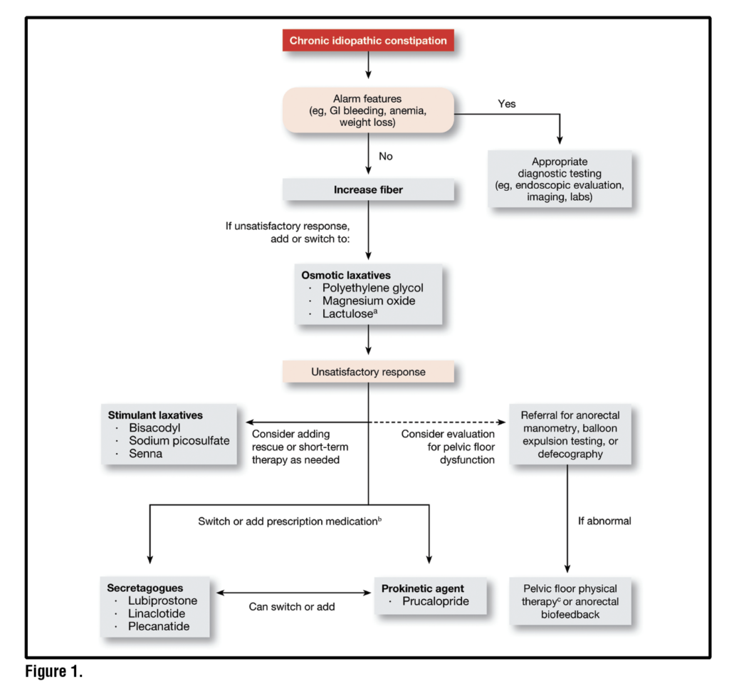
Chronic constipation (CC), also referred to as chronic idiopathic constipation (CIC) or functional constipation (FC), affects approximately 8–12% of adults and is associated with impaired quality of life, increased healthcare utilization, and substantial economic burden.1 CC is characterized by infrequent bowel movements, straining, sensation of anal blockage and/or a sensation of incomplete evacuation in the absence of structural or mucosal disease. CC is subtyped into normal transit constipation or slow transit constipation, with some overlap present. Lifestyle and behavioral measures including adequate hydration, regular physical activity, and dietary changes such as increased fiber intakeoften represent the initial step in treatment. However, a significant proportion of patients fail to achieve adequate relief and require pharmacologic therapy including osmotic and stimulant laxatives, secretagogues, and prokinetic agents.Thisreview follows the pragmatic, stepwise approach recommended by the AGA-ACG Clinical Practice Guideline on the pharmacological management of CC as outlined in Figure 1.2

Diagnosing Chronic Constipation

Chronic constipation is most often defined based on the Rome V Criteria (Table 1).3 While bowel symptoms may overlap with irritable bowel syndrome with constipation (IBS-C), the key differentiator is that abdominal pain and discomfort predominate in IBS-C. Notably many patients may transition between CC and IBS-C over time due to the spectrum and shared pathophysiology of these disorders of gut-brain interactions (DGBI).4

It is essential to exclude secondary causes of constipation. Clinicians should evaluate for alarm features including weight loss, rectal bleeding, anemia, new onset progressing worsening symptoms, family history of colon cancer or inflammatory bowel disease. These signs may warrant further investigation such as colonoscopy.  A thorough medication history should be obtained, with specific attention to medications known to cause or worsen constipation, such as opioids, calcium channel blockers, anticholinergics and certain antidepressants. Laboratory tests may be appropriate in select patients to screen for metabolic disorders, such as hypothyroidism, diabetes mellitus and hypercalcemia. A digital rectal examination is an essential part of the evaluation and can help identify a defecatory disorder, such as pelvic floor dyssynergia.5,6 This is best performed by asking the patient to bear down as if attempting to defecate while palpating the anal sphincter and puborectalis muscle during digital examination. Paradoxical contraction or failure to relax these muscles and/or lack of relaxation of the pelvic floor suggests a defecatory disorder. In patients with a suspected defecatory disorder further testing can be considered with anorectal manometry with balloon expulsion or defecography may be appropriate. Evaluation for a defecatory disorder is particularly important since these patients are best managed with anorectal biofeedback therapy.7 All patients should undergo age-appropriate colorectal screening in accordance with established guidelines. 

General Principles of Pharmacologic Therapy

Pharmacologic treatment of CC is best approached in a stepwise individualized manner.2 Therapy typically begins with widely available agents and progressing to prescription therapies for patients who do not respond adequately. Factors which may influence treatment selection include stool frequency and consistency, abdominal symptoms (e.g., bloating), patient age, comorbidities, and prior treatment experience. In more complex or refractory cases, combination therapy is often appropriate.

Table 1. Rome V Criteria for Chronic Constipation

Functional constipation is defined by the presence of ≥2 of the following symptoms for the last 3 months,
with symptom onset at least 6 months before diagnosis:
Straining during more than 25% of defecations Lumpy or hard stools (Bristol Stool Form Scale types 1–2) in more than 25% of defecations Sensation of incomplete evacuation in more than 25% of defecations Sensation of anorectal obstruction or blockage in more than 25% of defecations Manual maneuvers to facilitate defecation (e.g., digital evacuation, pelvic floor support) in more than 25% of defecations Fewer than three spontaneous bowel movements per week Additional requirements: Loose stools are rarely present without the use of laxatives Criteria for IBS are not met (i.e., abdominal pain is absent or does not meet IBS frequency/severity thresholds)


Bulk Forming Agents

Bulk forming laxatives are often considered first-line therapy for CC. These include commercially available fibers such as psyllium, methylcellulose, and calcium polycarbophil, which increase stool bulk and water content. Among these, psyllium is the best studied and has the strongest evidence particularly at doses exceeding 10 g/day.8 Notably, psyllium is soluble, gel-forming fiber that retains water, which can improve stool consistency in patients with constipation as well as in patients with loose stools. Gas and bloating are common. Therefore, gradual dose titration is recommended to minimize these side effects. Bulk agents are most effective in patients with mild constipation and normal colonic transit and may not provide relief in patients with slow colonic transit or defecatory dysfunction.9

Osmotic Laxatives

Osmotic laxatives are commonly used firstline pharmacologic agents.

Polyethylene Glycol (PEG)

PEG is a nonabsorbable hydrophilicpolymer that retains water in the lumen thereby softening stool and promoting transit. PEG is FDA approved for occasional constipation though it is now available over the counter and widely used for chronic constipation. Multiple randomized trials have shown consistent efficacy of PEG in improving bowel movement frequency, stool consistency, and global relief of constipation,10,11 though it does not appear to significantly improve abdominal pain in patients with IBS-C.12

PEG has been shown to have superior efficacy compared to lactulose with less bloating,13 and similar outcomes to prucalopride with fewer reports of headaches and nausea.14 PEG is generally well-tolerated though bloating, flatulence and diarrhea is sometimes reported. Due to its safety profile, wide availability and affordability, PEG is a preferred agent among osmotic laxatives.

Lactulose 

Lactulose is a non-absorbable synthetic disaccharide that functions as an osmotic laxative. Although older studies showed modest improvements in bowel frequency and global symptom relief, the quality of evidence is low due to small sample sizes, risk of bias and lack of modern diagnostic criteria and endpoints. Adverse events such as bloating and flatulence are common and often limit dose escalation. Lactulose is FDA approved for CIC and may be a reasonable option in patients who have failed fiber and other OTC laxatives. Lactulose is considered to be safe for use in pregnancy.2 

Magnesium Containing Agents

Magnesium hydroxide and magnesium citrate are effective osmotic laxatives and are commonly used as OTC therapies. Because magnesium can be absorbed from the gastrointestinal tract, high doses (i.e.,>1.5-2 grams per day) should be avoided to avoid hypermagnesemia. In a trial conducted in Japan, magnesium hydroxide (1.5 grams per day) improved symptoms in patients with chronic constipation.15 Likewise, in older individuals or patients with renal insufficiency, chronic use of magnesium agents should be used cautiously and monitored. 

Stimulant Laxatives

Stimulant laxatives, including senna and bisacodyl, work by stimulating colonic motility and increasing water secretion in the bowel lumen.16 They are typically used as short-term rescue or adjunctive agents when osmotic agents like polyethylene glycol (PEG) and fiber are not effective. Stimulant laxatives are widely available and are relatively inexpensive. They appear to be efficacious in improving constipation, though only a limited number of studies have been conducted.15,17 No high-quality long-term efficacy or safety data exist. While abdominal cramping and diarrhea are frequent, long-term mucosal damage or dependence does not appear to occur.18

Intestinal Secretagogues

These agents promote intestinal fluid secretion and accelerate transit. They are typically used in patients with moderate to severe symptoms or inadequate response to over-the-counter therapies.

Lubiprostone

Lubiprostone is a chloride channel (ClC-2) activator that increases intestinal fluid secretion and improves stool form and consistency. It is FDA approved for CIC in adult men and women at a dose of 24 mcg twice daily, and for IBS with constipation (IBS-C) in adult women only at a dose of 8 mcg twice daily. Nausea is the most common adverse effect occurring in approximately 30% of patients.19 However, it is generally transient and may be mitigated by taking lubiprostone with food.

Linaclotide

Linaclotide is a guanylate cyclaseC agonist that increases chloride and bicarbonate secretion into the lumen by opening cystic fibrosis transmembrane conductance regulator (CFTR) through an intracellular secondary messenger (cyclic GMP). Linaclotide also appears to reduce visceral pain signaling. It is approved for CIC at a dose of 72 mcg and 145 mcg once daily and IBS-C at a dose of 290 mcg once daily. Linaclotide should be taken on an empty stomach 30 minutes before breakfast.

In large phase 3 clinical trials linaclotide has shown efficacy across multiple endpoints, including stool frequency, consistency, bloating and abdominal pain. Diarrhea is the most common adverse event and resulted in 4.7% of patients withdrawing from the phase 3 clinical trials.20

Plecanatide

Plecanatide is another guanylate cyclaseC agonist similar to linaclotide. However, unlike linaclotide, plecanatide is pH sensitive. It is FDA approved for both CIC and IBS-C at a dose of 3 mg once daily, taken with or without food. The efficacy of plecanatide is similar to linaclotide, improving stool frequency, consistency, and patient-reported global constipation relief. Diarrhea is also the most common adverse event and resulted in 2.7% of patients withdrawing from the phase 3 clinical trials.21

Prokinetic Agents

Prucalopride

Prucalopride is a selective 5HT4 receptor agonist that stimulates colonic motility and accelerates intestinal transit. It is approved for CIC at a dose of 2 mg (1 mg for those with severe renal insufficiency). It may be taken with or without food. Prucalopride is not approved for IBS-C. Because of prokinetic properties, prucalopride may be particularly effective in patients with slow transit constipation. Headache, nausea, and diarrhea are the most common side effects, typically occurring early in therapy.  In phase 3 trials, withdrawals from these side effects were low (<3%).22 In a network meta-analysis, among studies with trials of at least 12 weeks in duration, prucalopride appeared to be the most efficacious of the medications for chronic constipation.10

Combination Therapy

Combining agents with complementary mechanisms (e.g., PEG plus a stimulant laxative or secretagogue) is often effective in refractory constipation and reflects real-world clinical practice.

Complementary and Novel Therapies

Nontraditional interventions such as electroacupuncture and mechanical devices have been investigated for CC. A large multicenter trial conducted in China involving over 1,000 patients with severe CC found 8 weeks of electroacupuncture (28 sessions) significantly increased in mean weekly number of spontaneous and complete spontaneous bowel movements (CSBMs) compared to baseline (1.76 vs. 0.87 CSBMs per week) than sham acupuncture.  Electroacupuncture also improved stool form and quality of life.23 More recently, an orally vibrating colon stimulating capsule was cleared by the FDA for CC. In a Phase III trial, patients receiving the vibrating capsule achieved significantly higher rates of CSBM response compared with placebo (e.g., ~39.3% vs 22.1% for >1 CSBM increase per week, and ~22.7% vs 11.4% for >2 CSBM per week), along with improvements in straining, stool consistency, and quality of life. Adverse events were generally mild and gastrointestinal in nature, with reports of a vibrating sensation in about 11% of treated patients.24

Practical Treatment Algorithm for Chronic Constipation

  1. Exclude secondary causes including medications potentially
  2. Initiate gradual titration of fiber supplementation (e.g. psyllium) > 10 g per day
  3. Add PEG 17-34 grams per day
  4. Add or substitute stimulant laxatives as needed
  5. Escalate to secretagogues (lubiprostone, linaclotide, or plecanatide) or prokinetic agents (prucalopride) for persistent symptoms
  6. Consider combination therapy and reassess diagnosis in refractory cases

Conclusion

Pharmacologic management of CC requires an individualized, stepwise approach that balances efficacy, tolerability, and patientcentered outcomes. A growing array of therapeutic options allows clinicians to tailor treatment based on symptom profile and underlying pathophysiology. Familiarity with mechanisms of action and practical use of these agents can substantially improve symptom control and quality of life for patients with chronic constipation. 


References

1. Sperber AD, Bangdiwala SI, Drossman DA, et al. Worldwide Prevalence and Burden of Functional Gastrointestinal Disorders, Results of Rome Foundation Global Study. Gastroenterology 2021;160:99-114 e3.

2. Chang L, Chey WD, Imdad A, et al. American Gastroenterological Association-American College of Gastroenterology Clinical Practice Guideline: Pharmacological Management of Chronic Idiopathic Constipation. Gastroenterology 2023;164:1086-1106.

3. Corsetti M, Shin A, Lacy BE, Cash BD, Simrén M, Schmulson MJ, Hou X, Lembo A. Bowel Disorders. Gastroenterology. 2026 May;170(6):1261-1282. doi: 10.1053/j.gastro.2026.02.003. Epub 2026 Feb 17. PMID: 41713703.

4. Heidelbaugh JJ, Stelwagon M, Miller SA, et al. The spectrum of constipation-predominant irritable bowel syndrome and chronic idiopathic constipation: US survey assessing symptoms, care seeking, and disease burden. Am J Gastroenterol 2015;110:580-7.

5. Rao SSC. Digital Rectal Examination: An Invaluable Clinical Tool. Gastro Hep Adv 2024;3:592-593.

6. Brandler J, Camilleri M. Pretest and Post-test Probabilities of Diagnoses of Rectal Evacuation Disorders Based on Symptoms, Rectal Exam, and Basic Tests: a Systematic Review. Clin Gastroenterol Hepatol 2020;18:2479-2490.

7. Rao SS, Benninga MA, Bharucha AE, et al. ANMS-ESNM position paper and consensus guidelines on biofeedback therapy for anorectal disorders. Neurogastroenterol Motil 2015;27:594-609.

8. Rao SSC, Brenner DM. Efficacy and Safety of Over-the-Counter Therapies for Chronic Constipation: An Updated Systematic Review. Am J Gastroenterol 2021;116:1156-1181.

9. Bijkerk CJ, de Wit NJ, Muris JW, et al. Soluble or insoluble fibre in irritable bowel syndrome in primary care? Randomised placebo controlled trial. BMJ 2009;339:b3154.

10. Luthra P, Camilleri M, Burr NE, et al. Efficacy of drugs in chronic idiopathic constipation: a systematic review and network meta-analysis. Lancet Gastroenterol Hepatol 2019;4:831-844.

11. Menees SB, Lembo AJ, Chey WD. Polyethylene Glycol 3350 in the Treatment of Chronic Idiopathic Constipation: Post hoc Analysis Using FDA Endpoints. Can J Gastroenterol Hepatol 2022;2022:3533504.

12. Chapman RW, Stanghellini V, Geraint M, Halphen M. Randomized clinical trial: macrogol/PEG 3350 plus electrolytes for treatment of patients with constipation associated with irritable bowel syndrome. Am J Gastroenterol 2013;108:1508-15.

13. Lee-Robichaud H, Thomas K, Morgan J, Nelson RL. Lactulose versus Polyethylene Glycol for Chronic Constipation. Cochrane Database Syst Rev 2010:CD007570.

14. Cinca R, Chera D, Gruss HJ, Halphen M. Randomised clinical trial: macrogol/PEG 3350+electrolytes versus prucalopride in the treatment of chronic constipation – a comparison in a controlled environment. Aliment Pharmacol Ther 2013;37:876-86.

15. Morishita D, Tomita T, Mori S, et al. Senna Versus Magnesium Oxide for the Treatment of Chronic Constipation: A Randomized, Placebo-Controlled Trial. Am J Gastroenterol 2021;116:152-161.

16. Corsetti M, Landes S, Lange R. Bisacodyl: A review of pharmacology and clinical evidence to guide use in clinical practice in patients with constipation. Neurogastroenterol Motil 2021;33:e14123.

17. Kamm MA, Mueller-Lissner S, Wald A, et al. Oral bisacodyl is effective and well-tolerated in patients with chronic constipation. Clin Gastroenterol Hepatol 2011;9:577-83.

18. Whorwell P, Lange R, Scarpignato C. Review article: do stimulant laxatives damage the gut? A critical analysis of current knowledge. Therap Adv Gastroenterol 2024;17:17562848241249664.

19. Johanson JF, Morton D, Geenen J, Ueno R. Multicenter, 4-week, double-blind, randomized, placebo-controlled trial of lubiprostone, a locally-acting type-2 chloride channel activator, in patients with chronic constipation. Am J Gastroenterol 2008;103:170-7.

20. Lembo AJ, Schneier HA, Shiff SJ, et al. Two randomized trials of linaclotide for chronic constipation. N Engl J Med 2011;365:527-36.

21. Miner PB, Jr., Koltun WD, Wiener GJ, et al. A Randomized Phase III Clinical Trial of Plecanatide, a Uroguanylin Analog, in Patients With Chronic Idiopathic Constipation. Am J Gastroenterol 2017;112:613-621.

22. Shin A, Camilleri M, Kolar G, et al. Systematic review with meta-analysis: highly selective 5-HT4 agonists (prucalopride, velusetrag or naronapride) in chronic constipation. Aliment Pharmacol Ther 2014;39:239-53.

23. Chakraborty S, Bharucha AE. In chronic severe functional constipation, electroacupuncture increased complete spontaneous bowel movements. Ann Intern Med 2016;165:JC69.

24. Rao SSC, Quigley EMM, Chey WD, et al. Randomized Placebo-Controlled Phase 3 Trial of Vibrating Capsule for Chronic Constipation. Gastroenterology 2023;164:1202-1210 e6.

Download Tables, Images & References

Nutrition Reviews in Gastroenterology, SERIES #33

The Role of the Crohn’s Disease Exclusion Diet in Treatment and Management of Crohn’s Disease

Read Article

The increasing incidence of inflammatory bowel disease underscores the importance of environmental factors, particularly nutrition, in disease development. Diet plays a key role in shaping the gut microbiome, modulating immune responses, and maintaining intestinal barrier integrity, making nutritional therapy a valuable approach in management. Exclusive enteral nutrition is effective for inducing remission in pediatric Crohn’s disease, with outcomes comparable to corticosteroids and added benefits for growth, nutritional status, and mucosal healing. However, its limited palatability, restrictive nature, and scarce adult data hinder widespread use. The Crohn’s Disease Exclusion Diet, especially when combined with partial enteral nutrition, has demonstrated efficacy in inducing remission in mild-to-moderate Crohn’s disease and may help maintain remission, although long-term data remain limited. This review outlines current advances in the diet in treatment of both pediatric and adult Crohn’s disease.

Background

The dramatic increase in inflammatory bowel disease (IBD) in industrialized countries and those adopting a Western diet suggest that environmental factors, including diet, are key contributors to the etiology of this disease. It is well established that dietary intake drives the gut microbiome, the intestinal immune response, and the intestinal barrier function. Nutritional interventions can play a significant role in managing Crohn’s disease (CD) by targeting inflammation and thereby alleviating symptoms. 

Exclusive enteral nutrition (EEN) was the first nutritional therapy shown to be effective in the treatment of CD. It involves the use of a nutritionally complete liquid formula as the sole source of nutrition for a defined period, typically 6–12 weeks, to induce remission.¹ Although  introduced in the 1970s, it was not until the late 1990s and early 2000s that randomized controlled trials and subsequent meta-analyses established its efficacy as a first-line therapy for children with active CD. These studies demonstrated that EEN is at least as effective as corticosteroids for inducing clinical remission, with additional benefits of improved growth, nutritional status, and mucosal healing, and without the steroid-related adverse effects.²

Although effective, EEN adherence is often limited to its poor palatability and strict regimen. This prompted further studies leading to the development of partial enteral nutrition (PEN), a nutritional therapy where a patient receives from 35% to up to 80% of their daily caloric needs via a nutrient-rich formula. Early attempts in the early 2000s to use PEN in conjunction with whole foods failed to induce remission, highlighting that the benefits of EEN arise not only from the formula’s composition but also from the exclusion of specific inflammatory dietary components.3,4 Recent evidence supports PEN as an adjunct therapy in CD, particularly for maintenance of remission and in alongside combination therapies. It demonstrated a dose-dependent benefit in reducing relapse risk, with higher caloric intake associated with greater efficacy. Emerging data also suggests PEN may enhance outcomes when combined with biologics, including improved short-term clinical response and sustained endoscopic remission with anti-tumor necrosis factor (TNF) therapy.5,6 

More recently, research has shifted toward combining PEN with anti-inflammatory dietary interventions. Compared with EEN, the Crohn’s Disease Exclusion Diet (CDED) with PEN offers a structured anti-inflammatory approach that incorporates whole foods that is less restrictive for inducing remission in pediatric and adult patients with mild-to-moderate CD.7 This review summarizes the diet phases and the evidence supporting the use of CDED in both pediatric and adult populations.

The Phases of the CDED

CDED is indicated for induction and maintenance of remission in pediatric and adult patients with mild-to-moderate luminal CD, particularly those with relatively short disease duration.8,9  The diet may also be useful in patients with loss of response to biologic therapy as a bridge, rescue, or adjunctive therapy (See Table 1). Caution is advised in patients who are underweight. Relative contraindications to CDED include strictures, severe malnutrition, and patients with avoidant/restrictive food intake disorder (ARFID) or other eating disorders.7,10

Phase 1 (Week 0-6)7,11-13

Is the induction phase

50% of total energy needs are provided by whole foods and the remaining 50% of energy and protein requirements are supplied through PEN

In this phase, there is an increased risk of weight loss, malnutrition, and micronutrient deficiencies if patients are not properly monitored

Phase 2 (Week 7-12)

It is the transition phase with gradual diet liberalization

Intake of whole foods is increased to provide 75% of total energy and protein requirements, while PEN is reduced to 25%

Phase 3 (Week >12)10,14

Is the maintenance phase. Some patients may continue to use PEN, but the overall goal is full transition to a long-term whole-food diet. 

This phase allows up to four “free meals” per week; ongoing studies are evaluating outcomes in both pediatric and adult populations

Table 1. Phases of Crohn’s Disease Exclusion Diet


Phase 1 (Induction Phase): Weeks 0-6Phase 2 (Transition Phase): Weeks 7-12Phase 3: Maintenance Phase
Phase OverviewIncrease soluble fiber Increase prebiotic fiber Increase resistant starch Decrease insoluble fiber Decrease fermentable fiber  Gluten/dairy-free   Rice/rice flour is the only grain allowedIncrease soluble fiber Increase insoluble fiber Increase fermentable fiber Continues to be dairy-free Gradual reintroduction of legumes, gluten as whole grain bread, and other grains such as oatmeal and quinoa Increase in variety of other fruits and vegetablesExpand and diversify intake of recommended foods based on tolerance and preference Reintroduction of yogurt as primary form of dairy All fibers allowed Mandatory foods by choice now Diet is for 5 days per week, allows 2 “skip days”* *Encourage homemade food over processed foods for the skip days 
Foods AllowedMandatory Chicken breast Egg Two potatoes (boiled/cooled,
ok to reheat) Two bananas One peeled apple Allowed Lean fish, rice (white) avocado, strawberries, melon, tomatoes, cucumbers, carrot, spinach, lettuce Condiments Olive/canola oil, fresh herbs (parsley, basil, etc.), lemon juice, garlic, ginger, salt, pepper, cumin, turmeric Sweeteners Honey, table sugar Drinks Water, sparkling water, home-squeezed orange juice, herbal teas (lemon, mint)
Additions from Phase 1 Tuna, lean beef (up to 7 oz) per week (1 slice/day) whole-grain bread daily, oats, quinoa, legumes, other fruits and vegetables (e.g., red pepper, blueberry, sweet potato, broccoli, kiwi) nuts- walnuts and almonds (small quantity, unsalted, unroasted, unprocessed) All foods are allowed except: Processed, precooked or smoked meats and fish (sausages, luncheon meats, salami, fish sticks), soy products, persimmons, pomegranate, cactus fruits, passion fruit, dried fruits (sulphate-free dried fruits allowed on weekends), canned fruit, large portions of kale, leeks, asparagus, artichokes, soft drinks, instant coffee, alcohol
Partial Enteral Nutrition50% of calorie needs25% of calorie needsPEN is optional or can be used to promote weight gain

The standard CDED protocol requires mandatory intake of two boiled and cooled potatoes, two bananas, and one peeled apple as sources of resistant starches and chicken breast, fish, and eggs as protein sources during the first two phases of the diet. In vegetarian and vegan patients, maintaining adequate intake of protein and key micronutrients (e.g., vitamin B12) is essential. The standard CDED protocol has not been formally studied or validated in these populations, and adaptations would require substituting the animal protein sources, which are limited by the exclusion of plant protein sources in Phase 1 of the diet. The use of PEN can help meet protein and caloric needs and plant-based formulas can be used as alternatives.

Enteral nutrition formulas used in CDED (See Table 2) are available over the counter or by prescription, with potential insurance coverage. These formulas may be purchased without a prescription from pharmacies, grocery stores, and online retailers; however, this represents a potential significant out-of-pocket expense for many. For insurance coverage, formulas may be reimbursed when prescribed by a physician for medical nutrition therapy in CD, particularly in pediatric patients receiving EEN and who may require enteral feeding tube support. As coverage varies widely across insurance plans, patients should work with their gastroenterologist to obtain a prescription with appropriate diagnostic codes and the provider’s office must submit medical documentation that highlights and justifies the formula’s therapeutic role in treatment of CD. 

The Role of the Gut Microbiome

Several dietary components influence the impact of the complex regulation of intestinal permeability, which is influenced by mucosal integrity, microbiome disbalances, and inflammatory changes in the epithelium.

In patients treated with CDED combined with PEN, diet-induced remission was associated with a correction of dysbiosis and change in both, the composition of the microbiome, as well as its metabolic pathways towards that of the microbial community in healthy controls.15

Table 2. Options for Enteral Formulas

Formula TypeMacronutrientsProsConsExamples*
Polymeric
(intact nutrients)
Milk-based (whey, lactose free) or plant-based (pea protein)Ready to drink Variety of flavors Caloric density 0.6-2.4 calories per milliliter Cost Some brands not available in retail stores Plant-based contain fiberPediatric PediaSure®, Boost Kids Essentials®; Kate Farms Pediatric Standard 1.2® Adult Ensure Plus®, Boost Plus®, Kate Farms Standard®
Semi-elemental (hydrolyzed peptide-based)Milk-based (whey, lactose free) or plant-based (pea protein) Maltodextrin, corn starch Medium-chain triglycerides, soybean oilReady to drink Caloric density 1.0-1.5 calories per milliliter Cost Access/availability Less flavor options Taste Plant-based contain fiberPediatric PediaSure® Peptide®, Peptamen Jr®; Kate Farms Pediatric Peptide Standard 1.2®
Adult Vital®, Peptamen®, Kate Farms Peptide®
Elemental
(amino acid-based)
Free amino acids Maltodextrin, modified corn starch Medium-chain triglycerides, soybean oilCaloric density is 1.0 calorie per milliliter – can be adjusted for higher calorie needsCost Access/availability Most brands only have powder Taste Micronutrient needs may not be met in older patientsPediatric  Elecare Jr®, Alfamino Junior®; Neocate Junior® Adult Vivonex RTF®

CDED has been shown to improve intestinal barrier integrity and induce distinct microbiome modifications, characterized by a reduction in Proteobacteria, and an increase in Firmicutes and Bacteroidetes—changes that correlate with clinical remission.11,15-17

CDED has also been associated with a more diverse gut microbiome compared with EEN, which may confer greater long-term benefit.¹¹ Despite these microbial shifts, diet-induced remission has not been consistently associated with sustained changes in short-chain fatty acids (SCFAs) or bile acids (BAs).15

A broader metabolomic analysis has shown that both the CDED combined with PEN, and EEN, are associated with significant changes in IBD-related metabolites, including kynurenine, ceramides, and amino acids. Reductions in specific kynurenine pathway metabolites (the primary route of tryptophan metabolism) and increases in serotonin pathway metabolites, have been associated with diet-induced and sustained remission.18,19 Importantly, in samples from patients who failed to maintain remission, these metabolic changes were not observed.

Table 3. Comparison of EEN and CDED with PEN Therapies


EEN29,30CDED with PEN11,26,27
PopulationPediatric and adult Crohn’s disease
Disease ActivityMild to moderate disease activity
Disease PhenotypeInflammatory: Primary treatment in mild to moderate disease Fistulizing: Not recommended as primary treatment Stricturing: EEN used as preoperative nutrition optimization; CDED with PEN is not used as primary treatment
Therapeutic UsePrimary therapy: Yes Adjunctive therapy: Yes Salvage therapy: Yes, but not well-established or routinely used
Evidence SourceRandomized controlled trials  Systematic reviews  Meta-analyses  Cohort studies  Clinical guidelines  Randomized controlled trials  Systematic reviews 
ResultsClinical remission: up to 80% pediatric cases  Endoscopic remission: 89% at week 8 Clinical remission 75-80% at 25 weeks  Endoscopic remission 35% at 24 weeks 
Structure100% liquid diet   100% energy and protein intake from enteral formula   Polymeric (intact nutrient formula) can be used. Elemental (hydrolyzed) formulas are not indicated unless polymeric formulas are not tolerated well  A whole-food diet with enteral formula to supplement the diet
PhasesUse for 4-12 weeks for induction of remission  Phase 1: Week 0-6
50% CDED whole foods + 50% PEN  Phase 2: Week 7-12
75% CDED whole foods + 25% PEN  Phase 3: Week >12
75% CDED whole foods + 25% PEN 

Pediatric Evidence with the CDED

There is strong evidence that CDED, particularly in combination with PEN, can induce remission in pediatric CD (See Table 3).

A pivotal study by Sigall-Boneh et al. found that 70% of children with active, mild-to-moderate luminal CD achieved clinical remission after six weeks with CDED plus 50% of calories coming from PEN.8 Notably, six of seven patients adhering to CDED without PEN also achieved clinical remission, underscoring the significant therapeutic potential of diet alone. These findings were further validated by Levine et al. in the first multinational randomized controlled trial comparing CDED plus PEN with EEN in 74 children with mild-to-moderate luminal CD.¹¹ Although both approaches were similarly effective in inducing remission at week 6 (75% vs. 59%, p = 0.38), use of the CDED plus PEN resulted in significantly higher sustained remission rates than EEN (76% vs. 45%, p = 0.01). A subsequent study by Sigall-Boneh et al. showed that both EEN and CDED plus PEN lead to a rapid clinical response by week 3, which may help identify patients likely to achieve remission by week 6 with continued adherence.20 In addition, CDED plus PEN, has been shown to lead to a significant decrease in fecal calprotectin levels in children with active mild to moderate CD.21

Beyond induction, several studies also support the role of CDED with PEN for maintaining remission. In the randomized controlled trial by Levine et al., children assigned to the CDED plus PEN maintained significantly higher rates of remission at 12 weeks compared to those who transitioned from EEN to a free diet with PEN.¹¹ Observational studies from Israel and Italy have also reported sustained remission beyond one year with CDED plus PEN, along with evidence of mucosal healing. Positive outcomes have been observed in both biologic-naïve and biologic-exposed patients.22,23 A retrospective Croatian study of 61 pediatric patients showed a significantly higher weight gain (p= 0.002) and increases in body mass index (BMI) z-score (p=0.001) in patients receiving CDED and PEN compared to patients who received EEN alone.24

CDED with PEN may also serve as a useful salvage regimen for patients with inadequate response to biologic therapy. In a prospective cohort study of pediatric and adult patients with loss of response to biologics despite dose escalation or combination therapy, treatment with CDED with PEN for 12 weeks resulted in clinical remission at 6 weeks in 61.9% of patients.17 Additional case series from Israel and Italy have also documented the benefits of CDED with PEN in refractory CD, including fistulizing and perianal phenotypes.22, 23

Adult Evidence with the CDED

Several observational studies and randomized controlled trials support the use of CDED in adults.9,25 

The efficacy of CDED with PEN versus CDED monotherapy has been examined in biologic-naïve patients with early, mild-to-moderate CD. At weeks 6 and 12, a greater proportion of patients in the combination therapy arm achieved remission compared with those on diet alone. By week 24, nearly half of the cohort maintained both clinical and endoscopic remission, with a numerical advantage for CDED in combination with PEN (63% vs. 38%; p = 0.11). Improvements were also observed in inflammatory markers and quality-of-life indices, supporting the feasibility of CDED, either alone or with PEN, as a strategy for remission induction and mucosal healing.26 CDED has also been studied in patients with limited therapeutic options. In a mixed pediatric–adult cohort that included 11 adults who had failed at least two biologics or combination regimens, remission was achieved most frequently in those with isolated ileal disease (83.3%).26 

CDED has also demonstrated higher remission rates compared with a Mediterranean diet control in a randomized trial of 24 adults with mild-to-moderate disease.27 This open-label study assessed symptomatic remission using the Harvey-Bradshaw Index (HBI), alongside fecal calprotectin and serum inflammatory indices at baseline, 12 weeks, and 24 weeks. The CDED demonstrated significantly higher remission rates at both time points: 70.8% vs. 38.1% at 12 weeks (p = .027) and 79.2% vs. 42.9% at 24 weeks (p < .0001). In addition, the CDED was associated with favorable changes in body composition, including a decrease in fat mass while maintaining or increasing fat-free mass and cellular mass.

In pouchitis, CDED has been shown to improve clinical, biochemical, and endoscopic manifestations. Clinical remission rates were as high as 66.7% at week 6 and were maintained at week 24 among 46.7% of participants despite addition of diverse variety of foods and free meals as allowed in the maintenance phase of the diet. By week 12, patients experienced significant reductions in symptoms—particularly stool frequency and urgency—as well as decreases in serum CRP levels, with the most pronounced improvements observed in those with full dietary adherence.28

Role of the Dietitian 

The involvement of an IBD-specialized Registered Dietitian (RD) is crucial. RDs evaluate indications and contraindications to determine patient suitability for the CDED and provide guidance for its safe and effective implementation, helping to minimize the risk of malnutrition while optimizing clinical outcomes.

With specialized training in nutritional assessment and management, RDs tailor macronutrient and micronutrient intake to individual needs, and monitor for deficiencies that may arise during dietary restriction or progression through the CDED phases.7,29The effectiveness of the CDED should be evaluated over time through assessment of gastrointestinal symptoms, nutritional status, and laboratory markers. In addition, RDs play a central role in tailoring the diet to cultural food practices by offering individualized recipes and counseling, while also helping patients manage limited food access, navigate social settings, and enhance family eating patterns.

Adherence to the CDED and any necessary adjustments can be assessed during follow-up appointments using tools such as dietary recalls. If remission is not achieved or sustained, or if nutritional deficiencies develop, the diet should be re-evaluated, and alternative therapies considered. Close coordination among the RD, the gastroenterologist, and patient is also essential to ensure alignment of nutrition care. 

Table 4. Micronutrient Monitoring

TestRecommendations  Considerations 
The following screening recommendations are adapted from American Gastroenterological Association (AGA)29 and North American Society for Pediatric Gastroenterology, Hepatology and Nutrition (NASPGHAN) guidelines
for IBD
31
Complete blood count with iron studiesAt diagnosis and every 6–12 months in patients in remission; every 3 months in those with active diseaseConsider IV iron infusions when inflammation is active 
Vitamin B12At diagnosis and annually in all patients; every 3–6 months in higher-risk groupsHigher-risk groups include patients with ileal disease or resection and those following a vegan diet
Vitamin B9 At diagnosis and annuallyPatients receiving methotrexate or sulfasalazine should receive folic acid supplementation
Vitamin D (25-OH)At diagnosis and every 6–12 monthsSupplementation based on level and age 
Calcium Assess dietary intake and supplements as neededPatients on CDED without PEN may need calcium supplementation, particularly during Phases 1 and 2 
Vitamin CConsider monitoring in selected patientsDeficiency is rare but may occur in patients consuming highly restrictive or poor-quality diets, including some patients on CDED without PEN
Zinc At diagnosis and annuallyAttempt for higher levels as it is involved in wound healing, tissue repair and immune function
Magnesium At diagnosis and depending on clinical symptomsRecommended in patients with chronic or severe diarrhea, extensive small bowel disease, or high-output states

Table 5. Resources for CDED Tools

ModuLife www.mymodulife.comThe ModuLife website and its app focus on CDED food lists and recipes, but access is only available through a healthcare provider who has been trained in the ModuLife program.
Crohn’s and Colitis Foundation www.crohnscolitisfoundation.orgA platform called “Gut Friendly Recipes” is available where CDED recipes for all diet phases can be accessed using filters.
GI Nutrition Foundation www.ginutritionfoundation.comCDED recipes for all phases of the diet are accessible through its online recipe database. The platform also provides updates on research related to the CDED.


Laboratory Monitoring

Routine screening for nutritional deficiencies is essential. As no CDED-specific micronutrient monitoring guidelines currently exist, the following recommendations are adapted from standard IBD guidelines, with CDED-specific considerations noted where applicable (See Table 4).

The American Gastroenterological Association (AGA) recommends that all patients with IBD be monitored for vitamin D and iron deficiency, and that patients with extensive ileal disease or prior ileal resection be monitored for vitamin B12 deficiency.29Serum micronutrient levels should ideally be assessed during periods of disease quiescence, as many are acute-phase reactants that may fluctuate in the setting of active inflammation.

CDED with PEN has been shown to be nutritionally adequate, meeting more than 80% of the recommended daily intake for key micronutrients in adults. Patients receiving CDED without PEN, or those who are malnourished, may be at increased risk for micronutrient deficiencies and warrant closer monitoring. For patients on CDED for 12 weeks or with specific risk factors, monitoring and repletion of zinc, copper, folate, and fat-soluble vitamins should be considered. Magnesium, zinc, selenium, and folate deficiencies are particularly common due to reduced intake and increased gastrointestinal losses. In contrast, selenium, vitamin B12, vitamin D, and vitamin B9 levels may be affected by both dietary restriction and impaired absorption, particularly in patients with ileal involvement or prior intestinal surgery.29

Additional considerations apply to patients receiving CDED in combination with aminosalicylates (5-ASAs) or immunomodulators. Those treated with methotrexate or sulfasalazine should receive folic acid supplementation and be monitored for anemia. Monitoring of vitamin A, vitamin C, vitamin B6, and β-carotene may also be appropriate in selected patients, as low intake and serum levels have been reported in adults with active CD on exclusion diets.26,29

Strengths, Barriers, and Opportunities

The CDED offers several distinct strengths as a non-pharmacologic approach to CD management. Unlike pharmacologic therapies, the CDED directly targets nutrition, with the potential to improve overall dietary quality, supports growth in pediatric patients, and promotes beneficial changes in the gut microbiome. The structured phase of the diet also encourages family engagement, as dietary changes are often implemented at the household level, fostering shared investment in the patient’s care.7,29

Implementation of the CDED may pose challenges due to hunger, social aspects of eating, dietary monotony, and the psychological strain of restriction. Adherence may be improved through support from healthcare teams, family, and peers and use of palatable supplements.12,23 

Strategies such as offering different formula options, varying temperatures, or using overnight nasogastric feeding can also help reduce formula fatigue and improve tolerability. Tools, such as recipes to support implementation of the CDED are available, although they remain limited (See Table 5). 

Although use of the CDED in clinical practice is growing, implementation remains inconsistent and further education is needed for patients and healthcare providers. When used as a treatment strategy, either alone or alongside other therapies, clinicians should clearly explain the monitoring plan over a defined period, including expected response targets and when to reassess effectiveness or begin reintroducing foods or food groups. The strongest evidence supports its use in children and adults with mild-to-moderate Crohn’s disease. Expanding the evidence base in more complex disease phenotypes and underrepresented populations, such as pregnant individuals, patients with stricturing or penetrating disease, and J-pouch patients, remains an important area for future research.

Conclusion

CDED is an established dietary treatment for mild-moderate CD in the pediatric and adult population. Its three-phase structured approach emphasizes whole, anti-inflammatory foods that may aid to restore microbiome balance, reduce inflammation, support epithelial function and therefore mucosal immune response. Evidence demonstrates that CDED, particularly when combined with PEN, can induce and sustain remission, improve inflammatory biomarkers and enhance quality of life, and may be beneficial in complex settings such as biologic failure, pregnancy, and pouchitis. While generally better tolerated than EEN and associated with improved adherence in some studies, adherence to the CDED can be limited by dietary monotony, psychosocial burden, financial challenges, and nutritional deficiency risks. Success is improved with structured healthcare and social support, palatable supplements, and access to an IBD-specialized dietitian who can tailor the diet to lifestyle and cultural practices. Further research is needed to clarify its role in complex disease phenotypes and long-term outcomes. 

References

  1. Kansal S, Wagner J, Kirkwood CD, Catto-Smith AG. Enteral nutrition in Crohn’s disease: an underused therapy. Gastroenterol Res Pract. 2013;2013:482108. 
  2. Urlep D, Benedik E, Orel R. Exclusive and partial enteral nutrition in Crohn’s disease. In: Mahdi BM, ed. New Concepts in Inflammatory Bowel Disease. London, UK: IntechOpen; 2018. 
  3. González-Torres L, Moreno-Álvarez A, Fernández- Lorenzo AE, Leis R, Solar-Boga A. The role of partial enteral nutrition for induction of remission in Crohn’s disease: a systematic review of controlled trials. Nutrients. 2022;14(24):5263. 
  4. Kerbiriou C, Dickson C, Nichols B, et al. Treatment of active Crohn’s disease with exclusive enteral nutrition diminishes the immunostimulatory potential of fecal microbial products. Inflamm Bowel Dis. 2024;30(12):2457-2466. 
  5. Jatkowska A, White B, Gkikas K, Seenan J, Macdonald J, Gerasimidis K. Partial enteral nutrition in the management of Crohn’s disease: a systematic review and meta-analysis. J Crohns Colitis. 2024;19. 
  6. Huang C, Chen C, Wu H, et al. The efficacy of infliximab combined with partial enteral nutrition in the treatment of Crohn’s disease: a cohort study. Front Nutr. 2025;12:1591954. 
  7. Sigall Boneh R, Westoby C, Oseran I, et al. The Crohn’s disease exclusion diet: a comprehensive review of evidence, implementation strategies, practical guidance, and future directions. Inflamm Bowel Dis. 2024;30(10):1888- 1902. 
  8. Sigall Boneh R, Pfeffer-Gik T, Segal I, Zangen T, Boaz M, Levine A. Partial enteral nutrition with a Crohn’s disease exclusion diet is effective for induction of remission in children and young adults with Crohn’s disease. Inflamm Bowel Dis. 2014;20(8):1353-1360. 
  9. Fliss-Isakov N, Cohen NA, Bromberg A, et al. Crohn’s disease exclusion diet for the treatment of Crohn’s disease: real-world experience from a tertiary center. J Clin Med. 2023;12(16):5428. 
  10. Walia CLS, Cerezo CS, Smith A, et al. North American Society for Pediatric Gastroenterology, Hepatology, and Nutrition position on the role of the registered dietitian nutritionist in the care of the pediatric patient with chronic gastrointestinal diseases. J Pediatr Gastroenterol Nutr. 2023;76(3):390-399. 
  11. Levine A, Wine E, Assa A, et al. Crohn’s disease exclusion diet plus partial enteral nutrition induces sustained remission in a randomized controlled trial. Gastroenterology. 2019;157(2):440-450.e8. 
  12. Russell EE, Day AS, Dimitroff C, et al. Practical application of the Crohn’s disease exclusion diet as therapy in an adult Australian population. J Gastroenterol Hepatol. 2024;39(3):446-456. 
  13. Correia I, Oliveira PA, Antunes ML, Raimundo MDG, Moreira AC. Is there evidence of Crohn’s disease exclusion diet in remission of active disease in children and adults? A systematic review. Nutrients. 2024;16(7):987. 
  14. Mutsekwa RN, Edwards JT, Angus RL. Exclusive enteral nutrition in the management of Crohn’s disease: a qualitative exploration of experiences, challenges and enablers in adult patients. J Hum Nutr Diet. 2021;34(2):440-449. 
  15. Verburgt CM, Dunn KA, Ghiboub M, et al. Successful dietary therapy in paediatric Crohn’s disease is associated with shifts in bacterial dysbiosis and inflammatory metabotype towards healthy controls. J Crohns Colitis. 2023;17(1):61-72. 
  16. Verburgt CM, Sigall Boneh R, Dunn KA. Intestinal permeability improvement with nutritional therapy is associated with specific microbiome profiles and functional pathways in pediatric Crohn’s disease. Gastroenterology. 2023;164(6):1025-1026. 
  17. Hart L, Verburgt CM, Wine E, et al. Nutritional therapies and their influence on the intestinal microbiome in pediatric inflammatory bowel disease. Nutrients. 2022;14(1):4. 
  18. Ghiboub M, Penny S, Verburgt CM, et al. Metabolome changes with diet-induced remission in pediatric Crohn’s disease. Gastroenterology. 2022;163(4):922-936.e15. 19. Ghiboub M, Sigall Boneh R, Sovran B, et al. Sustained diet-induced remission in pediatric Crohn’s disease is associated with kynurenine and serotonin pathways. Inflamm Bowel Dis. 2023;29(5):684-694. 

    20. Sigall Boneh R, Van Limbergen J, Wine E, et al. Dietary therapies induce rapid response and remission in pediatric patients with active Crohn’s disease. Clin Gastroenterol Hepatol. 2021;19(4):752-759. 

    21. Matuszczyk M, Meglicka M, Wiernicka A, et al. Effect of the Crohn’s disease exclusion diet on the fecal calprotectin level in children with active Crohn’s disease. J Clin Med. 2022;11(14):4146. 

    22. Levine A, El-Matary W, Van Limbergen J. A case-based approach to new directions in dietary therapy of Crohn’s disease: food for thought. Nutrients. 2020;12(3):880. 

    23. Scarallo L, Banci E, Pierattini V, Lionetti P. Crohn’s disease exclusion diet in children with Crohn’s disease: a case series. Curr Med Res Opin. 2021;37(7):1115-1120. 

    24. Niseteo T, Sila S, Trivić I, Mišak Z, Kolaček S, Hojsak I. Modified Crohn’s disease exclusion diet is equally effective as exclusive enteral nutrition: real-world data. Nutr Clin Pract. 2022;37(2):435-441. doi:10.1002/ncp.10752. 

    25. Szczubełek M, Pomorska K, Korólczyk-Kowalczyk M, et al. Effectiveness of Crohn’s disease exclusion diet for induction of remission in adult patients with Crohn’s disease. Nutrients. 2021;13(11):4112. 

    26. Yanai H, Levine A, Hirsch A, et al. The Crohn’s disease exclusion diet for induction and maintenance of remission in adults with mild-to-moderate Crohn’s disease (CDED-AD): an open-label, pilot, randomised trial. Lancet Gastroenterol Hepatol. 2022;7(1):49-59. 

    27. Pasta A, Formisano E, Calabrese F, et al. The use of the Crohn’s disease exclusion diet (CDED) in adults with Crohn’s disease: A randomized controlled trial. Eur J Clin Invest. 2025;55(6):e14389. 

    28. Fliss-Isakov N, Kornblum J, Zemel M, et al. The effect of the Crohn’s disease exclusion diet on patients with pouch inflammation: an interventional pilot study. Clin Gastroenterol Hepatol. 2023;21(6):1654-1656.e3. 

    29. Hashash JG, Elkins J, Lewis JD, Binion DG. AGA clinical practice update on diet and nutritional therapies in patients with inflammatory bowel disease: expert review. Gastroenterology. 2024;166(3):521-532. 

    30. Pigneur B, Lepage P, Mondot S, et al. Mucosal Healing and Bacterial Composition in Response to Enteral Nutrition Vs Steroid-based Induction Therapy-A Randomised Prospective Clinical Trial in Children with Crohn’s Disease. J Crohns Colitis. 2019;13(7):846-855. 

    31. Goyal A, Zheng Y, Albenberg LG, et al. Anemia in Children with Inflammatory Bowel Disease: A Position Paper by the IBD Committee of the North American Society of Pediatric Gastroenterology, Hepatology and Nutrition. J Pediatr Gastroenterol Nutr. 2020;71(4):563- 582. 

Download Tables, Images & References

Frontiers in Endoscopy, Series #107

Endoscopic Management of Achalasia

Read Article

Introduction

Achalasia is a rare, chronic motility disorder of the esophagus with a worldwide prevalence of 1.8 to 12.6 per 100,000 people. In the US, a study published in 2022 reported incidence and prevalence rates of 10.5 and 18.0 per 100,000 people, respectively. Individuals of all races and sex are equally affected, but incidence increases with advancing age.1 This review examines the efficacy and safety of endoscopic treatment options for achalasia.

Etiology

The pathogenesis of achalasia is poorly understood, complex, and likely multifactorial. Affected patients have impaired function of inhibitory neurons in the distal esophageal myenteric plexus leading to a decrease in inhibitory vasoactive intestinal peptide (VIP) and nitric oxide (NO) and unopposed excitatory acetylcholine within the lower esophageal sphincter (LES) and distal esophagus. This results in impaired relaxation of the LES and diminished, or absent, esophageal peristalsis.1,,

Achalasia is most commonly idiopathic with the leading hypothesis suggesting an autoimmune process in genetically predisposed patients or those with a preceding viral illness., Secondary etiologies include malignancy, specifically small cell lung cancer via paraneoplastic secretion of antineuronal antibodies, and Chagas disease from Trypanosoma cruzi infection.5 Chagas disease is highly common in endemic regions such as Central and South America with secondary achalasia occurring in 7-15.5% of patients with Chagas disease in these regions.

Symptoms

Patients present with progressive solid and liquid dysphagia, often accompanied by chest pain (50% of patients), regurgitation (75%), weight loss (60%), and/or heartburn (40%) which leads many patients to first be diagnosed with gastroesophageal reflux disease (GERD).1,3,, Symptoms are quantified using the Eckardt symptom scoring system with composite scores ranging from 0-12 based on frequency of dysphagia (0-3), regurgitation (0-3), retrosternal pain (0-3), and weight loss (0-3). In most studies, post-treatment composite scores ≤3 indicate clinical remission whereas composite scores >3 indicate treatment failure.

Diagnosis

Many patients undergo upper endoscopy which is important to rule out pseudoachalasia from mechanical obstruction or malignancy and may reveal a tight, puckered gastroesophageal junction (GEJ) with saliva and/or food retention.1 Barium esophagram may be performed revealing esophageal dilation with a classic “bird beak” appearance.1 However, the gold standard diagnostic modality which provides important prognostic and treatment implications is high-resolution esophageal manometry.1

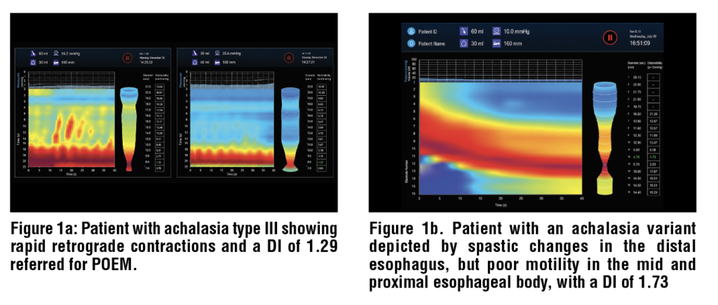
Using high-resolution esophageal manometry, patients are classified into 3 subtypes according to the Chicago Classification. All three achalasia subtypes share the disorder’s hallmark of aperistalsis with impaired GEJ relaxation but vary by their dominant pattern of esophageal body contractility.11 Type I achalasia occurs in 20-40% of cases and is the most severe form characterized by low or absent esophageal pressurization.1,11, Type II achalasia occurs in 50-70% of cases and is characterized by rapid panesophageal pressurization >30mmHg.1,11,12 Type III achalasia occurs in 5-10% of cases and is characterized by rapid spastic contractions.1,11,12 While all the same disorder, this classification is helpful when determining the best treatment approach.

Botulinum Toxin Injections

Technique

Botulinum toxin injections (BTI) induce local, transient, and partial paralysis of the LES to decrease sphincter tone and improve esophageal emptying. Botulinum toxin (BTX) works by inhibiting the release of acetylcholine into neuromuscular junctions, thereby preventing muscle contraction at the LES.13 The three A1 BTX subtypes, abobotulinum, incobotulinum, and onabotulinum, are the most studied and, therefore, most commonly administered BTX formulations.13

To perform BTI, a standard endoscopic injection needle is used to inject 80-100U (20-25U in each quadrant) of BTX approximately 1cm proximal to the Z-line under endoscopic visualization.1,13, (Figure 1) In general, injections of BTX produce symptom relief for several months at a time. BTX injections can be repeated every 3-6 months as needed. The procedure is usually performed on an outpatient basis, taking minutes to complete on average.14

Efficacy

Pasricha et al. performed the first RCT in 1995 with 21 patients showing a significant decrease in symptom score (5.4 vs 0.5; p=0.001) and LES pressure (33% decrease vs 12% increase; p=0.02) between BTX and placebo groups, respectively. Subsequent studies have shown good short-term efficacy with clinical success observed in 73.7-90.5% of patients at 1 month.15,,,, Unfortunately, symptom recurrence remains a major problem. Campos et al. highlighted this in a meta-analysis of 9 studies with 315 patients which revealed a progressive decline in symptomatic improvement with 78.7%, 70%, 53.3%, and 40.6% of patients in remission at 1, 3, 6, and 12 months, respectively.17 Because of this, BTI is only recommended as first-line treatment in older patients who have decreased life expectancy and/or are poor candidates for more invasive endoscopic techniques and surgery.1

Adverse Events

No serious adverse events were reported across multiple studies.15,18,19,, Patients may experience minor adverse events such as transient post-procedural chest or epigastric pain as reported by D’Onofrio et al. and Annese et al.16,19

There is potential for fibrosis to develop at the GEJ following BTI with potentially important implications for future therapeutic interventions. According to Patti et al., patients who did not respond to BTI have a lower risk for developing fibrosis and responded well to subsequent surgical myotomy. Conversely, patients who initially responded to BTI but underwent subsequent surgical myotomy for relapse of symptoms experienced an increased number of perforations and failure rates, presumably due to BTI-induced scarring at the GEJ.22, Interestingly, recent studies suggest efficacy and safety are not impacted by post-BTI fibrosis following subsequent peroral endoscopic myotomy (POEM).,,

Pneumatic Dilation

Technique

Pneumatic dilation (PD) utilizes single-use balloon dilators to mechanically disrupt LES fibers, resulting in a widened LES orifice and improved esophageal emptying.8 Becker et al.’s RCT with 35 patients compared high compliance balloons made of latex that expand with lower inflation pressures and low compliance balloons made of polyethylene that require higher pressures to expand and found no significant difference in outcomes. Despite similar outcomes, low compliance balloon dilators have overtaken high compliance balloon dilators on the market because they allow for a more controlled dilation process. Commercially available options in the US include the Rigiflex II single use balloon dilator (Boston Scientific, Natick, MA, United States), Achalasia Over the Wire Balloon Dilator (Hobbs Medical, Stafford Springs, CT, United States), and Achalasia Balloon (Cook Medical, Winston Salem, NC, United States) with fully inflated balloon diameters ranging 30-40mm.

To perform PD, a guidewire is positioned across the GEJ, and the uninflated balloon is passed over the wire until the midpoint of the balloon aligns with the LES.18 If fluoroscopy is used, the radiopaque ends of the balloon can be visualized to ensure adequate placement across the level of the diaphragm prior to inflation.28, The next steps are operator-dependent, but the balloon is gradually inflated to 10-15 PSI and held in place for 15-60 seconds.18,29,,, Khan et al. and Gideon et al. compared shorter inflation durations (6 and 15 seconds, respectively) to a 60-second inflation time and found no significant differences in efficacy or adverse events between groups.29, In general, most patients are initially dilated using a 30mm diameter ballon, but serial dilations with 35mm or 40mm balloons may be required every few weeks until symptomatic relief is achieved.31

Efficacy

Leyden et al. performed a meta-analysis revealing symptom resolution in 80.7% (46/57) and 73.3% (55/75) of patients undergoing PD at 6 and 12 months, respectively. van Hoeij et al.’s larger meta-analysis of 10 studies with 643 patients undergoing PD compared clinical success over time following single dilation with different balloon diameters (30mm, 35mm, and 40mm). At 6 months, clinical success was observed in 81%, 79%, and 90% of patients following PD with 30mm, 35mm, or 40mm balloons, respectively.35 At 12 months, clinical success was sustained in 77%, 70%, and 87% of patients, respectively.35 This contrasts a single-institution prospective study published previously by Farhoomand et al. which showed dilation using a 30mm balloon dilator alone had a significantly lower 3-year success rate (37%; 95% CI 26-53) compared to serial dilation using 30mm followed by 35mm (76%; 95% CI 65-88) or 30mm followed by 35mm and 40mm (88%; 95% CI 80-97).

Remission is more likely to be achieved in patients who are female, >40-45-years-old, and those with post-PD LES pressures <10mmHg, though this measurement is not commonly performed.1,36, Farhoomand et al. identified high early failure rates (88% within 3 months) in young men treated with 30mm balloons.36 Therefore, younger men may benefit from initial dilation with a larger balloon (35mm or 45mm) and/or serial PD.

Adverse Events

Minor adverse events include chest pain or reflux symptoms, but the major, life-threatening, and well-documented adverse event associated with PD is esophageal perforation. Perforation occurs in approximately 1.0-1.6% of patients undergoing PD.30,34,35 van Hoeij et al. compared the risk of perforation based on balloon diameter and found a significantly (p<0.027) higher rate of perforation following use of 35mm balloons (3.2%; 9/282) compared to 30mm balloons (1.0%; 6/588).35 However, the use of 40mm balloons surprisingly did not result in any adverse events, including esophageal perforation (0%; 0/62).35 The reason behind this lower rate of perforation following use of the largest balloon size is unclear. Perforations occurred significantly more after the initial PD compared to subsequent PD procedures (9.3% vs 0.97%, p=0.0017), and there was no correlation between inflation time or maximum balloon pressure and perforations.35

Because of the high risk for perforation, routine testing after PD has been debated, but the most recent ACG guidelines discourage performing post-PD gastrograffin esophagrams unless there is clinical concern for perforation.1

Esophageal Functional Luminal Imaging Probe Dilation

Technique

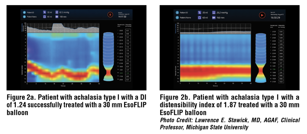
Esophageal functional luminal imaging probe (EsoFLIP) dilation is a novel method of mechanically disrupting LES fibers in a more controlled manner than PD. This FDA-approved device utilizes advanced technology to provide real-time measurements of intraluminal pressure and diameter at multiple points along the balloon catheter before, during, and after LES dilation.38, The EsoFLIP ES-330 balloon catheter (Medtronic, Minneapolis, MN, United States) is commercially available in the US with a maximum inflated balloon diameter of 30mm.39 Baumann et al. outline various step-by-step techniques to successfully perform EsoFLIP dilation.38

Efficacy

Very few prospective and retrospective cohort studies have been performed to date with short-term clinical success achieved in 52.9-85% of patients.,,,, However, no individual study included more than 28 patients with achalasia. Therefore, larger prospective studies, including studies aimed at assessing long-term symptom relief, and randomized controlled trials are needed.

Adverse Events

Similar to post-PD patients, reflux symptoms (10.7-25% of patients) and transient chest pain (15%) were the most common adverse events encountered after EsoFLIP dilation.40,41 Esophageal perforation occurred in 1.2% (1/85) of patients in these small studies.40,41,42,43,44 

Peroral Endoscopic Myotomy

Technique

Peroral endoscopic myotomy (POEM) utilizes third-space endoscopy to treat achalasia. The concept of POEM was first outlined by Pasricha et al. in 2007 through experimentation on pigs and later introduced as a treatment option for individuals with achalasia by Inoue et al. in 2009.,

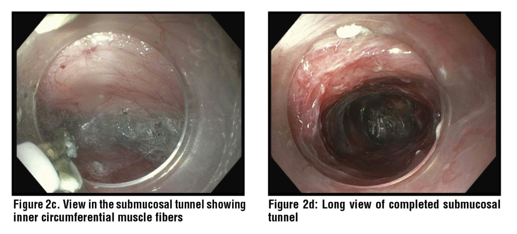
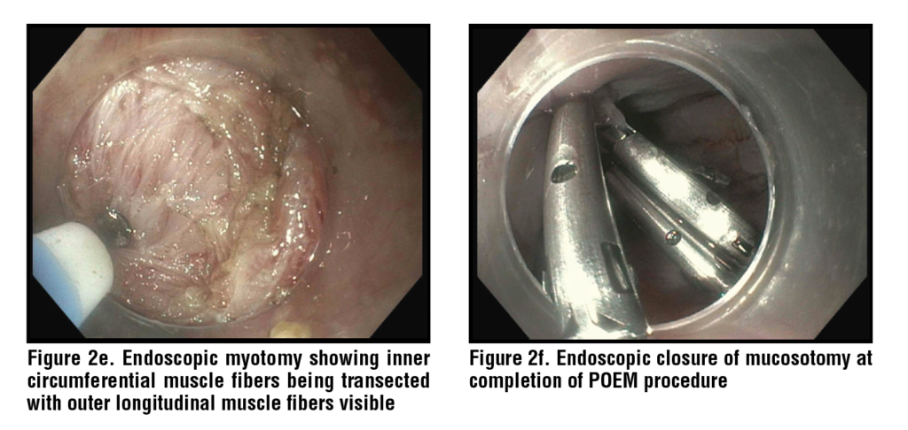
There are many variations to POEM, but in general, a mucosal entry site is made within the esophagus to expose the submucosal space. Once inside, a submucosal tunnel is created, extending a short distance past the GEJ. Then, circular LES muscle fibers are dissected until the endoscope can be easily passed through the GEJ to confirm successful myotomy. Finally, the mucosal entry site is closed. (Figures 2a-f)

Efficacy

Inoue et al. published the first prospective cohort study in 2010 which showed a significant reduction in symptom score (10 to 1.3; p=0.0003) and LES pressure (52.4mmHg to 19.9mmHg; p=0.0001).47 Since then, many meta-analyses have demonstrated high clinical success in 90-97% of patients.,,,, Vespa et al. performed one of the largest meta-analyses of 31 studies with 3,023 patients revealing a clinical success rate of 91% at a median follow-up of 29-months. Zhang et al. analyzed symptom resolution over time and reported clinical success rates of  91.3%, 90.4%, 89.8%, and 82.2% at 2, 3, 4, and 5 years, respectively. Post-POEM quality of life also significantly (p<0.05) improved according to a meta-analysis by Zhong et al.

Shou et al. identified risk factors for POEM failure in a meta-analysis of 27 studies with 9,371 patients. Worse outcomes occurred in patients with a sigmoid esophagus (a manifestation of advanced achalasia) (OR 1.90, 95% CI 1.45-2.47), type I achalasia (OR 1.30, 95% CI 1.04-1.63), type III achalasia (OR 1.26, 95% CI 0.89-1.78), prior Heller myotomy (OR 5.75, 95% CI 3.97-8.34), and prior PD (OR 1.18, 95% CI 1.07-1.29).56 Better outcomes occurred in patients with type II achalasia (OR 0.59, 95% CI 0.47-0.75).56 In contrast, there was no significant difference in symptom resolution between achalasia subtypes according to meta-analyses by Zhang et al. and Andolfi et al.54, POEM was examined in the elderly patient population by Hayat et al. who found no significant difference in success among patients ≥65-years-old (95.74% vs 95.31%) or ≥75-years-old (99.01% vs 97.97%) compared to their younger counterparts.

When initial POEM fails, repeat POEM may be attempted with good outcomes. Hayat et al. analyzed 6 studies with 123 patients who underwent repeat POEM 12.2-13.5 months on average after their initial failed POEM. 82.69% of patients achieved clinical success after repeat POEM.59

To optimize outcomes, many variations in POEM technique have been studied. Dhoop et al. compared full-thickness and circular myotomy and found no significant difference in clinical success at 1 or 3 years. However, full-thickness myotomy had an increased risk for post-POEM GERD (RR 3.03, 95% CI 1.53-6.02; p=0.002) and esophagitis (RR 1.79, 95% CI 1.09-2.76; p=0.02) compared to circular myotomy.61 Rodriguez de Santiago et al. and Mohan et al. performed meta-analyses comparing anterior and posterior wall tunneling, each finding no significant difference between myotomy approaches.52, Procedure time was shorter with posterior (62.1 minutes) compared to anterior (82.7 minutes) wall tunneling.62 In practice, individual operators are free to perform POEM as they see fit.

Optimal myotomy length has been extensively studied, comparing short and long myotomy lengths. Zhang et al., Weng et al., and Ghazaleh et al. performed meta-analyses which showed shorter myotomy resulted in shorter procedure times, as expected, with the same or less risk of reflux but no significant difference in clinical success.,, 

POEM has also been successfully used as rescue therapy following failed improvement with other treatment modalities for achalasia. Tan et al. and Zhong et al. demonstrated clinical success in 90.8% and 91.0% of these patients, respectively.,

Adverse Events

Symptomatic reflux and reflux esophagitis are two of the most common adverse events seen following POEM with rates ranging from 22.0-26.2% and 5.9-19.0%, respectively.47,48,49,53,54,, Mota et al. performed a meta-analysis of 25 studies and found no statistical difference in post-POEM GERD rates when comparing full-thickness, partial-thickness and circular myotomy, anterior and posterior myotomy, or subtypes of achalasia. This differs from Dhoop et al.’s more recent meta-analysis which showed an increased risk of post-POEM GERD and esophagitis with full-thickness myotomy.61 POEM combined with fundoplication (POEM-F) has been trialed in an attempt to reduce post-POEM reflux. The rate of post-POEM-F reflux reported by Gopakumar et al.’s meta-analysis of 7 studies with 127 patients was 13.8% which is lower than most reflux rates after POEM alone but comes with far higher costs.

Serious adverse events include subcutaneous emphysema   (21.8-31.6%    of    patients), pneumoperitoneum (16.2-30.6%), pneumothorax (8.7-11.0%), and in very rare instances, mediastinal emphysema/leak (0.3-4.9%), esophageal/gastric perforation (2.6%), bleeding (0.9-1.1%), and death (0.09%).48,69

Conclusion

Achalasia is a complex and debilitating esophageal motility disorder, but there are many efficacious endoscopic treatment options available, enabling individualized treatment approaches. Botulinum toxin injections have shown decent short-term clinical success with few adverse events and are best suited for older patients and/or those who are not candidates for more invasive endoscopic modalities. Pneumatic dilation has shown better short-term outcomes but is limited by high rates of symptom relapse and a small risk of serious adverse events, most notably esophageal perforation. Peroral endoscopic myotomy offers the highest long-term success rates but may be accompanied by post-procedural reflux symptoms and/or reflux esophagitis. Treatment should be tailored to patient preferences and institutional expertise. 

References

1 Vaezi MF, Pandolfino JE, Yadlapati RH, Greer KB, Kavitt RT. ACG Clinical Guidelines: Diagnosis and Management of Achalasia. Am J Gastroenterol. 2020;115(9):1393-1411. doi:10.14309/ajg.0000000000000731 

2 Gaber CE, Eluri S, Cotton CC, et al. Epidemiologic and Economic Burden of Achalasia in the United States. Clin Gastroenterol Hepatol. 2022;20(2):342-352.e5. doi:10.1016/j.cgh.2021.02.035 

3 Savarino E, Bhatia S, Roman S, et al. Achalasia. Nat Rev Dis Primers. 2022;8(1):28. Published 2022 May 5. doi:10.1038/s41572-022-00356-8 

4 Park W, Vaezi MF. Etiology and pathogenesis of achalasia: the current understanding. Am J Gastroenterol. 2005;100(6):1404-1414. doi:10.1111/j.1572- 0241.2005.41775.x 

5 Francis DL, Katzka DA. Achalasia: update on the disease and its treatment. Gastroenterology. 2010;139(2):369-374. doi:10.1053/j.gastro.2010.06.024 

6 Kahrilas PJ, Boeckxstaens G. The spectrum of achalasia: lessons from studies of pathophysiology and high-resolution manometry. Gastroenterology. 2013;145(5):954-965. doi:10.1053/j.gastro.2013.08.038 

7 Panesso-Gómez S, Pavia P, Rodríguez-Mantilla IE, et al. Trypanosoma cruzi Detection in Colombian Patients with a Diagnosis of Esophageal Achalasia. Am J Trop Med Hyg. 2018;98(3):717-723. doi:10.4269/ajtmh.17-0417 

8 Khashab MA, Vela MF, Thosani N, et al. ASGE guideline on the management of achalasia. Gastrointest Endosc. 2020;91(2):213-227.e6. doi:10.1016/j.gie.2019.04.231 

9 Spechler SJ, Souza RF, Rosenberg SJ, Ruben RA, Goyal RK. Heartburn in patients with achalasia. Gut. 1995;37(3):305- 308. doi:10.1136/gut.37.3.305 

10 Eckardt VF, Aignherr C, Bernhard G. Predictors of outcome in patients with achalasia treated by pneumatic dilation. Gastroenterology. 1992;103(6):1732-1738. doi:10.1016/0016-5085(92)91428-7 

11 Pandolfino JE, Kwiatek MA, Nealis T, Bulsiewicz W, Post J, Kahrilas PJ. Achalasia: a new clinically relevant classification by high-resolution manometry. Gastroenterology. 2008;135(5):1526-1533. doi:10.1053/j.gastro.2008.07.022 

12 Vespa E, Barchi A, Mandarino FV, et al. Standard length of peroral endoscopic myotomy (POEM) for achalasia: a systematic review and meta-analysis. Dis Esophagus. 2024;37(12):doae069. doi:10.1093/dote/doae069 

13 Nassri A, Ramzan Z. Pharmacotherapy for the management of achalasia: Current status, challenges and future directions. World J Gastrointest Pharmacol Ther. 2015;6(4):145- 155. doi:10.4292/wjgpt.v6.i4.145 

14 Storr M, Born P, Frimberger E, et al. Treatment of achalasia: the short-term response to botulinum toxin injection seems to be independent of any kind of pretreatment [published correction appears in BMC Gastroenterol. 2003 Nov 7;3(1):32]. BMC Gastroenterol. 2002;2:19. doi:10.1186/1471-230x-2-19 

15 Pasricha PJ, Ravich WJ, Hendrix TR, Sostre S, Jones B, Kalloo AN. Intrasphincteric botulinum toxin for the treatment of achalasia [published correction appears in N Engl J Med 1995 Jul 6;333(1):75]. N Engl J Med. 1995;332(12):774- 778. doi:10.1056/NEJM199503233321203 

16 D’Onofrio V, Annese V, Miletto P, et al. Long-term follow-up of achalasic patients treated with botulinum toxin. Dis Esophagus. 2000;13(2):96-103. doi:10.1046/j.1442- 2050.2000.00094.x 

17 Campos GM, Vittinghoff E, Rabl C, et al. Endoscopic and surgical treatments for achalasia: a systematic review and meta-analysis. Ann Surg. 2009;249(1):45-57. doi:10.1097/ SLA.0b013e31818e43ab 

18 Zhu Q, Liu J, Yang C. Clinical study on combined therapy of botulinum toxin injection and small balloon dilation in patients with esophageal achalasia. Dig Surg. 2009;26(6):493-498. doi:10.1159/000229784 

19 Annese V, Bassotti G, Coccia G, et al. A multicentre randomised study of intrasphincteric botulinum toxin in patients with oesophageal achalasia. GISMAD Achalasia Study Group. Gut. 2000;46(5):597-600. doi:10.1136/gut.46.5.597 

20 D’Onofrio V, Annese V, Miletto P, et al. Long-term follow-up of achalasic patients treated with botulinum toxin. Dis Esophagus. 2000;13(2):96-103. doi:10.1046/j.1442- 2050.2000.00094.x 

21 Leyden JE, Moss AC, MacMathuna P. Endoscopic pneumatic dilation versus botulinum toxin injection in the management of primary achalasia. Cochrane Database Syst Rev. 2014;2014(12):CD005046. doi:10.1002/14651858. CD005046.pub3 

22 Patti MG, Feo CV, Arcerito M, et al. Effects of previ

ous treatment on results of laparoscopic Heller myotomy for achalasia. Dig Dis Sci. 1999;44(11):2270-2276. doi:10.1023/a:1026660921776 

23 Smith CD, Stival A, Howell DL, Swafford V. Endoscopic therapy for achalasia before Heller myotomy results in worse outcomes than heller myotomy alone. Ann Surg. 2006;243(5):579-586. doi:10.1097/01. sla.0000217524.75529.2d 

24 Kadiyala J, Canakis A, Lee DU, Xue P, Fan GH, Kim RE. Comparing clinical success and procedural difficulty between treatment-naïve and treatment-refractory patients with esophageal motility disorders during peroral endoscopic myotomy. Gastrointest Endosc. 2023;98(1):19-27. e11. doi:10.1016/j.gie.2023.01.052 

25 Khan SZ, Benson J, Boutros C, Lyons J, Hashimoto DA, Marks JM. Do preoperative endoscopic interventions predict symptom recurrence or reintervention after peroral endoscopic myotomy?. Surg Endosc. 2023;37(9):7153-7158. doi:10.1007/s00464-023-10196-6 

26 Jones EL, Meara MP, Pittman MR, Hazey JW, Perry KA. Prior treatment does not influence the performance or early outcome of per-oral endoscopic myotomy for achalasia. Surg Endosc. 2016;30(4):1282-1286. doi:10.1007/s00464-015- 4339-y 

27 Becker K, Biesenbach S, Erckenbrecht JF, Frieling T. Effect of balloon compliance on symptomatic success of pneumatic dilation in achalasia patients. Z Gastroenterol. 2001;39(10):831-836. doi:10.1055/s-2001-17868 

28 Uppal DS, Wang AY. Update on the endoscopic treatments for achalasia. World J Gastroenterol. 2016;22(39):8670- 8683. doi:10.3748/wjg.v22.i39.8670 

29 Khan AA, Shah SW, Alam A, Butt AK, Shafqat F, Castell DO. Pneumatic balloon dilation in achalasia: a prospective comparison of balloon distention time. Am J Gastroenterol. 1998;93(7):1064-1067. doi:10.1111/j.1572- 0241.1998.00330.x 

30 Ghoshal UC, Chaudhuri S, Pal BB, Dhar K, Ray G, Banerjee PK. Randomized controlled trial of intrasphincteric botulinum toxin A injection versus balloon dilatation in treatment of achalasia cardia. Dis Esophagus. 2001;14(3-4):227- 231. doi:10.1046/j.1442-2050.2001.00189.x 

31 Ponds FA, Fockens P, Lei A, et al. Effect of Peroral Endoscopic Myotomy vs Pneumatic Dilation on Symptom Severity and Treatment Outcomes Among Treatment-Naive Patients With Achalasia: A Randomized Clinical Trial. JAMA. 2019;322(2):134-144. doi:10.1001/jama.2019.8859 

32 Bansal R, Nostrant TT, Scheiman JM, et al. Intrasphincteric botulinum toxin versus pneumatic balloon dilation for treatment of primary achalasia. J Clin Gastroenterol. 2003;36(3):209-214. doi:10.1097/00004836-200303000- 00005 

33 Gideon RM, Castell DO, Yarze J. Prospective randomized comparison of pneumatic dilatation technique in patients with idiopathic achalasia. Dig Dis Sci. 1999;44(9):1853- 1857. doi:10.1023/a:1018898824135 

34 Leyden JE, Moss AC, MacMathuna P. Endoscopic pneumatic dilation versus botulinum toxin injection in the management of primary achalasia. Cochrane Database Syst Rev. 2014;2014(12):CD005046. doi:10.1002/14651858. CD005046.pub3 

35 van Hoeij FB, Prins LI, Smout AJPM, Bredenoord AJ. Efficacy and safety of pneumatic dilation in achalasia: A systematic review and meta-analysis. Neurogastroenterol Motil. 2019;31(7):e13548. doi:10.1111/nmo.13548 

36 Farhoomand K, Connor JT, Richter JE, Achkar E, Vaezi MF. Predictors of outcome of pneumatic dilation in achalasia. Clin Gastroenterol Hepatol. 2004;2(5):389-394. doi:10.1016/s1542-3565(04)00123-5 

37 Eckardt VF, Aignherr C, Bernhard G. Predictors of outcome in patients with achalasia treated by pneumatic dilation. Gastroenterology. 1992;103(6):1732-1738. doi:10.1016/0016-5085(92)91428-7 

38 Baumann AJ, Carlson DA. EsoFLIP for esophageal dilation: proposed advantages. Curr Opin Gastroenterol. 2020;36(4):329-335. doi:10.1097/MOG.0000000000000639 

39 Iqbal U, Yodice M, Ahmed Z, et al. Safety and efficacy of EsoFLIP dilation in patients with esophageal dysmotility: a systematic review. Dis Esophagus. 2024;37(8):doae036. doi:10.1093/dote/doae036 

40 Schnurre L, Murray FR, Schindler V, et al. Short-term outcome after singular hydraulic EsoFLIP dilation in patients with achalasia: A feasibility study. Neurogastroenterol Motil. 2020;32(9):e13864. doi:10.1111/nmo.13864 

41 Kappelle WF, Bogte A, Siersema PD. Hydraulic dilation with a shape-measuring balloon in idiopathic achalasia: a feasibility study. Endoscopy. 2015;47(11):1028-1034. doi:10.1055/s-0034-1392481 

42 Chan B, Ding J, Bilal M et al. Safety and efficacy of EsoFLIP hydraulic dilation for achalasia. Gastrointest Endosc 2020; 91(6): AB136–7. https://doi.org/10.1016/j. gie.2020.03.1055 

43 Carlson D A, Atluri S, Balla M, Listernick Z, Kahrilas P, Pandolfino J E. 871 hydraulic dilation for achalasia: a series utilizing a 30-mm functional lumen imaging probe dilator, the EsoFLIP. Gastrointest Endosc 2016; 83(5): AB173. https://doi.org/10.1016/j.gie.2020.03.1055 

44 Tsai L, Kunkel D. Treatment outcomes from EsoFLIP dilaiton in patients with achalasia vary by subtype, Digestive Disease Week 2021, AGA Abstracts, Supplemental Su207, S-649. 2021. 

45 Pasricha PJ, Hawari R, Ahmed I, et al. Submucosal endoscopic esophageal myotomy: a novel experimental approach for the treatment of achalasia. Endoscopy. 2007;39(9):761- 764. doi:10.1055/s-2007-966764 

46 Inoue, H., Minami, H., Satodate, H., & Kudo, S. (2009). First Clinical Experience of Submucosal Endoscopic Esophageal Myotomy for Esophageal Achalasia with No Skin Incision. Gastrointestinal Endoscopy, 69. doi:10.1016/j. gie.2009.03.133 

47 Inoue H, Minami H, Kobayashi Y, et al. Peroral endoscopic myotomy (POEM) for esophageal achalasia. Endoscopy. 2010;42(4):265-271. doi:10.1055/s-0029-1244080 

48 Talukdar R, Inoue H, Nageshwar Reddy D. Efficacy of peroral endoscopic myotomy (POEM) in the treatment of achalasia: a systematic review and meta-analysis. Surg Endosc. 2015;29(11):3030-3046. doi:10.1007/s00464-014-4040-6 

49 Barbieri LA, Hassan C, Rosati R, Romario UF, Correale L, Repici A. Systematic review and meta-analysis: Efficacy and safety of POEM for achalasia. United European Gastroenterol J. 2015;3(4):325-334. doi:10.1177/2050640615581732 

50 Evensen H, Kristensen V, Larssen L, Sandstad O, Hauge T, Medhus AW. Outcome of peroral endoscopic myotomy (POEM) in treatment-naive patients. A systematic review. Scand J Gastroenterol. 2019;54(1):1-7. doi:10.1080 /00365521.2018.1549271 

51 Li H, Peng W, Huang S, et al. The 2 years’ long-term 

efficacy and safety of peroral endoscopic myotomy for the treatment of achalasia: a systematic review. J Cardiothorac Surg. 2019;14(1):1. Published 2019 Jan 3. doi:10.1186/ s13019-018-0811-9 

52 Rodríguez de Santiago E, Mohammed N, Manolakis A, Shimamura Y, Onimaru M, Inoue H. Anterior versus posterior myotomy during poem for the treatment of achalasia: systematic review and meta-analysis of randomized clinical trials. J Gastrointestin Liver Dis. 2019;28(1):107-115. doi:10.15403/jgld.2014.1121.281.pom 

53 Vespa E, Barchi A, Mandarino FV, et al. Standard length of peroral endoscopic myotomy (POEM) for achalasia: a systematic review and meta-analysis. Dis Esophagus. 2024;37(12):doae069. doi:10.1093/dote/doae069 

54 Zhang H, Zeng X, Huang S, et al. Mid-Term and Long- Term Outcomes of Peroral Endoscopic Myotomy for the Treatment of Achalasia: A Systematic Review and Meta- Analysis. Dig Dis Sci. 2023;68(4):1386-1396. doi:10.1007/ s10620-022-07720-4 

55 Zhong C, Tan S, Ren Y, et al. Quality of Life Following Peroral Endoscopic Myotomy for Esophageal Achalasia: A Systematic Review and Meta-Analysis. Ann Thorac Cardiovasc Surg. 2020;26(3):113-124. doi:10.5761/atcs. ra.19-00273 

56 Shou Y, Wang X, Liu D. Factors associated with peroral endoscopic myotomy for achalasia outcomes: systematic review and meta-analysis. Surg Endosc. 2024;38(7):3503- 3519. doi:10.1007/s00464-024-10862-3 

57 Andolfi C, Fisichella PM. Meta-analysis of clinical outcome after treatment for achalasia based on manometric subtypes. Br J Surg. 2019;106(4):332-341. doi:10.1002/ bjs.11049 

58 Hayat U, Kamal F, Memon A, et al. Peroral Endoscopic Myotomy for Achalasia Among the Elderly Population: A Systematic Review and Meta-Analysis. J Clin Gastroenterol. Published online February 10, 2025. doi:10.1097/ MCG.0000000000002103 

59 Hayat U, Kamal F, Rana UI, et al. Efficacy and Safety of Repeat Per-oral Endoscopic Myotomy After Failed POEM for Achalasia: A Systematic Review and meta-analysis. J Clin Gastroenterol. Published online December 5, 2024. doi:10.1097/MCG.0000000000002117 

60 Mota RCL, de Moura EGH, de Moura DTH, et al. Risk factors for gastroesophageal reflux after POEM for achalasia: a systematic review and meta-analysis. Surg Endosc. 2021;35(1):383-397. doi:10.1007/s00464-020-07412-y 

61 Dhoop S, Abu-Rumaileh M, Sayeh W, et al. Efficacy and safety of full-thickness versus circular peroral endoscopic myotomy for treatment of achalasia: a systematic review and meta-analysis. Ann Gastroenterol. 2025;38(2):143-155. doi:10.20524/aog.2025.0946 

62 Mohan BP, Ofosu A, Chandan S, et al. Anterior versus posterior approach in peroral endoscopic myotomy (POEM): a systematic review and meta-analysis. Endoscopy. 2020;52(4):251-258. doi:10.1055/a-1090-0788 

63 Zhang H, Zeng X, Huang S, et al. Safety and Efficacy of Peroral Endoscopic Shorter Myotomy versus Longer Myotomy for Patients with Achalasia: A Systematic Review and Meta-analysis. Gastroenterol Res Pract. 2022;2022:6770864. Published 2022 Mar 30. doi:10.1155/2022/6770864 

64 Weng CY, He CH, Zhuang MY, Xu JL, Lyu B. Peroral endoscopic longer vs shorter esophageal myotomy for achalasia treatment: A systematic review and meta-analysis. World J Gastrointest Surg. 2022;14(3):247-259. doi:10.4240/wjgs. v14.i3.247 

65 Ghazaleh S, Beran A, Khader Y, et al. Short versus standard peroral endoscopic myotomy for esophageal achalasia: a systematic review and meta-analysis. Ann Gastroenterol. 2021;34(5):634-642. doi:10.20524/aog.2021.0644 

66 Tan S, Zhong C, Ren Y, et al. Efficacy and Safety of Peroral Endoscopic Myotomy in Achalasia Patients with Failed Previous Intervention: A Systematic Review and Meta- Analysis. Gut Liver. 2021;15(2):153-167. doi:10.5009/ gnl19234 

67 Zhong C, Ni B, Liu S, et al. The Effect of Peroral Endoscopic Myotomy in Achalasia Patients with Prior Endoscopic Intervention: A Systematic Review and Meta-Analysis. Dig Surg. 2021;38(2):136-148. doi:10.1159/000512627 

68 Vespa E, Pellegatta G, Chandrasekar VT, et al. Long-term outcomes of peroral endoscopic myotomy for achalasia: a systematic review and meta-analysis. Endoscopy. 2023;55(2):167-175. doi:10.1055/a-1894-0147 

69 Patel K, Abbassi-Ghadi N, Markar S, Kumar S, Jethwa P, Zaninotto G. Peroral endoscopic myotomy for the treatment of esophageal achalasia: systematic review and pooled analysis. Dis Esophagus. 2016;29(7):807-819. doi:10.1111/ dote.12387 

70 Mota RCL, de Moura EGH, de Moura DTH, et al. Risk factors for gastroesophageal reflux after POEM for achalasia: a systematic review and meta-analysis. Surg Endosc. 2021;35(1):383-397. doi:10.1007/s00464-020-07412-y 

71 Gopakumar H, Annor E, Vohra I, et al. Peroral endoscopic myotomy with fundoplication (POEM-F) for achalasia: Systematic review and meta-analysis [published correction appears in Endosc Int Open. 2025 Mar 19;13:a25624787. doi: 10.1055/a-2562-4787.]. Endosc Int Open. 2025;13:a25368132. Published 2025 Mar 14. doi:10.1055/a-2536-8132 

Download Tables, Images & References

Dispatches from the GUILD Conference, Series #74

Fertility and Use of Assisted Reproductive Technology among Women with Inflammatory Bowel Disease: A Practical Guide

Read Article

Women with Inflammatory Bowel Disease (IBD) increasingly utilize assisted reproductive technologies (ART), such as in vitro fertilization (IVF) and oocyte cryopreservation. Women with active IBD or a history of pelvic surgery may have higher rates of infertility compared to the general population. Currently, data regarding ART and induction of IBD flare are limited. However, recent evidence demonstrates that ART is safe in women with well-controlled IBD, with the rate of disease flare during ART procedures remaining low. This article reviews the current evidence on fertility in IBD, when to refer patients for evaluation, safety and efficacy of ART, and cost considerations. Physicians should counsel patients that achieving disease remission prior to conception is strongly recommended and reassure patients that most IBD medications do not appear to impair the efficacy of ART. With a multidisciplinary approach, we can improve fertility rates and pregnancy outcomes among women with IBD. 

Introduction

The incidence of IBD, comprising Crohn’s disease (CD) and Ulcerative Colitis (UC), peaks between the ages of 15–30 years across Western Europe and North America, with a second smaller peak occurring between the ages of 60–79 years. As 25% of all IBD patients are diagnosed before the age of twenty, the disease burden is often concentrated during a woman’s reproductive years.1

Caring for a young woman with IBD often involves addressing concerns regarding medication safety and risk of flares during pregnancy. Women with IBD face higher rates of voluntary childlessness compared to the general population, a phenomenon caused by misconceptions rather than physical inability.2,4

This article addresses practical questions providers will face behind closed doors. Could I ever have children? Should I see a fertility specialist? Is IVF safe for me? Will medications hurt my baby? Will I flare during IVF treatment? By using up-to-date literature, we offer a roadmap for optimizing reproductive health in this population. 

Understanding Fertility in IBD

It is a common misconception that a diagnosis of IBD means infertility. Up to 18% of women with CD and 14% women with UC may experience infertility.12 However, this increased risk of infertility is not generalizable across all women with IBD. In fact, multiple systematic reviews and national cohort studies suggest there are similar fertility rates in women with UC and slightly decreased fertility rates in women with CD compared to the general population.2 The primary drivers of reduced fertility are active disease at the time of conception, prior pelvic surgery (particularly ileal pouch-anal anastomosis), and active peri-anal disease.1,2 Decreased fertility is most often seen after ileal-pouch-anal anastomosis (J-pouch) surgery from pelvic adhesions and tubal obstruction while active disease can lead to a dysfunctional placenta and reduced ovarian reserve, reflected by decreased anti-Mullerian hormone levels. Fortunately, systematic reviews have reported that women with IBD in remission have similar ovarian reserves to healthy controls.2,10 This data supports the key clinical target of maintaining 3-6 months of remission prior to conception. 

For gastroenterologists, it is also important to screen for non-biological causes of infertility. Decreased libido and dyspareunia resulting from active perianal or pelvic disease are frequently overlooked. Additionally, misinformation regarding medication teratogenicity, heritability, and poor pregnancy outcomes also contributes to voluntary infertility.2 A review of sexual function and common misconceptions is the best first step in identifying patients who need gastroenterology or fertility support. 

Combating Misinformation

Patients often rely on unverified online sources for health information. Gastroenterologists can use the evidence-based talking points in Table 1 to address some common myths. Additionally, they can refer them to the PIANOstudy.org website for a patient education video in seven languages.

When to Refer for Fertility Evaluation

Gastroenterologists should have a lower threshold for referral to a reproductive endocrinologist in patients with IBD compared to the general population.  While standard guidelines recommend referral after 12 months of unsuccessful conception, women with IBD, particularly those with CD or prior pelvic surgery, should be referred after 6 months of trying. For patients older than 40 years or those with extensive pelvic surgeries (such as J-pouch), referral after 4 months of unsuccessful conception is appropriate.3,4

Early referral to a fertility expert is important for three reasons:

Anatomical complications from prior surgeries may create a challenging pathway to natural conception.3 

If a patient has active disease, the path to remission may require medication changes and imaging/endoscopic evaluation, a process that can take several months.2,3

Natural age-related fertility decline compounds the existing fertility challenges in those with chronic inflammatory conditions such as IBD.2,3,5

Table 1. Myths and Facts About IBD And Pregnancy

MYTHSFACTS
If I have IBD, my child will get itWhile genetics can play a role, the absolute risk to a child is lower than expected.  If one parent has IBD, the risk of a child developing the disease is around 6-9%
(compared to ~1% in the general population).  If both parents have IBD, the risk increases to 30%.1,2 
There is nothing I can do to lower the risk for my child getting IBDEnvironmental exposures during pregnancy may play a role in IBD pathogenesis, and many are modifiable.  Smoking: Maternal smoking is associated with a 1.5-fold increased risk of IBD in the offspring. Smoking cessation is the single most effective action a patient can take.2,3 Antibiotics: Prenatal exposure to antibiotics, especially multiple courses or use during the third trimester, is associated with increased risk of IBD in the offspring, likely due to maternal microbiome disruption. While antibiotics should be used for necessary indications, antibiotic stewardship is encouraged.1,2 Diet and Food Additives: The MOMMY-IBD study, a prospective birth cohort from China, found that mothers with IBD consumed higher amounts of food additives compared to non-IBD mothers. Higher intake of food additives was associated with depletion of beneficial gut Bacteroides species and proliferation of Streptococcus species. Fecal calprotectin levels were significantly higher in infants born to mothers with higher food additive intake, regardless of parental IBD status. Common dietary emulsifiers have been shown to directly increase pro-inflammatory potential and promote dysbiosis. Encouraging a diet rich in natural, whole foods while limiting ultra-processed foods containing emulsifiers and additives may support the development of a healthy infant microbiome and potentially reduce IBD risk in offspring.17
I need to stop my IBD medications before getting pregnantDiscontinuing effective therapy is rarely necessary and often dangerous. The risk of disease flare poses a far greater risk of poor outcomes to both the pregnancy and the fetus than the medications themselves. Maintenance therapies, including 5-ASAs and biologics, should be continued throughout gestation. Thiopurines are also considered low risk, as current data show no increased risk of congenital malformations.1,2,4  The only exception is methotrexate (stop 1-3 months prior to conception). JAK inhibitors (tofacitinib, upadacitinib, filgotinib), and S1P modulators should be discontinued 4 weeks prior to attempting conception unless there is no other viable option for the mother.1,2,13
IBD medications will cause birth defectsMultiple systematic reviews, including the PIANO registry covering > 1,400 live births, found no increased risk of congenital malformations associated with anti-TNFs, thiopurines, or combination therapy.  Biologics consist of large monoclonal antibodies that cannot cross the placenta passively during first trimester during key fetal organogenesis (weeks 2-8), making teratogenicity unlikely. Most monoclonal antibody transfers actively in the third trimester (~80%). However, small molecules (JAK inhibitors, S1P receptor modulators) can cross the placenta throughout pregnancy, raising concerns about potential teratogenicity.2  Overall, the rate of congenital anomalies in exposed infants is similar to the general population.  Conversely, while corticosteroids are not teratogenic, they increase the risk of preterm birth and gestational diabetes; therefore, providers should strive to get patients on steroid-sparing agents and into remission.1,2,3,5
I cannot breastfeed while on IBD medicationsBreastfeeding is safe and encouraged for able mothers with IBD.  Monoclonal antibodies (biologics) have very poor transfer into breast milk, with less than 1% of maternal serum levels.2  Any amount ingested is largely broken down by the infant’s digestive system before it can be absorbed.1,2  Clinical data from the PIANO registry confirms that breastfed infants exposed to biologics meet developmental milestones at the same rates as unexposed or non-breastfed infants and do not have an increased risk of infection or other complications.1,2 While global guidelines advocate for breastfeeding for at least 6-12 months, a clear distinction must be made for small molecule therapies. Women treated with methotrexate, cyclosporine, allopurinol, JAK inhibitors, and S1P modulators should avoid breastfeeding, as these smaller compounds can readily cross into breast milk.2
All women with IBD need a C-sectionAn IBD diagnosis does not always require a C-section. For many patients, vaginal delivery is safe and appropriate.2,5 C-sections are recommended in women with active perianal Crohn’s disease (including complex perianal fistulas, rectovaginal fistulas, active rectal inflammation, rectal abscesses) and a history of ileal pouch-anal anastomosis (IPAA).1,2,5 
My baby cannot receive vaccines if I was on biologics during pregnancyInfants exposed to biologics in utero can and should receive most vaccinations on schedule. The Bacillus Calmette-Guérin (BCG) vaccine and live oral polio (U.S. uses inactive polio vaccine) should be avoided for the first 6 months of life among infants exposed to biologics in utero.2  New data indicate that the live rotavirus vaccination is safe in this population, with studies showing no serious adverse events in infants with in utero biologic exposure.2 Regarding breastfeeding, there should be minimal to no transfer of biologic medications into the breastmilk, and thus, do not cause systemic immunosuppression in the infant. Live vaccines should therefore be given on the standard schedule regardless of breastfeeding.1,2 
I cannot take aspirin during pregnancy because it will cause an IBD flareWhile IBD patients are typically counseled to avoid NSAIDs to prevent disease flare, low-dose aspirin is an exception. There is no evidence of increased IBD disease activity while on low-dose aspirin (150–162 mg). Recent studies have confirmed that flare rates were similar between women taking low-dose aspirin compared to those who were not.1,2,5 Low-dose aspirin can reduce the risk of preterm preeclampsia by over 60%. Because women with IBD are at increased risk for preeclampsia, it is recommended to initiate low-dose aspirin (~150-162 mg) between 12 and 16 weeks of gestation and continue throughout pregnancy.2 

Preconception/Pre-ART Optimization

All women with IBD of childbearing age should be offered pre-conception counseling. The goal should be made clear: endoscopic and steroid-free clinical remission for 3-6 months prior to pursuing ART or natural conception.2,5

The rationale for this recommendation is that disease activity within six months of conception is associated with a 5-fold increased risk of disease activity during pregnancy. Active IBD increases the risk of adverse outcomes, including pre-term birth, low fetal birth weight, pre-eclampsia, and C-section delivery.2,4,5

Confirmation of remission is more than the lack of clinical symptoms. Gastroenterologists should assess for:

  • Fecal calprotectin <150 μg/g
  • Normal C-Reactive Protein (CRP)
  • Mucosal healing on colonoscopy or flexible sigmoidoscopy or transmural healing on intestinal ultrasound
  • Adequate drug levels for thiopurines and anti-TNFs

If disease activity is present, the patient should ideally optimize therapy and delay conception until optimal control is achieved.2 For those pursuing ART, remission is also recommended to optimize tolerance of hormonal therapies and maximize the success of implantation.

Pre-conception/Pre-ART Management 

The safety of IBD medication during pregnancy planning is a frequent source of confusion. The current data suggest that most IBD medications have no negative effects on egg harvesting, ART efficacy, or pregnancy rates. 

  1. Safe to continue: Anti-TNFs, 5-ASA (mesalamine), thiopurines (azathioprine), corticosteroids, integrin blockers (vedolizumab), and IL-12/23 and IL-23 inhibitors (ustekinumab, risankizumab, mirikizumab, guselkumab).2,13
  2. Must discontinue:
  • a. Methotrexate: A known teratogen and abortifacient, this should be discontinued at least 1 month prior to attempting conception.1,2,13
  • b. Small Molecules: JAK inhibitors (e.g., upadacitinib, tofacitinib) and S1P receptor modulators (e.g., ozanimod) should be discontinued at least 4 weeks prior to conception due to limited safety data unless there is no other viable option for the mother.1,13 While there is growing, but small, data on use in pregnancy, there is no data on use during cryopreservation. However, there is currently no evidence or theoretical reason to think a risk may exist. A risk-to-benefit discussion should be had with the patient on these medications. In many cases, given disease severity, the drug is continued. 

Safety and Efficacy of ART in IBD

Flares occurred in 3.4% of post-ART encounters with minimal IBD-related hospitalization (0.7%), steroid use post-ART (2.7%), and medication escalation (1.3%). (Figure 1B)

Efficacy was also high, with egg retrieval rates exceeding 97% and an embryo transfer rate of approximately 92% occurring without an IBD flare. (Figure 1A)

Multiple cohort studies confirm these findings, showing that women with medically managed IBD achieve live birth rates comparable to healthy controls.8 These findings suggest that ART is low risk for flare among women with IBD and is effective.8,11,14 

However, those with prior surgeries have different outcomes. Women with a J-pouch have a 64% lower live birth rate after IVF compared to those who have their UC medically managed. Similarly, women with CD who have had prior pelvic surgeries have a 49%-71% lower live birth rate after ART compared to those with medically managed CD.3 

Options and Financial Considerations

Women with IBD should be aware of the many ways to have a family – natural conception, ART, surrogacy, and adoption. For those with significant prior or current disease burden or with medication concerns, these options should be discussed. For women undergoing colectomy for UC, many centers offer the option of a subtotal proctocolectomy with ileostomy and rectal stump during childbearing to avoid scarring in the pelvis.  

The financial burden of surrogacy and ART represents a significant barrier for many patients struggling to conceive naturally. The average cost of one cycle of IVF ranges from $15,000-$30,000 when medications are used.6,16 Since many patients require multiple cycles to achieve pregnancy, the costs can quickly escalate. A financing industry survey noted that 70% of women who underwent IVF went into debt, and 34% of respondents reported cessation of treatment due to high treatment costs.15

Insurance coverage for ART is not standardized. As of 2025, 25 states have some form of fertility insurance by law, but coverage often includes spending caps or cycle limits. Patients should understand their insurance benefits and plan accordingly. Financial stress can add to the emotional toll of fertility treatment.15,16

Summary

  • Fertility among women with IBD in remission is similar to the general population. Infertility is usually driven by active disease or prior pelvic surgery. 
  • Misinformation often drives voluntary childlessness. Reassure patients that the risk of passing IBD to a child is low and that most medications are safe to use.  
  • Stop methotrexate during conception, pregnancy, and breastfeeding. 
  • JAK inhibitors and S1P modulators should be stopped unless there is no viable option for maternal health
  • Continue biologics and thiopurines. 
  • Referral to a fertility expert should be made early – after 6 months of concerted attempts or 4 months if the patient is older than 40 and/or has had prior pelvic surgery. 
  • Pre-conception optimization requires 3-6 months of steroid-free remission. Active disease at conception increases the risk of flares by 5-fold and increases risk for pre-term birth, low birth weight, C-section deliveries, and NICU admission. 
  • ART is safe in IBD patients, with low rates of flares (~3%), steroid use (2.7%), and hospitalizations (<1%). 
  • Discuss the various options to have a family and prepare patients for the high cost of IVF ($15,000-$30,000 per cycle) with variable insurance coverage.

With proper counseling and multidisciplinary support, we can even the scales, giving women with IBD a safe path to a successful pregnancy and a healthy family. 

References

1. Nielsen OH, Gubatan JM, Kolho KL, Streett SE, Maxwell
C. Updates on the management of inflammatory bowel
disease from periconception to pregnancy and lactation.
Lancet. 2024;403(10433):1291-1303. doi:10.1016/S0140-
6736(24)00052-7
2. Mahadevan U, Seow CH, Barnes EL, et al. Global consensus
statement on the management of pregnancy in inflammatory
bowel disease. Am J Gastroenterol. Published
online 2025. doi:10.14309/ajg.0000000000003651
3. Laube R, Tran Y, Paramsothy S, Leong RW. Assisted
reproductive technology in Crohn’s disease and ulcerative
colitis: a systematic review and meta-analysis. Am
J Gastroenterol. 2021;116(12):2334-2344. doi:10.14309/
ajg.0000000000001537
4. Shmidt E, Dubinsky MC. Inflammatory bowel disease
and pregnancy. Am J Gastroenterol. 2022;117(10S):60-68.
doi:10.14309/ajg.0000000000001963
5. Kothari S, Afshar Y, Friedman LS, Ahn J. AGA clinical
practice update on pregnancy-related gastrointestinal
and liver disease: expert review. Gastroenterology.
2024;167(5):1033-1045. doi:10.1053/j.gastro.2024.06.014
6. Wu AK, Odisho AY, Washington SL, Katz PP, Smith JF. Outof-
pocket fertility patient expense: data from a multicenter
prospective infertility cohort. J Urol. 2014;191(2):427-432.
doi:10.1016/j.juro.2013.08.083
7. Chambers GM, Sullivan EA, Ishihara O, Chapman MG,
Adamson GD. The economic impact of assisted reproductive
technology: a review of selected developed countries.
Fertil Steril. 2009;91(6):2281-2294. doi:10.1016/j.fertnstert.
2009.04.029
8. Smith ADAC, Tilling K, Nelson SM, Lawlor DA. Livebirth
rate associated with repeat in vitro fertilization treatment
cycles. JAMA. 2015;314(24):2654-2662. doi:10.1001/
jama.2015.17296
9. Katz P, Showstack J, Smith JF, et al. Costs of infertility
treatment: results from an 18-month prospective cohort
study. Fertil Steril. 2011;95(3):915-921. doi:10.1016/j.
fertnstert.2010.11.026
10. Phillips K, Olanrewaju RA, Omole F. Infertility: evaluation
and management. Am Fam Physician. 2023;107(6):623-
630.
11. Murali S, Suh D, Sriram S, Kirkland A, Dupree JM.
Willingness to pay for in vitro fertilization: results from a
national sample of reproductive-aged adults in the US. J
Assist Reprod Genet. Published online 2025. doi:10.1007/
s10815-025-03539-w
12. Tandon P, Govya P, McCurdy JG, et al. Pregnancy and
live birth rates over time in women with inflammatory
bowel disease: a population-based cohort study. Am
J Gastroenterol. 2023;118(1):127-136. doi:10.14309/
ajg.0000000000001960
13. Sousa P, Gisbert JP, Julsgaard M, Selinger CP, Chaparro M.
Navigating reproductive care in patients with inflammatory
bowel disease: a comprehensive review. J Crohns Colitis.
2024;18(Suppl 2):ii16-ii30. doi:10.1093/ecco-jcc/jjae048
14. Khanna R, Garcia N, Fenton C, Long M, Mahadevan
U. Impact of in vitro fertilization on disease activity and
pregnancy outcomes in women with inflammatory bowel
disease. Abstract submitted to: Digestive Disease Week
(DDW); 2025.
15. Ethics Committee of the American Society for Reproductive
Medicine. Access to fertility treatment irrespective of marital
status, sexual orientation, or gender identity: an Ethics
Committee opinion. Fertil Steril. 2021;116(2):326-330.
doi:10.1016/j.fertnstert.2021.02.019
16. Peterson SK, Jennings Mayo-Wilson L, Spigel L, Morgan I,
Parker A. Health care experiences of individuals accessing
or undergoing in vitro fertilization (IVF) in the U.S.: a narrative
review of qualitative studies. Front Reprod Health.
2025;7:1490917. doi:10.3389/frph.2025.1490917
17. Zhang L, Liu Y, Lin L, et al. Impact of maternal food additive
on gut microbiome and vertical bacteria strain sharing
between women with IBD and their infants (MOMMY-IBD
study). Gastroenterology. 2024;166:S156-S157.

Download Tables, Images & References

Nutrition Reviews in Gastroenterology, SERIES #32

The Gastrointestinal Side Effectsof GLP-1 Therapies: Understanding the Physiology to Prevent Malnutrition

Read Article

Glucagon-like peptide-1 (GLP-1) receptor agonists (RAs) have rapidly transformed the management of type 2 diabetes and obesity. While their cardiovascular and metabolic benefits are well established, their effects on the gastrointestinal (GI) system are often underappreciated in routine care. GLP-1 RAs modulate appetite, gastric emptying, and intestinal motility, making the GI tract both the therapeutic target and the primary site of adverse events. Nausea, vomiting, gastroesophageal reflux, diarrhea, and constipation are common, and in susceptible patients these effects may progress to short-term gastroparesis, dehydration, sarcopenia, or malnutrition. This review summarizes current evidence on GLP-1 RA-induced GI effects, examines mechanisms underlying appetite suppression and dysmotility, and highlights strategies for identifying at-risk patients. Included in this review are a comparison of GLP-1 RAs tolerability, a summary of micronutrient deficiencies often found in patients receiving GLP-1 RAs, and recommendations for managing complications.

Introduction

The introduction of Glucagon-like peptide-1 (GLP-1) receptor agonists (RAs) has reshaped the therapeutic landscape of metabolic disease. Their benefits extend beyond glycemic control and include weight reduction, improved cardiometabolic risk factors, and even renal protection.1,2 However, the enthusiasm for their use has at times overshadowed the recognition of their gastrointestinal (GI) effects. For healthcare providers, this often creates challenges in clinical practice: they are among the most effective pharmacologic tools for obesity and diabetes, but they may disrupt normal GI physiology and nutrition. Understanding the mechanisms by which GLP-1 RAs alter appetite, motility, and nutrient handling is critical for clinicians tasked with balancing efficacy against tolerability, especially with additional single, dual, and triple agonists in development. This article reviews the physiology of GLP-1 RAs, explores the GI side effects commonly encountered, identifies patient populations at increased risk, and outlines evidence-based management strategies to mitigate these adverse effects.

Physiological Role of GLP-1 in Appetite Regulation and Motility

Endogenous GLP-1 is primarily secreted by enteroendocrine L-cells of the distal small bowel and colon at low levels during fasting. Levels rise rapidly within minutes in response to nutrient exposure with additional modulation by neural and hormonal signals. Once released, it has a very short half-life (1-2 minutes) due to rapid degradation by dipeptidyl peptidase-4 (DPP-4).3 GLP-1 RAs mimic the action of endogenous GLP-1, resist degradation by DPP-4, and thereby achieve extended bioavailability. At a molecular level, GLP-1 RAs exert their effects by binding to GLP-1 receptors distributed throughout the body, including the central nervous system (CNS), vagal afferents, enteric neurons, pancreatic islets, stomach, small bowel, and gallbladder.4 Such widespread expression of receptors reflects GLP-1’s diverse physiological roles including CNS control of appetite, satiety signaling, coordination of gut hormones, and delayed gastric emptying. 

CNS Control of Appetite

Appetite is controlled by the brain through a balance of signals that either maintain energy needs (homeostatic feeding) or drive eating for pleasure (hedonic feeding). GLP-1 RAs influence both systems by acting on the brainstem, hypothalamus, and reward pathways.5–8 In the brainstem, they amplify satiety signals from the gut, reinforcing post-prandial fullness. In the hypothalamus, they promote appetite-suppressing signals while reducing hunger-related messengers, shifting the balance toward reduced intake and higher energy expenditure.5 In the mesolimbic reward system, GLP-1 RAs reduce dopamine-driven reward responses, lowering the pleasure response to high-calorie foods. Together, these actions limit overeating by curbing both physiological hunger and reward-driven eating.

Satiety Signaling

Leptin, a hormone produced by adipocytes, helps signal the brain to reduce appetite and increase energy expenditure. Although patients with obesity have higher circulating levels of leptin due to increased adipose tissue, resistance often blunts its response. GLP-1 RAs may improve leptin sensitivity, allowing the brain to respond more effectively to leptin’s satiety signals leading to stronger appetite suppression.5,9

Gut Hormone Regulation

Gut hormones such as ghrelin, cholecystokinin (CCK), and peptide YY (PYY) are key regulators of appetite and digestion, working in coordination with GLP-1 signaling.4–6 Ghrelin, produced primarily by the stomach during fasting, stimulates hunger, increases food intake, and promotes fat deposition. GLP-1 RAs may reduce circulating ghrelin levels, thereby decreasing hunger signals.10 CCK is released from the small bowel in response to fat and protein intake and promotes digestion by stimulating pancreatic enzyme secretion and gallbladder contraction while slowing gastric emptying. GLP-1 RAs enhance these CCK-mediated effects, reinforcing satiety. PYY, secreted from the distal small bowel and colon after eating, also acts as a potent satiety hormone that slows gastric emptying and reduces appetite.11 GLP-1 RAs augment the release and activity of PYY, prolonging fullness and contributing to better control of food intake.12,13 Through the hypothesized combined modulation of these hormones, suppressing ghrelin while enhancing CCK and PYY activity, GLP-1 RAs coordinate multiple peripheral and central pathways that promote satiety and regulate energy balance.

Gastric Emptying

GLP-1 and GLP-1 RAs (dose-dependently) slow gastric emptying through coordinated effects on the smooth muscles and neural pathways of the GI tract.4,5 They relax the gastric fundus, increase gastric compliance, inhibit antral contractility, and increase pyloric tone, all of which delay the passage of food from the stomach to the small bowel. Activation of the vagus nerve further reduces gastric contractions and prolongs food retention in the stomach. The effects of delayed gastric emptying enhance satiety, decrease overall food intake, and contribute to the reduction of postprandial glucose elevations, supporting both glycemic control and weight management.6

Nutritional Risks of Therapy 

Common side-effects alter nutrient intake and fluid balance  

GLP-1 RAs are commonly associated with adverse GI effects. These effects include nausea, diarrhea, vomiting, constipation, and abdominal pain, with respective incidences of 25-44%, 19-30%, 8-24%, 17-24%, and 9-20% (Table 1).14–16 Patients generally tolerate these symptoms, with discontinuation rates due to adverse effects of less than 10% in many of the clinical trials.17 If these symptoms persist, they can impact individuals’ feeding habits and thus nutrient intake along with fluid balance.  

While GI side-effects are often transient, persistent symptoms can reduce oral intake, leading to unintentional nutritional deficits. In the context of a severely restricted diet, as noted in malnourished anorexia nervosa patients, there is a high prevalence of deficiencies of zinc, vitamin D, copper, selenium, vitamin B1, vitamin B12, and vitamin B9.18 Individuals with obesity may already be deficient in vitamin D, vitamin B12, folate, zinc, iron, selenium, and thiamine, in addition to having inadequate intake of vitamins A, E, and C plus calcium and magnesium,19,20 meaning further restriction could worsen pre-existing deficiencies. In individuals with obesity, these deficiencies are thought to result from a combination of altered metabolism, tissue distribution, and/or inadequate oral intake.14,21 The most common micronutrients of concern for patients taking GLP-1 RAs and supplementation recommendations are found in Table 2.22 Interestingly, while clinical trials suggest treatment emergent adverse events related to nutrient deficiencies are uncommon,23 real-world evidence notes over 20% of patients develop nutritional deficiencies within a year of starting GLP-1 RAs.24 

Persistent GI intolerance can further precipitate clinically significant hypovolemia and electrolyte derangements. If patients experience significant vomiting and/or diarrhea, they may develop a negative fluid and electrolyte balance. Additionally, GLP-1 RAs have been demonstrated to independently suppress water intake in rodents.25 Poor hydration is associated with several adverse outcomes across multiple organ systems.26 These adverse outcomes are particularly concerning for older adults, who may be at increased risk for falls and resultant serious injury in the setting of orthostatic hypotension.27 

Therapeutic Mechanisms Further Drive Reduced Caloric Intake

Peripherally, GLP-1 RAs slow gastric emptying, leading to potential over-restriction in oral intake. At the extreme end of the spectrum, GLP-1 RAs are associated with an elevated risk of gastroparesis,28 which is associated with an increased incidence of avoidant/restrictive food intake disorder symptoms.29 Failing to improve oral food tolerance can lead to many of the nutritional and electrolyte disturbances previously discussed in addition to unhealthy caloric restriction. 

Centrally, GLP-1 RAs lead to reduced hunger and increased satiety, further promoting reduced caloric intake. While this mechanism promotes healthy weight loss, there are cases where accelerated weight loss can negatively impact health, even with higher BMIs.30 Various malnutrition diagnostic criteria can help identify nutritional risk with weight loss, such as consuming less than 50% of estimated needs for one week, or less than 75% of estimated needs for one month.31 Of note, minimum energy intake recommendations for individuals taking GLP-1 medications are generally set at 1,200 kcal/day for females and 1,500 kcal/day for males with an appropriate calorie deficit of 500-1,000 kcal/day,19,32 but because individuals with an elevated BMI may require more energy than the population on average,33,34 they still may be at risk of malnutrition even when meeting the minimum energy intake guidelines.

Elevated Likelihood of Skeletal Muscle Wasting  

Lastly, a key concern with GLP-1 RA weight loss is a loss of lean body mass. Trials suggest loss of fat-free mass, of which skeletal muscle accounts for 50% of by mass, drives 25-40% of the weight loss associated with GLP-1 RAs.35 The literature is heterogenous related to if these body changes are substantially different from lifestyle measures or bariatric surgery.36 Nonetheless, given a lesser percentage reduction in fat-free mass with some lifestyle measures, this has led to claims of treatment-induced “frailty” that have yet to be consistently documented.30

Table 1. Comparison of GLP-1 RA Tolerability14–16,21

GLP-1 RABrand Name(s)Indication(s)Route(s) of AdministrationFrequency of AdministrationMechanism of ActionIncidence of Nausea
LiraglutideVictoza® Saxenda®T2DM, weight management  SQDaily (short-acting)GLP-115%
SemaglutideOzempic® Rybelsus® Wegovy®T2DM, weight management PO SQDaily for PO, Weekly (long-acting) for SQGLP-122%
DulaglutideTrulicity®T2DMSQWeekly GLP-110%
ExenatideBydureon® Byetta®T2DMSQWeekly GLP-132%
Tirzapetide Mounjaro® Zepbound®T2DM, weight managementSQWeekly GLP-1/GIP25%
Abbreviations:
T2DM, type 2 diabetes mellitus; PO, per os; SQ, subcutaneous injection;
GIP, glucose-dependent insulinotrophic polypeptide

Identifying At-Risk Patients

Not all patients are equally vulnerable to nutritional complications. Those at highest risk include:37

Older adults and frail patients with low muscle mass and increased risk of sarcopenia

Patients with pre-existing gastroparesis, motility disorders, or dyspepsia in whom GLP-1 RAs may further disrupt motility

Post-bariatric surgery patients, already predisposed to malabsorption and restricted intake. Given many post-op patients are often prescribed GLPs if there is weight recurrence or lack of response, surgical candidacy for patients with nutrient deficiencies or poor intake at baseline should be carefully reviewed

Patients with a history of foregut surgery given increased risk for vagal nerve dysfunction

Patients with chronic kidney disease (CKD) or congestive heart failure (CHF), for whom dehydration from vomiting or diarrhea is poorly tolerated. Given management of these co-morbidities can benefit from GLP-1s, these benefits should be balanced against the risks of nutritional complications.

Patients with cancer whose protein and energy needs are elevated while appetite is often suppressed (of note, GLP-1 RAs are contraindicated in patients with medullary thyroid carcinoma and/or MEN2)

Patients on opioids, anticholinergics,
and/or diuretics, which can compound GLP-1–related motility slowing or fluid loss

Patients with a current or past history of eating disorders

Management Strategies

Clinical evaluation in conjunction with multidisciplinary care is imperative to exclude alternative causes of symptoms and optimize management in patients experiencing unwanted GI adverse effects. Management requires a combination of patient education, dietary modification, pharmacologic support, and dose adjustment.38,39 If weight loss is greater than expected, clinicians should rule out secondary etiologies including endocrine, psychiatric, and oncological disorders. Weight loss that exceeds expectations may also be related to nutrition knowledge deficits and a lack of awareness of food intake. In some cases, patients are not attuned to hunger cues and may require intentional reminders to consume regular meals. Other contributing factors include unrealistic weight loss expectations and the presence of disordered eating behaviors or eating disorders. 

Of note, the multidisciplinary team should include a registered dietitian (RD) specializing in weight management. Dietitians can support patients with individualized nutrition and lifestyle strategies to help minimize symptoms, support weight loss, and promote sustainable, long-term eating habits. In the clinical trials for both semaglutide and tirzepatide, nutrition counseling by a nutrition expert was provided. 

Table 2. Micronutrient Deficiencies Associated with GLP-1 RA, Obesity and a Restricted Diet 22

Nutrient DeficiencyCommon Supplementation 
CalciumChronic deficiency: 1.2-2.4 g daily in split doses Prophylaxis: 1.2-1.5 g daily in split doses
IronDeficiency: 150-300 mg elemental iron two to three times daily 
Vitamin ASevere Deficiency: 100,000 units for 3 days, then 50,000 units daily for 2 weeks, then 10,000-20,000 units daily for 2 months
Vitamin B1 (thiamine)Treatment: 100-500 mg daily for 5-7 days Prophylaxis: 100 mg daily
Vitamin B9 (folate)Maintenance: 0.4 mg daily Pregnancy: 0.8 mg daily
Vitamin B12Treatment: 1,000 mcg SQ daily for 7days, then every 7 days for 4-8 weeks, then 1,000 mcg monthly for life Prophylaxis: 1,000 mcg by mouth daily 
Vitamin CTreatment: 200 mg IV at 33 mg/min daily for 7 days.
Oral (off-label): 1 to 2 g daily for 2 days, then 500 mg daily for 1 week.
Vitamin DDeficiency in people without obesity: 50,000 IU weekly for 8-12 weeks followed by 1500-2000 IU daily maintenance dose Deficiency in individuals with obesity: 6000-10,000 IU daily for 8 weeks followed by 3000-6000 IU daily maintenance dose Prophylaxis (off-label): 1000-2000 IU daily
Vitamin ETreatment: 15-25 IU daily
ZincTreatment: 0.5-1 mg/kg/day elemental zinc daily (up to 60 mg two to three times daily)
*Oral doses unless otherwise stated
Abbreviations: SQ, subcutaneous injection; IU, international units; IV, intravenous

Nutrition and Hydration

For patients experiencing post-prandial symptoms and/or loss of appetite, adaptation of gastroparesis dietary strategies is recommended (Table 3).19,40 Patients may benefit from small, frequent, low-fat meals to minimize gastric stasis. Liquids and soft-textured foods are often better tolerated than solids. Protein intake should be emphasized to help preserve muscle mass and protein powders, shakes, or bars considered for those unable to meet daily targets. Keeping a food diary may be useful in identifying foods or meal timings that exacerbate symptoms.  In severe cases, medication should be stopped.

In those with diarrhea, avoidance of dairy products, laxative juices, coffee, alcoholic drinks, high-sugar drinks and products with sweeteners ending in “ol” (sorbitol, mannitol, xylitol, maltitol) can be helpful with the addition of soluble fiber. Monitoring urine output and orthostatic symptoms can also help detect early dehydration. Encourage structured fluid goals of 1.5–2 L/day, adjusted for comorbidities (i.e., CKD/CHF). Oral rehydration solutions are useful for those with vomiting or diarrhea. Isotonic drinks (i.e., sports drinks) should be avoided.

Combining medication use with exercise may mitigate the effects of reduced muscle mass.41 As such, in addition to nutritional interventions focusing on protein content, patients may benefit from starting an exercise program when initiating these medications.

Furthermore, a well-balanced high quality (e.g., nutrient rich) diet can prevent some adverse effects of GLP-1 RAs. There is also the consideration of micronutrient supplementation like a multivitamin when clinically indicated and/or the addition of fiber, both in its soluble and insoluble forms.30

Pharmacological Support

Short courses of antiemetics can control nausea. Ondansetron is effective in reducing nausea but also slows down motility and may worsen constipation. Other antiemetics (i.e., promethazine, aprepitant, scopolamine, trimethobenzamide) can also be considered depending on availability, versus prokinetics such as metoclopramide or erythromycin. Laxatives or soluble fiber supplementation can be effective for constipation, whereas anti-diarrheal medications such as loperamide can be used for diarrhea. 

Table 3. Dietary Recommendations for GLP-1 RA-Related GERD and Gastroparesis19,40


GERDGastroparesis
Overall AimReduce reflux and discomfort without compromising overall intake.Reduce nausea, early satiety, and fullness while preserving calories and protein.
Meal PatternSmaller, more frequent meals; avoid very large evening meals or “feast” days.Small, frequent meals or snacks (about 5–6 per day); avoid large portions at any one time.
Texture/Particle SizeEncourage thorough chewing; avoid very large, tough or dry pieces of food.Prefer soft, small-particle foods (minced, mashed, puréed, soups, smoothies) to ease gastric emptying.
Fat ContentLimit very high-fat meals (fried foods, heavy cream sauces, fast food), especially at night.Choose low-fat options at each meal; use small amounts of added fats spread across the day.
FiberIf symptoms worsen with big salads or very coarse fiber, trial more cooked/soft vegetables and fruits.Limit coarse, high-fiber foods (large salads, peels, nuts, seeds); choose cooked, peeled, soft fruits and vegetables.
HydrationPromote regular fluids; avoid very large volumes with meals and reflux-trigger beverages (carbonated, very acidic, or alcoholic drinks).Take frequent small sips of non-carbonated fluids through the day; consider small volumes of oral nutrition drinks if solid intake is poor.
Protein FocusLean, softer proteins (fish, eggs, yogurt, tofu, ground meats) to support satiety and weight maintenance.Soft, easily digested proteins (eggs, yogurt, milk or protein shakes, soft tofu, tender fish); spread protein across multiple small meals.
Position/Timing
& GLP-1 Dosing Days
Stay upright after meals and avoid lying down within 3–4 hours of eating; be extra cautious with meal size and fat content on injection days. Walk for 15 minutes after eating. Remain upright after meals; consider gentler, softer, lower-fat meals on GLP-1 injection days and during dose increases when symptoms peak.
Abbreviation: GERD, gastroesophageal reflux disease

Dose Adjustment

GI adverse events are most common during dose escalation. Extending each titration step by two to four weeks often helps with tolerability. If symptoms persist despite the measures above, temporary dose reductions or drug holidays (1-2 weeks off therapy) allow symptoms to resolve before cautiously restarting at a lower dose. For refractory cases, switching to a more tolerable agent can be considered. If nutritional compromise persists, discontinuation is warranted to prevent complications from malnutrition. 

Conclusions

GLP-1 RAs are among the most effective therapies for diabetes and obesity, but their GI side effects may compromise nutrition and hydration. Their physiological mechanisms, central satiety and delayed gastric emptying, are the same forces driving intolerance. For healthcare providers, recognizing at-risk patients, applying nutrition-centered management, and engaging in multidisciplinary care are the keys to balancing efficacy with safety.  

References

1. Gerstein HC, Colhoun HM, Dagenais GR, et al. Dulaglutide and cardiovascular outcomes in type 2 diabetes (REWIND): a double-blind, randomised placebo-controlled trial. The Lancet. 2019;394(10193):121-130. 

2. Wilding JPH, Batterham RL, Calanna S, et al. Once-Weekly Semaglutide in Adults with Overweight or Obesity. N Engl J Med. 2021;384(11):989-1002. 

3. Holst JJ. The Physiology of Glucagon-like Peptide 1. Physiol Rev. 2007;87(4):1409-1439. 

4. Zheng Z, Zong Y, Ma Y, et al. Glucagon-like peptide-1 receptor: mechanisms and advances in therapy. Signal Transduct Target Ther. 2024;9(1):234. 

5. Moiz A, Filion KB, Tsoukas MA, Yu OHy, Peters TM, Eisenberg MJ. Mechanisms of GLP-1 Receptor Agonist-Induced Weight Loss: A Review of Central and Peripheral Pathways in Appetite and Energy Regulation. Am J Med. 2025;138(6):934-940. 

6. Jalleh RJ, Marathe CS, Rayner CK, et al. Physiology and Pharmacology of Effects of GLP-1-based Therapies on Gastric, Biliary and Intestinal Motility. Endocrinology. 2024;166(1):bqae155.

7. Van Bloemendaal L, IJzerman RG, Ten Kulve JS, et al. GLP-1 Receptor Activation Modulates Appetite- and Reward-Related Brain Areas in Humans. Diabetes. 2014;63(12):4186-4196. 

8. Drucker DJ. Mechanisms of Action and Therapeutic Application of Glucagon-like Peptide-1. Cell Metab. 2018;27(4):740-756. 

9. Elmquist JK, Maratos-Flier E, Saper CB, Flier JS. Unraveling the central nervous system pathways underlying responses to leptin. Nat Neurosci. 1998;1(6):445-450.

10. Ronveaux CC, Tomé D, Raybould HE. Glucagon-like peptide 1 interacts with ghrelin and leptin to regulate glucose metabolism and food intake through vagal afferent neuron signaling. J Nutr. 2015;145(4):672-680. 

11. Alhabeeb H, AlFaiz A, Kutbi E, et al. Gut Hormones in Health and Obesity: The Upcoming Role of Short Chain Fatty Acids. Nutrients. 2021;13(2):481. 

12. Boland BB, Laker RC, O’Brien S, et al. Peptide-YY3-36/glucagon-like peptide-1 combination treatment of obese diabetic mice improves insulin sensitivity associated with recovered pancreatic β-cell function and synergistic activation of discrete hypothalamic and brainstem neuronal circuitries. Mol Metab. 2022;55:101392. 

13. Linnemann AK, Neuman JC, Battiola TJ, Wisinski JA, Kimple ME, Davis DB. Glucagon-Like Peptide-1 Regulates Cholecystokinin Production in β-Cells to Protect From Apoptosis. Mol Endocrinol. 2015;29(7):978-987. 

14. Mozaffarian D, Agarwal M, Aggarwal M, et al. Nutritional priorities to support GLP-1 therapy for obesity: a joint Advisory from the American College of Lifestyle Medicine, the American Society for Nutrition, the Obesity Medicine Association, and The Obesity Society. Am J Clin Nutr. 2025;122(1):344-367. 

15. Xie X, Yang S, Deng S, Liu Y, Xu Z, He B. Comparative gastrointestinal adverse effects of GLP-1 receptor agonists and multi-target analogs in type 2 diabetes: a Bayesian network meta-analysis. Front Pharmacol. 2025;16:1613610. 

16. Aronne LJ, Horn DB, Le Roux CW, et al. Tirzepatide as Compared with Semaglutide for the Treatment of Obesity. N Engl J Med. 2025;393(1):26-36. 

17. Tomlinson B, Hu M, Zhang Y, Chan P, Liu ZM. Investigational glucagon-like peptide-1 agonists for the treatment of obesity. Expert Opin Investig Drugs. 2016;25(10):1167-1179. 

18. Hanachi M, Dicembre M, Rives-Lange C, et al. Micronutrients Deficiencies in 374 Severely Malnourished Anorexia Nervosa Inpatients. Nutrients. 2019;11(4):792. 

19. Almandoz JP, Wadden TA, Tewksbury C, et al. Nutritional considerations with anti besity medications. Obesity. 2024;32(9):1613-1631.

20. Damms-Machado A, Weser G, Bischoff SC. Micronutrient deficiency in obese subjects undergoing low calorie diet. Nutr J. 2012;11(1):34. 

21. Astrup A, Bügel S. Overfed but undernourished: recognizing nutritional inadequacies/deficiencies in patients with overweight or obesity. Int J Obes. 2019;43(2):219-232. 

22. Roberts KM, Estes-Doetsch H, Nahikian-Nelms M, (editors). Academy of Nutrition and Dietetics Pocket Guide to Micronutrient Management. Academy of Nutrition and Dietetics; 2025.

23. Almandoz JP, Pickett-Blakely O, Tewksbury C, et al. Nutritional status with tirzepatide in obesity: A post hoc analysis of the SURMOUNT-1-4 randomized clinical trials. Obes Pillars. 2026;17:100248.

24. Scott Butsch W, Sulo S, Chang AT, et al. Nutritional deficiencies and muscle loss in adults with type 2 diabetes using GLP-1 receptor agonists: A retrospective observational study. Obes Pillars. 2025;15:100186. 

25. McKay NJ, Kanoski SE, Hayes MR, Daniels D. Glucagon-like peptide-1 receptor agonists suppress water intake independent of effects on food intake. Am J Physiol-Regul Integr Comp Physiol. 2011;301(6):R1755-R1764.

26. El-Sharkawy AM, Sahota O, Lobo DN. Acute and chronic effects of hydration status on health. Nutr Rev. 2015;73(suppl 2):97-109. 

27. Soriano T. Falls in the community-dwelling older adult: A review for primary-care providers. Clin Interv Aging. 2008;Volume 2:545-553.

28. Niu C, Sun K, Zhang J, et al. Gastrointestinal and Hepatobiliary Safety of Glucagon-Like Peptide-1 Receptor Agonists in Patients With Type 2 Diabetes. Am J Gastroenterol. Published online September 3, 2025. 

29. Burton Murray H, Jehangir A, Silvernale CJ, Kuo B, Parkman HP. Avoidant/restrictive food intake disorder symptoms are frequent in patients presenting for symptoms of gastroparesis. Neurogastroenterol Motil. 2020;32(12):e13931.

30. Mehrtash F, Dushay J, Manson JE. Integrating Diet and Physical Activity When Prescribing GLP-1s—Lifestyle Factors Remain Crucial. JAMA Intern Med. 2025;185(9):1151. 

31. White JV, Guenter P, Jensen G, et al. Consensus statement: Academy of Nutrition and Dietetics and American Society for Parenteral and Enteral Nutrition: characteristics recommended for the identification and documentation of adult malnutrition (undernutrition). JPEN J Parenter Enteral Nutr. 2012;36(3):275-283. 

32. Gigliotti L, Warshaw H, Evert A, et al. Incretin-Based Therapies and Lifestyle Interventions: The Evolving Role of Registered Dietitian Nutritionists in Obesity Care. J Acad Nutr Diet. 2025;125(3):408-421.

33. Ravussin E. Twenty-four-hour energy expenditure and resting metabolic rate in obese, moderately obese, and control subjects. Am J Clin Nutr. 1982;35(3):566-573. 

34. DeLany JP, Kelley DE, Hames KC, Jakicic JM, Goodpaster BH. High energy expenditure masks low physical activity in obesity. Int J Obes. 2013;37(7):1006-1011. 

35. Conte C, Hall KD, Klein S. Is Weight Loss–Induced Muscle Mass Loss Clinically Relevant? JAMA. 2024;332(1):9. 

36. Dubin RL, Heymsfield SB, Ravussin E, Greenway FL. Glucagon-like peptide-1 receptor agonist-based agents and weight loss composition: Filling the gaps. Diabetes Obes Metab. 2024;26(12):5503-5518. 

37. Drucker DJ. Efficacy and Safety of GLP-1 Medicines for Type 2 Diabetes and Obesity. Diabetes Care. 2024;47(11):1873-1888. 

38. Gorgojo-Martínez JJ, Mezquita-Raya P, Carretero-Gómez J, et al. Clinical Recommendations to Manage Gastrointestinal Adverse Events in Patients Treated with Glp-1 Receptor Agonists: A Multidisciplinary Expert Consensus. J Clin Med. 2022;12(1):145. 

39. World Health Organization (WHO) Department of Nutrition and Food Safety (NFS). WHO guideline on the use of glucagon-like peptide-1 (GLP-1) therapies for the treatment of obesity in adults. Published online 2025. https://app.magicapp.org/#/guideline/LrRxrL. Accessed November 1, 2025.

40. Olausson EA, Störsrud S, Grundin H, Isaksson M, Attvall S, Simrén M. A small particle size diet reduces upper gastrointestinal symptoms in patients with diabetic gastroparesis: a randomized controlled trial. Am J Gastroenterol. 2014;109(3):375-385. 

41. Bagherzadeh-Rahmani B, Marzetti E, Karami E, et al. Tirzepatide and exercise training in obesity. Clin Hemorheol Microcirc. 2024;87(4):465-480.

Download Tables, Images & References

Frontiers in Endoscopy, Series #106

Functional Lumen Imaging Probe (EndoFLIP)and EsoFLIP: Practical Indications, Interpretation, and Limitations

Read Article

Introduction

Advances in gastrointestinal functional testing have highlighted the limitations of relying on high-resolution manometry (HRM) alone to characterize esophageal pathology, especially with regards to manometric disturbances in patients with dysphagia. While HRM remains the gold standard for evaluating esophageal motor disorders, it provides a pressure-based assessment that may not fully reflect tissue compliance during bolus transit. The functional lumen imaging probe (FLIP) (EndoFLIP developed by Crospon, now Medtronic, Minneapolis, MN, USA) is an impedance planimetry-based technology that provides real-time assessment of luminal geometry and distensibility during volumetric distension.

EndoFLIP serves as an adjunctive diagnostic tool during upper endoscopy to clarify esophageal physiology/pathology when standard testing such as HRM and/or Timed Barium Esophagram (TBE) is inconclusive or discordant with the patient’s symptoms. It is especially helpful in patients with dysphagia, chest pain, or refractory reflux when standard manometry appears normal or equivocal, as FLIP can reveal inadequate esophagogastric junction (EGJ) opening or reduced distensibility that may otherwise be missed.

In addition, EndoFLIP can be used before and after intervention to assess changes in esophagogastric junction opening and distensibility following pneumatic dilation, esophageal or gastric peroral endoscopic myotomy (POEM or G-POEM), laparoscopic Heller myotomy, fundoplication, transoral incisionless fundoplication (TIF), and endoscopic dilation of benign esophageal strictures.

EsoFLIP (Medtronic, Minneapolis, MN, USA) is a therapeutic version of FLIP designed for controlled endoscopic dilation. It uses a stiff, noncompliant balloon that dilates the lumen while measuring diameter in real time. Unlike standard through-the-scope balloons, EsoFLIP allows direct three-dimensional visualization of luminal opening during dilation, adjustment to a target diameter, and dilation without fluoroscopy.

This manuscript summarizes current evidence on EndoFLIP and EsoFLIP with an emphasis on practical indications, clinical interpretation, and recognized limitations. Using common clinical scenarios including Achalasia, Esophagogastric Junction Outlet Obstruction (EGJOO), Esophageal strictures, and anti-reflux or myotomy based therapies, we aim to provide clinicians with a practical guide for applying these technologies in routine clinical practice. 

INTRODUCTION

Principle of Impedance

Compliance refers to how easily a hollow organ stretches and increases in volume when pressure is applied, which reflects how well ingested contents can pass through the segment. Impedance planimetry measures this by calculating compliance (volume change relative to pressure) and distensibility (cross-sectional area relative to pressure).1 EndoFLIP was first developed by Hans Gregersen in the 1980s to measure EGJ compliance and distensibility in conditions like GERD and achalasia.,,, 

Eventually, in 2009, the first commercially available device of the functional lumen imaging probe (EndoFLIP developed by Crosson, now Medtronic, Minneapolis, MN, USA) was introduced. The EndoFLIP device helps to assess the biomechanical properties of a sphincter or a tubular organ in the gastrointestinal (GI) tract by providing a three-dimensional image of said structure.6 It uses a balloon catheter with impedance electrodes to calculate luminal diameter, cross-sectional area, and distension pressure, allowing for the evaluation of luminal distensibility and compliance. This provides a more detailed understanding of sphincter function compared to HRM and also helps to evaluate esophageal wall stiffness.7

EndoFLIP was initially used to evaluate the EGJ in patients with gastroesophageal reflux disease (GERD) and achalasia.7 Over the past decade, its use has expanded to other esophageal conditions like eosinophilic esophagitis (EOE), and other locations of the GI tract, mostly to the stomach for pylorus measurements in patients with gastroparesis undergoing evaluation and therapy.7 It can also be used before or after foregut procedures (POEM, G-POEM, TIF, surgical myotomy, and fundoplication) to assess treatment response and guide management.6

EsoFLIP (Medtronic, Minneapolis, MN, USA) is a variant of EndoFLIP which combines the advantage of both a diagnostic and a therapeutic tool.6 It is being used as an alternative device for dilation of functional and structural stenoses in the GI tract.6

Technology Overview: What These Tools Actually Measure

EndoFLIP

The EndoFLIP system compromises a 24 cm long catheter with a 3-mm outer diameter and a highly compliant balloon integrated into its tip.7 The catheter is available in two lengths: EF-322 (16 cm) and EF-325 (8 cm).7 The 16 cm catheter consists of 16 electrode pairs spaced 1 cm apart and measures both EGJ metrics and esophageal body contractile response, whereas the 8 cm catheter consists of 16 electrode pairs spaced 0.5 cm apart and primarily measures EGJ metrics.,,,

FLIP measurements utilize impedance planimetry, a technique that is based on Ohm’s Law (voltage is proportional to the impedance which increases with the filling of the balloon) to determine luminal cross-sectional area (CSA), distensibility of the EGJ and/or esophageal body, and esophageal body contractile response to volumetric distention. The equipment consists of a FLIP catheter with impedance planimetry electrodes, and a pressure transducer located inside of polyurethane bag, as well as a digital data acquisition and display system.8

The Dallas Consensus (2025) is an expert statement that standardizes how FLIP panometry is to be performed and interpreted during endoscopy in patients with suspected esophageal motility disorders. This is important because it brings consistency to a test that has been used variably, making FLIP findings easier to interpret and compare across centers.13

EndoFLIP is performed after a standard upper endoscopy to inspect the mucosa and clear luminal contents, typically under monitored anesthesia care (MAC) or general anesthesia.7 The deflated balloon catheter is zeroed to atmospheric pressure and advanced transorally or transnasally into the esophagus or stomach.7 After positioning is confirmed, the balloon is inflated stepwise, starting at 30 mL and increasing in 10 mL increments up to 60 mL for the 16-cm balloon, or from 20 mL up to 50 mL for the 8-cm balloon.7 At each volume, measurements of diameter, cross-sectional area (CSA), intraballoon pressure (IBP), distensibility index (DI), maximum EGJ diameter, and esophageal body contractile pattern are recorded during stable distension for 15–30 seconds to account for respiration and esophageal contractions.7 The balloon is deflated before removal.7

Clinical Indications: When EndoFLIP Adds Value

Use of EndoFLIP in the Esophagus

As per American Gastroenterological Association (AGA) clinical practice update guidelines 2025, clinicians should perform a high-quality upper endoscopy evaluating for esophageal pathology immediately prior to considering FLIP to evaluate for any structural and mucosal abnormalities.8 Physicians should document any visible pathology such as strictures, Eosinophilic Esophagitis (EoE), post operative surgical anatomy, hiatal hernia, as it can impact FLIP catheter placement.,, FLIP can be performed when alternate investigations like HRM or TBE are inconclusive in patients with symptoms of esophageal obstruction such as dysphagia, esophageal-type regurgitation and/or meal-related chest pain.8

Discordance between esophageal test findings is not unusual in patients with symptoms of esophageal dysmotility or dysphagia.8 In a study of 126 symptomatic patients who underwent HRM, TBE, and FLIP, concordance among all the 3 tests assessing lower esophageal sphincter was only 57%. When initial tests are discordant, clinicians should consider using more than one physiologic test to improve diagnostic confidence before proceeding with irreversible lower esophageal sphincter (LES) disruption.,

By measuring distensibility, EndoFLIP helps to diagnose both obstructive and reflux-related processes at the LES and complements the assessment of gastrointestinal motility disorders, particularly achalasia.7 

Recent studies involving healthy controls have defined normal EGJ distensibility as EGJ-DI > 2.8 mm2/mm Hg and a maximum EGJ diameter >18 mm. The EGJ opening is described using two parameters, EGJ-DI and maximum EGJ diameter as normal EGJ opening (NEO), reduced EGJ opening (REO), or inconclusive EGJ opening (IEO).13 Esophageal body contractile response patterns can be normal (multiple distinct antegrade contractions and/or repetitive antegrade contractions) or abnormal (absent, diminished, disordered, or spastic contractile response patterns).13 FLIP pressure measurements discriminate diminished ( <40 mm Hg) from disordered ( >40 mm Hg) contractility when distinct antegrade contractions or spastic contractile response are not seen.

Use of FLIP Findings in Making Obstructive Structural and Motor Diagnoses

FLIP panometry aids in distinguishing obstructive structural and motor disorders by combining EGJ opening metrics with esophageal body contractile patterns.8 

Normal EGJ opening (EGJ-DI ≥ 2.0 mm²/mm Hg and maximum EGJ diameter ≥ 16 mm) with normal antegrade contractions suggests that a major motor disorder is unlikely.8

Reduced EGJ opening (EGJ-DI < 2.0 mm²/mm Hg and maximum EGJ diameter < 12 mm) supports a structural or motor obstruction in the presence of compatible symptoms and endoscopy findings.8

Absent or weak esophageal contractions, defined as no contractions or a pressure increase < 40 mm Hg, suggest non-spastic achalasia.8 Spastic contractile patterns, such as sustained LES contraction or sustained occluding contractions, are more consistent with spastic achalasia.8 If EGJ opening is reduced but contractility is preserved, a mechanical obstruction should be considered.8

Inconclusive EGJ opening refers to isolated EGJ-DI < 2.0 mm²/mm Hg or a maximum EGJ diameter < 16 mm that does not meet criteria for reduced opening.8 This can be seen with any contractile pattern, and both motor and mechanical obstruction remain possible, requiring further evaluation.8

Role of FLIP in Evaluating Esophageal Motility Disorders

Achalasia

Achalasia is characterized by LES dysfunction and abnormal esophageal peristalsis.7 HRM remains the gold standard test to diagnose achalasia, with EndoFLIP providing supplementary information in some patients.7 HRM has high sensitivity and can identify three distinct achalasia subtypes but may yield inconclusive results in patients with borderline pressure values.7

Patients with clinically suspected achalasia who do not meet the HRM criteria of the Chicago Classification v 4.0 (Integrated Relaxation Pressure (IRP) >15 mm Hg in supine position) can be recognized by the FLIP by decreased EGJ-DI or abnormal esophageal contractions., FLIP findings of REO and abnormal contractile response patterns are strongly associated with achalasia.8 (Figure 1)

Endoscopic findings of achalasia are quantified as the CARS (content, anatomy, resistance, and stasis) score. A CARS score ≥ 4 has a specificity of 99% and a sensitivity of 68-72% to identify achalasia. A CARS score of 0-1 combined with NEO on FLIP had a negative predictive value (NPV) of 100% for achalasia and a CARS >4 with REO on FLIP had a positive predictive value(PPV) of 97% for non-spastic achalasia. If upper endoscopy does not demonstrate findings suggestive of achalasia, abnormal EGJ opening on FLIP should prompt further testing with HRM and/or TBE to confirm the diagnosis.8

EGJOO

EndoFLIP plays an important role in evaluating non-achalasia EGJOO.7 EGJOO is a manometric diagnosis with elevated IRP and normal esophageal peristalsis and requires additional testing to confirm EGJ obstruction by either TBE or EndoFLIP to be considered clinically significant.6 

In a validation study by Carlson et al., FLIP panometry identified clinically relevant EGJOO in 93% (229/245) of patients when the EGJ-DI was ≥ 3.0 mm²/mm Hg and the maximum EGJ diameter was > 12 mm. These thresholds help to differentiate clinically relevant EGJOO from borderline or incidental findings.27

Role of FLIP in Eosinophilic Esophagitis (EoE)

Eosinophilic esophagitis (EoE) is characterized by chronic immune-mediated inflammation of the esophagus, leading to fibrous changes, stiffening of the esophageal wall with a decrease in esophageal compliance, luminal narrowing, and the formation of strictures., EoE surveillance normally involves upper endoscopy with biopsies and/or dilation as needed to assess histologic activity and guide treatment. 

EndoFLIP offers an alternative approach by providing an objective measurement to evaluate esophageal remodeling as well as detection and localization of esophageal narrowing and strictures. EndoFLIP measures esophageal distensibility by recording cross-sectional area and intraluminal pressure during controlled balloon distension.7 This helps to identify the distensibility plateau which corresponds to the luminal opening of the esophagus at its narrowest point.

Patients with EoE have significantly reduced esophageal compliance compared to healthy individuals, with lower distensibility plateaus associated with endoscopic findings such as rings, strictures, increased risk of food impaction and the need for periodic dilation.

The distensibility plateau is defined as a static esophageal diameter despite increasing distention volume and pressure.8 A distensibility plateau of >225 mm2 (diameter > 17 mm) was associated with a lower risk of food impaction and the need for dilation in EoE patients.33 The severity of reduced distensibility plateau correlates with symptom duration and diagnostic delay. In a study by Araujo et al., abnormal esophageal distensibility (≤17 mm) was present in 23% of patients with symptoms for less than 5 years, compared to 64% in those with symptoms for 25 years or more.34 This progressive decline in distensibility supports the concept of EoE as a fibrostenotic disease that worsens over time, particularly in patients with persistent mucosal eosinophilia (≥15 eosinophils per high-powered field).34 

Carlson et al. evaluated 215 patients with EoE using FLIP panometry and found abnormal composite measures of esophageal body compliance, contractile response, EGJ distensibility, and maximal diameter were associated with higher eosinophil counts and worse endoscopic severity.

Role of FLIP in Foregut Interventions

Clinicians can consider performing EndoFLIP interprocedurally during myotomy [per-oral endoscopic myotomy (POEM) or laparoscopic Heller Myotomy (LHM)].8

The LES can be evaluated while performing POEM or LHM and can help direct the length of myotomy and adequacy of LES disruption in real time., Intra-operative EGJ-DI <2-3 mm2/mm Hg
may predict persisting symptoms and may prompt additional myotomy.,, In contrast, high intraoperative EGJ-DI may be associated with increased postoperative reflux symptoms, and increase risk of erosive esophagitis after POEM or LHM.,, 

Role of FLIP in Symptom Evaluation after Foregut Interventions

Clinicians should consider performing EndoFLIP in patients who have persistent symptoms of esophageal obstruction after treatment of achalasia spectrum disorders by POEM or LHM.8 An EGJ-DI <2-3 mm2/mm Hg after LES disruption is associated with suboptimal esophageal emptying, suggesting ineffective LES disruption, and may warrant consideration of additional LES-directed therapy.,, In contrast, NEO on FLIP should prompt consideration of alternative explanation for symptoms, including abnormal esophageal body anatomy and gastroesophageal reflux disease (GERD).46

FLIP can also be used to assess symptoms after antireflux surgery (ARS), such as fundoplication or bariatric intervention.9 EGJ-DI has been used to tailor fundoplication in attempts to prevent postoperative dysphagia., Tightness of hiatal closure impacts EGJ-DI more than the actual fundoplication. Ideal EGJ-DI ranges have been proposed for Toupet ( EGJ-DI 2.6-3.7 mm2/ mm Hg) and Nissen ( >2.2 mm2/ mm Hg) fundoplication, which helps with intraoperative decision making such as altering the type of ARS, or loosening or tightening the wrap.48

Use of EndoFLIP in the Stomach

Pylorospasm (decreased pyloric distensibility) during gastric contraction plays a key role in the pathophysiology of gastroparesis. Pylorus targeted therapy such as G-POEM can help patients in reducing the symptoms of gastroparesis.6 While EndoFLIP can assess pyloric distensibility, its correlation with gastric emptying and symptoms in gastroparesis remains inconsistent, and the European Society of Gastrointestinal Endoscopy (ESGE) does not currently recommend routine use for pylorus-targeted therapy.

Malik et al. assessed pyloric distensibility in 54 patients with idiopathic and diabetic gastroparesis, the mean distensibility index was 10.7 ± 2.57 mm2/mm Hg, but a wide range of values for both distensibility (1–55 mm2/mm Hg) and diameter (5.6–22.1 mm) was observed. Snape et al. demonstrated that patients presenting with nausea and vomiting and confirmed delayed gastric emptying had decreased pyloric distensibility compared to patients with normal gastric emptying (8 mm2/mm Hg vs. 12.4 mm2/mm Hg). An upper cut-off of 9-10 mm2/mm Hg was used by most of the authors to define pylorospasm while using EndoFLIP.6 Lower pyloric distensibility index in gastroparesis has been linked to better outcomes after G-POEM.

EsoFLIP: The Therapeutic Dilation Catheter

Bougie and balloon dilation are widely used for benign gastrointestinal strictures but provide limited real-time feedback on luminal properties despite endoscopic or fluoroscopic guidance.6 EsoFLIP (Medtronic, Minneapolis, MN) is a more recently developed therapeutic device for esophageal hydraulic balloon dilation and uses the same hardware and technology platform as EndoFLIP, but comes with a stiffer balloon to generate sufficient pressure for dilation.6 The EsoFLIP system offers three catheter sizes: 10 mm (ES-310) and 20 mm (ES-320) balloons for esophageal strictures and a 30 mm (ES-330) balloon, mainly used for patients with achalasia and esophago-gastric junction outflow obstruction.7 

Indications for EsoFLIP

EsoFLIP balloons are used in adults for LES dilation in achalasia and for treatment of esophageal strictures due to surgery, GERD, or radiation as well as the pylorus. (Figure 2)

The inflation of the balloon is manually controlled via an electrohydraulic pump, allowing adjustments based on the target diameter.7 Initial measurements of the diameter or CSA at partial filling volume (20 mL for ES-320 and 30 mL for ES-330) are followed by gradual, stepwise filling to approximately 30 mL/50 mL, with additional increments of 1–3 mL to reach the target diameter. The maximum filling volume of 42 mL/75 mL (ES-320/ES-330) is used to achieve a target diameter of 20-30 mm.7

The advantages of using EsoFLIP over traditional methods of dilation include dilation without the need for fluoroscopy, reducing radiation exposure, precise control of the balloon filling to a desired diameter, and measurement of stricture size and assessment of dilation effect in real time.7 However, these benefits may come at the expense of higher procedural costs.7

Although clinical outcome studies on EsoFLIP are limited, small series have demonstrated its technical feasibility, safety, and short-term efficacy.7 In a feasibility study of 10 patients with achalasia, EsoFLIP achieved dilation to 28–30 mm without serious adverse events with improvement in esophageal symptoms assessed by the Eckardt score as well as improved esophageal emptying recorded with TBE.

Limitations of EndoFLIP and EsoFLIP

EndoFLIP

These include high costs, lack of real-time data processing software, and limited data storage solutions.7 CSA and pressure measurements can vary during stable distension volumes due to respiratory artifacts, vascular effects, and both spontaneous and balloon-induced esophageal contractions. There is limited penetrance outside specialized centers despite commercial availability since 2009, due to lack of data supporting utility in general practice.

EsoFLIP

Although EsoFLIP offers several advantages, it has practical limitations, including lack of through-the-scope capability, the need to remove the external guidewire before dilation, reliance on an external pressure monitor, additional time for balloon filling and emptying, and higher procedural costs.55

Conclusion

The EndoFLIP is an advanced diagnostic tool that helps with the evaluation of sphincters and GI luminal organs by providing real-time dynamic monitoring of distensibility, luminal geometry, and biomechanical properties. It was originally used to assess EGJ, but its application has expanded to other locations including the esophageal body and the pylorus. The device’s ability to measure distensibility can guide the tailoring of specific endoscopic and surgical therapies, such as dilation, fundoplication, and myotomy, to achieve desired outcomes during a single session.

The EsoFLIP is a therapeutic modification of the EndoFLIP catheter that provides real-time visualization and monitoring of dilation as it is being performed via controlled volumetric filling. EsoFLIP represents a useful addition to the treatment of esophageal disorders, although further studies are needed to better define its role and outcomes. 

References

1 Wu H, Attaar M, Wong HJ, Campbell M, Kuchta K, Denham
W, Linn J, Ujiki MB. Impedance planimetry (EndoFLIP™)
reveals changes in gastroesophageal junction compliance
during fundoplication. Surg Endosc. 2022 Sep;36(9):6801-
6808. doi: 10.1007/s00464-021-08966-1. Epub 2022 Jan 11.
PMID: 35015103.
2 Gregersen H, Stodkilde-Jorgensen H, Djurhuus JC,
Mortensen SO. The four-electrode impedance technique:
a method for investigation of compliance in luminal
organs. Clin Phys Physiol Meas. 1988;9 Suppl A:61-4. doi:
10.1088/0143-0815/9/4a/011. PMID: 3240652.
3 McMahon BP, Frøkjaer JB, Drewes AM, Gregersen H. A
new measurement of oesophago-gastric junction competence.
Neurogastroenterol Motil. 2004 Oct;16(5):543-6. doi:
10.1111/j.1365-2982.2004.00540.x. PMID: 15500510.
4 McMahon BP, Drewes AM, Gregersen H. Functional oesophago-
gastric junction imaging. World J Gastroenterol. 2006
May 14;12(18):2818-24. doi: 10.3748/wjg.v12.i18.2818.
PMID: 16718804; PMCID: PMC4087796.
5 McMahon BP, Frøkjaer JB, Kunwald P, Liao D, Funch-
Jensen P, Drewes AM, Gregersen H. The functional lumen
imaging probe (FLIP) for evaluation of the esophagogastric
junction. Am J Physiol Gastrointest Liver Physiol. 2007
Jan;292(1): G377-84. doi: 10.1152/ajpgi.00311.2006. Epub
2006 Aug 31. PMID: 16950760.
6 Vackova Z, Levenfus I, Pohl D. Interventional functional
diagnostics in gastrointestinal endoscopy: Combining diagnostic
and therapeutic tools in the endoscopy suite with the
functional lumen imaging probe. Curr Opin Pharmacol. 2023
Dec; 73:102414. doi: 10.1016/j.coph.2023.102414. Epub
2023 Nov 28. PMID: 38029641.
7 Nagl S, Ebigbo A, Messmann H. Use of Endoflip – Impedance
Planimetry System: For Which Indications? Visc Med. 2025
Jun;41(3):137-146. doi: 10.1159/000542545. Epub 2025 Jan
7. PMID: 40519765; PMCID: PMC12165644.
8 Nguyen AD, Carlson DA, Patel A, Gyawali CP. AGA
Clinical Practice Update on Incorporating Functional Lumen
Imaging Probe into Esophageal Clinical Practice: Expert
Review. Gastroenterology. 2025 Sep;169(4):726-736.e1.
doi: 10.1053/j.gastro.2025.05.011. Epub 2025 Jul 18. PMID:
40679455.
9 Kwiatek MA, Kahrilas K, Soper NJ, Bulsiewicz WJ,
McMahon BP, Gregersen H, Pandolfino JE. Esophagogastric
junction distensibility after fundoplication assessed with a
novel functional luminal imaging probe. J Gastrointest Surg.
2010 Feb;14(2):268-76. doi: 10.1007/s11605-009-1086-1.
PMID: 19911238; PMCID: PMC2877633.
10 Rohof WO, Hirsch DP, Kessing BF, Boeckxstaens GE.
Efficacy of treatment for patients with achalasia depends
on the distensibility of the esophagogastric junction.
Gastroenterology. 2012 Aug;143(2):328-35. doi: 10.1053/j.
gastro.2012.04.048. Epub 2012 May 2. PMID: 22562023.
11 Tucker E, Sweis R, Anggiansah A, Wong T, Telakis E,
Knowles K, Wright J, Fox M. Measurement of esophagogastric
junction cross-sectional area and distensibility by an
endolumenal functional lumen imaging probe for the diagnosis
of gastro-esophageal reflux disease. Neurogastroenterol
Motil. 2013 Nov;25(11):904-10. doi: 10.1111/nmo.12218.
Epub 2013 Aug 26. PMID: 23981175.
12 Carlson DA, Lin Z, Rogers MC, Lin CY, Kahrilas PJ,
Pandolfino JE. Utilizing functional lumen imaging probe topography to evaluate esophageal contractility during volumetric
distention: a pilot study. Neurogastroenterol Motil.
2015 Jul;27(7):981-9. doi: 10.1111/nmo.12572. Epub 2015
Apr 20. PMID: 25898916; PMCID: PMC4478241.
13 Carlson DA, Pandolfino JE, Yadlapati R, Vela MF, Spechler
SJ, Schnoll-Sussman FH, Lynch K, Lazarescu A, Khan A,
Katz P, Jain AS, Gyawali CP, Gupta M, Garza JM, Fass R,
Clarke JO, Chokshi RV, Chen J, Ravi K, Chan WW, Sultan
S, Konda VJA. A Standardized Approach to Performing and
Interpreting Functional Lumen Imaging Probe Panometry
for Esophageal Motility Disorders: The Dallas Consensus.
Gastroenterology. 2025 Jun;168(6):1114-1127.e5. doi:
10.1053/j.gastro.2025.01.234. Epub 2025 Feb 4. PMID:
39914779; PMCID: PMC12104001.
14 Kahrilas PJ, Kim HC, Pandolfino JE. Approaches to the
diagnosis and grading of hiatal hernia. Best Pract Res
Clin Gastroenterol. 2008;22(4):601-16. doi: 10.1016/j.
bpg.2007.12.007. PMID: 18656819; PMCID: PMC2548324.
15 Nguyen NT, Thosani NC, Canto MI, et al. The American
Foregut Society White Paper on the Endoscopic Classification
of Esophagogastric Junction Integrity. Foregut: The Journal
of the American Foregut Society. 2022;2(4):339-348.
doi:10.1177/26345161221126961.
16 Jailwala J, Massey B, Staff D, Shaker R, Hogan W. Postfundoplication
symptoms: the role for endoscopic assessment
of fundoplication integrity. Gastrointest Endosc. 2001
Sep;54(3):351-6. doi: 10.1067/mge.2001.117548. PMID:
11522977.
17 Reddy CA, Ellison A, Cipher DJ, Mendoza R, Souza RF,
Spechler SJ, Konda VJA, Nguyen AD. Frequent discrepancies
among diagnostic tests for detecting lower esophageal
sphincter-related obstruction. Neurogastroenterol Motil.
2024 Mar;36(3):e14729. doi: 10.1111/nmo.14729. Epub
2023 Dec 21. PMID: 38129627.
18 Yadlapati R, Kahrilas PJ, Fox MR, Bredenoord AJ, Prakash
Gyawali C, Roman S, Babaei A, Mittal RK, Rommel
N, Savarino E, Sifrim D, Smout A, Vaezi MF, Zerbib
F, Akiyama J, Bhatia S, Bor S, Carlson DA, Chen JW,
Cisternas D, Cock C, Coss-Adame E, de Bortoli N, Defilippi
C, Fass R, Ghoshal UC, Gonlachanvit S, Hani A, Hebbard
GS, Wook Jung K, Katz P, Katzka DA, Khan A, Kohn GP,
Lazarescu A, Lengliner J, Mittal SK, Omari T, Park MI,
Penagini R, Pohl D, Richter JE, Serra J, Sweis R, Tack J,
Tatum RP, Tutuian R, Vela MF, Wong RK, Wu JC, Xiao
Y, Pandolfino JE. Esophageal motility disorders on highresolution
manometry: Chicago classification version 4.0©.
Neurogastroenterol Motil. 2021 Jan;33(1):e14058. doi:
10.1111/nmo.14058. Erratum in: Neurogastroenterol Motil.
2024 Feb;36(2):e14179. doi: 10.1111/nmo.14179. PMID:
33373111; PMCID: PMC8034247.
19 Bredenoord AJ, Babaei A, Carlson D, Omari T, Akiyama J,
Yadlapati R, Pandolfino JE, Richter J, Fass R. Esophagogastric
junction outflow obstruction. Neurogastroenterol Motil.
2021 Sep;33(9):e14193. doi: 10.1111/nmo.14193. Epub
2021 Jun 12. PMID: 34120375.
20 Carlson DA, Kou W, Lin Z, Hinchcliff M, Thakrar A,
Falmagne S, Prescott J, Dorian E, Kahrilas PJ, Pandolfino JE.
Normal Values of Esophageal Distensibility and Distension-
Induced Contractility Measured by Functional Luminal
Imaging Probe Panometry. Clin Gastroenterol Hepatol. 2019
Mar;17(4):674-681.e1. doi: 10.1016/j.cgh.2018.07.042.
Epub 2018 Aug 3. PMID: 30081222; PMCID: PMC6360138.
21 Chambo L, Pandolfino JE, Halder S, Kahrilas PJ, Konda
V, Carlson DA. Discriminating “impaired” from “disordered”
contractile response on FLIP panometry by utilizing
pressure measurement. Neurogastroenterol Motil. 2024
Jul;36(7):e14818. doi: 10.1111/nmo.14818. Epub 2024 May
19. PMID: 38764235.
22 Carlson DA, Lin Z, Kahrilas PJ, Sternbach J, Donnan
EN, Friesen L, Listernick Z, Mogni B, Pandolfino JE.
The Functional Lumen Imaging Probe Detects Esophageal
Contractility Not Observed With Manometry in Patients
With Achalasia. Gastroenterology. 2015 Dec;149(7):1742-
51. doi: 10.1053/j.gastro.2015.08.005. Epub 2015 Aug 14.
PMID: 26278501; PMCID: PMC4663149.
23 Ponds FA, Bredenoord AJ, Kessing BF, Smout AJ.
Esophagogastric junction distensibility identifies achalasia
subgroup with manometrically normal esophagogastric
junction relaxation. Neurogastroenterol Motil. 2017
Jan;29(1). doi: 10.1111/nmo.12908. Epub 2016 Jul 25.
PMID: 27458129.
24 Ellison A, Peller M, Nguyen AD, Carlson DA, Keswani R,
Schauer JM, Reddy CA, Souza RF, Spechler SJ, Pandolfino
JE, Konda VJA. An endoscopic scoring system for achalasia:
the CARS score. Gastrointest Endosc. 2024 Sep;100(3):417-
428.e1. doi: 10.1016/j.gie.2024.02.020. Epub 2024 Feb 29.
PMID: 38431105.
25 Li M, Fass OZ, Carlson DA, Pitisuttithum P, Goudie E,
Kristinsdottir K, Kaklamanos E, Etemadi M, Keswani
RN, Ellison A, Konda VJA, Pandolfino JE. Endoscopic
Prediction of Achalasia: Putting the CART Before the
CARS. Neurogastroenterol Motil. 2025 Jul;37(7):e70024.
doi: 10.1111/nmo.70024. Epub 2025 Mar 17. PMID:
40096578; PMCID: PMC12163212.
26 Carlson DA, Li M, Fass O, Pitisuttithum P, Goudie E, Kou
W, Keswani RN, Pandolfino JE. A combined endoscopy
and functional lumen imaging probe panometry approach
can expedite diagnosis of esophageal motility disorders.
Gastrointest Endosc. 2025 Dec;102(6):811-821.e4. doi:
10.1016/j.gie.2025.03.1329. Epub 2025 Apr 8. PMID:
40210016; PMCID: PMC12354036.
27 Carlson DA, Prescott JE, Baumann AJ, Schauer JM, Krause A,
Donnan EN, Kou W, Kahrilas PJ, Pandolfino JE. Validation of
Clinically Relevant Thresholds of Esophagogastric Junction
Obstruction Using FLIP Panometry. Clin Gastroenterol
Hepatol. 2022 Jun;20(6):e1250-e1262. doi: 10.1016/j.
cgh.2021.06.040. Epub 2021 Jun 30. PMID: 34216821;
PMCID: PMC8716679.
28 Liacouras CA, Furuta GT, Hirano I, Atkins D, Attwood
SE, Bonis PA, Burks AW, Chehade M, Collins MH, Dellon
ES, Dohil R, Falk GW, Gonsalves N, Gupta SK, Katzka
DA, Lucendo AJ, Markowitz JE, Noel RJ, Odze RD,
Putnam PE, Richter JE, Romero Y, Ruchelli E, Sampson
HA, Schoepfer A, Shaheen NJ, Sicherer SH, Spechler S,
Spergel JM, Straumann A, Wershil BK, Rothenberg ME,
Aceves SS. Eosinophilic esophagitis: updated consensus
recommendations for children and adults. J Allergy Clin
Immunol. 2011 Jul;128(1):3-20.e6; quiz 21-2. doi: 10.1016/j.
jaci.2011.02.040. Epub 2011 Apr 7. PMID: 21477849.
29 Schoepfer AM, Safroneeva E, Bussmann C, Kuchen T,
Portmann S, Simon HU, Straumann A. Delay in diagnosis
of eosinophilic esophagitis increases risk for stricture formation
in a time-dependent manner. Gastroenterology. 2013
Dec;145(6):1230-6.e1-2. doi: 10.1053/j.gastro.2013.08.015.
Epub 2013 Aug 13. PMID: 23954315.
30 Dellon ES, Gonsalves N, Hirano I, Furuta GT, Liacouras CA, Katzka DA; American College of Gastroenterology.
ACG clinical guideline: Evidenced based approach to the
diagnosis and management of esophageal eosinophilia and
eosinophilic esophagitis (EoE). Am J Gastroenterol. 2013
May;108(5):679-92; quiz 693. doi: 10.1038/ajg.2013.71.
Epub 2013 Apr 9. PMID: 23567357.
31 Kwiatek MA, Hirano I, Kahrilas PJ, Rothe J, Luger D,
Pandolfino JE. Mechanical properties of the esophagus
in eosinophilic esophagitis. Gastroenterology. 2011
Jan;140(1):82-90. doi: 10.1053/j.gastro.2010.09.037. Epub
2010 Sep 19. PMID: 20858491; PMCID: PMC3008315.
32 Nicodème F, Hirano I, Chen J, Robinson K, Lin Z, Xiao
Y, Gonsalves N, Kwasny MJ, Kahrilas PJ, Pandolfino JE.
Esophageal distensibility as a measure of disease severity
in patients with eosinophilic esophagitis. Clin Gastroenterol
Hepatol. 2013 Sep;11(9):1101-1107.e1. doi: 10.1016/j.
cgh.2013.03.020. Epub 2013 Apr 13. PMID: 23591279;
PMCID: PMC3790569. Nicodème F, Hirano I, Chen J,
Robinson K, Lin Z, Xiao Y, Gonsalves N, Kwasny MJ, Kahrilas
PJ, Pandolfino JE. Esophageal distensibility as a measure of
disease severity in patients with eosinophilic esophagitis.
Clin Gastroenterol Hepatol. 2013 Sep;11(9):1101-1107.e1.
doi: 10.1016/j.cgh.2013.03.020. Epub 2013 Apr 13. PMID:
23591279; PMCID: PMC3790569.
33 Chen JW, Pandolfino JE, Lin Z, Ciolino JD, Gonsalves N,
Kahrilas PJ, Hirano I. Severity of endoscopically identified
esophageal rings correlates with reduced esophageal
distensibility in eosinophilic esophagitis. Endoscopy. 2016
Sep;48(9):794-801. doi: 10.1055/s-0042-107340. Epub 2016
May 20. PMID: 27200524; PMCID: PMC5757503.
34 Araujo IK, Shehata C, Hirano I, Gonsalves N, Kahrilas
PJ, Tetreault MP, Schauer JM, Farina D, Peterson S, Kou
W, Pandolfino JE, Carlson DA. The Severity of Reduced
Esophageal Distensibility Parallels Eosinophilic Esophagitis
Disease Duration. Clin Gastroenterol Hepatol. 2024
Mar;22(3):513-522.e1. doi: 10.1016/j.cgh.2023.04.027. Epub
2023 May 8. PMID: 37164112; PMCID: PMC10630533.
35 Carlson DA, Hirano I, Gonsalves N, Kahrilas PJ, Araujo
IK, Yang M, Tetreault MP, Pandolfino JE. Composite score
of physio mechanical esophageal function using functional
lumen imaging probe panometry in eosinophilic esophagitis.
Gastrointest Endosc. 2024 Apr;99(4):499-510.e1.
doi: 10.1016/j.gie.2023.10.048. Epub 2023 Oct 27. PMID:
37890596; PMCID: PMC11090643.
36 Yang D, Bechara R, Dunst CM, Konda VJA. AGA Clinical
Practice Update on Advances in Per-Oral Endoscopic
Myotomy (POEM) and Remaining Questions-What We
Have Learned in the Past Decade: Expert Review.
Gastroenterology. 2024 Dec;167(7):1483-1490. doi:
10.1053/j.gastro.2024.08.038. Epub 2024 Oct 16. PMID:
39425737.
37 Campagna RAJ, Carlson DA, Hungness ES, Holmstrom
AL, Pandolfino JE, Soper NJ, Teitelbaum EN. Intraoperative
assessment of esophageal motility using FLIP during myotomy
for achalasia. Surg Endosc. 2020 Jun;34(6):2593-
2600. doi: 10.1007/s00464-019-07028-x. Epub 2019 Aug 2.
PMID: 31376012; PMCID: PMC6995437.
38 Teitelbaum EN, Soper NJ, Pandolfino JE, Kahrilas PJ,
Hirano I, Boris L, Nicodème F, Lin Z, Hungness ES.
Esophagogastric junction distensibility measurements during
Heller myotomy and POEM for achalasia predict
postoperative symptomatic outcomes. Surg Endosc. 2015
Mar;29(3):522-8. doi: 10.1007/s00464-014-3733-1. Epub
2014 Jul 24. PMID: 25055891; PMCID: PMC4343529.
39 Ngamruengphong S, von Rahden BH, Filser J, Tyberg A,
Desai A, Sharaiha RZ, Lambroza A, Kumbhari V, El Zein
M, Abdelgelil A, Besharati S, Clarke JO, Stein EM, Kalloo
AN, Kahaleh M, Khashab MA. Intraoperative measurement
of esophagogastric junction cross-sectional area by impedance
planimetry correlates with clinical outcomes of peroral
endoscopic myotomy for achalasia: a multicenter study. Surg
Endosc. 2016 Jul;30(7):2886-94. doi: 10.1007/s00464-015-
4574-2. Epub 2015 Oct 20. PMID: 26487227.
40 Amundson JR, Wu H, VanDruff V, Campbell M, Kuchta K,
Hedberg HM, Ujiki MB. Esophagogastric junction compliance
on impedance planimetry (EndoFLIP™) following
peroral endoscopic myotomy (POEM) predicts improvement
in postoperative eckardt score. Surg Endosc. 2023
Feb;37(2):1493-1500. doi: 10.1007/s00464-022-09432-2.
Epub 2022 Jul 15. PMID: 35838832.
41 Holmstrom AL, Campagna RJ, Carlson DA, Pandolfino
JE, Soper NJ, Hungness ES, Teitelbaum EN. Comparison
of preoperative, intraoperative, and follow-up functional
luminal imaging probe measurements in patients undergoing
myotomy for achalasia. Gastrointest Endosc. 2021
Sep;94(3):509-514. doi: 10.1016/j.gie.2021.02.031. Epub
2021 Mar 1. PMID: 33662363; PMCID: PMC8380635.
42 Eke C, Greenberg I, Bushe B, Joelson A, Acharya P, Lee
D, Sethi A, Kedia P. Utility of Intraprocedural Luminal
Diameter and Distensibility Measurements During the
Esophageal Peroral Endoscopic Myotomy Procedure. J
Clin Gastroenterol. 2024 Nov-Dec 01;58(10):970-974. doi:
10.1097/MCG.0000000000001960. Epub 2024 Jan 15.
PMID: 38227843.
43 Attaar M, Su B, Wong HJ, Kuchta K, Denham W, Haggerty
SP, Linn J, Ujiki MB. Intraoperative impedance planimetry
(EndoFLIP™) results and development of esophagitis in
patients undergoing peroral endoscopic myotomy (POEM).
Surg Endosc. 2021 Aug;35(8):4555-4562. doi: 10.1007/
s00464-020-07876-y. Epub 2020 Aug 13. PMID: 32789722.
44 Rohof WO, Hirsch DP, Kessing BF, Boeckxstaens GE.
Efficacy of treatment for patients with achalasia depends
on the distensibility of the esophagogastric junction.
Gastroenterology. 2012 Aug;143(2):328-35. doi: 10.1053/j.
gastro.2012.04.048. Epub 2012 May 2. PMID: 22562023.
45 Pandolfino JE, de Ruigh A, Nicodème F, Xiao Y, Boris L,
Kahrilas PJ. Distensibility of the esophagogastric junction
assessed with the functional lumen imaging probe
(FLIP™) in achalasia patients. Neurogastroenterol Motil.
2013 Jun;25(6):496-501. doi: 10.1111/nmo.12097. Epub
2013 Feb 17. PMID: 23413801; PMCID: PMC3789137.
46 Jain AS, Carlson DA, Triggs J, Tye M, Kou W, Campagna
R, Hungness E, Kim D, Kahrilas PJ, Pandolfino JE.
Esophagogastric Junction Distensibility on Functional
Lumen Imaging Probe Topography Predicts Treatment
Response in Achalasia-Anatomy Matters! Am J
Gastroenterol. 2019 Sep;114(9):1455-1463. doi: 10.14309/
ajg.0000000000000137. PMID: 30741739; PMCID:
PMC6682473.jai.
47 Hoppo T, McMahon BP, Witteman BP, Kraemer SJ,
O’Rourke RW, Gravesen F, Bouvy ND, Jobe BA. Functional
lumen imaging probe to assess geometric changes in the
esophagogastric junction following endolumenal fundoplication.
J Gastrointest Surg. 2011 Jul;15(7):1112-20. doi:
10.1007/s11605-011-1562-2. Epub 2011 May 20. PMID:
21597882.

Download Tables, Images & References

Dispatches from the GUILD Conference, Series #74

Fertility and Use of Assisted Reproductive Technology among Women with Inflammatory Bowel Disease: A Practical Guide

Read Article


Women with Inflammatory Bowel Disease (IBD) increasingly utilize assisted reproductive technologies (ART), such as in vitro fertilization (IVF) and oocyte cryopreservation. Women with active IBD or a history of pelvic surgery may have higher rates of infertility compared to the general population. Currently, data regarding ART and induction of IBD flare are limited. However, recent evidence demonstrates that ART is safe in women with well-controlled IBD, with the rate of disease flare during ART procedures remaining low. This article reviews the current evidence on fertility in IBD, when to refer patients for evaluation, safety and efficacy of ART, and cost considerations. Physicians should counsel patients that achieving disease remission prior to conception is strongly recommended and reassure patients that most IBD medications do not appear to impair the efficacy of ART. With a multidisciplinary approach, we can improve fertility rates and pregnancy outcomes among women with IBD. 

Introduction

The incidence of IBD, comprising Crohn’s disease (CD) and Ulcerative Colitis (UC), peaks between the ages of 15–30 years across Western Europe and North America, with a second smaller peak occurring between the ages of 60–79 years. As 25% of all IBD patients are diagnosed before the age of twenty, the disease burden is often concentrated during a woman’s reproductive years.1

Caring for a young woman with IBD often involves addressing concerns regarding medication safety and risk of flares during pregnancy. Women with IBD face higher rates of voluntary childlessness compared to the general population, a phenomenon caused by misconceptions rather than physical inability.2,4

This article addresses practical questions providers will face behind closed doors. Could I ever have children? Should I see a fertility specialist? Is IVF safe for me? Will medications hurt my baby? Will I flare during IVF treatment? By using up-to-date literature, we offer a roadmap for optimizing reproductive health in this population. 

Understanding Fertility in IBD

It is a common misconception that a diagnosis of IBD means infertility. Up to 18% of women with CD and 14% women with UC may experience infertility.12 However, this increased risk of infertility is not generalizable across all women with IBD. In fact, multiple systematic reviews and national cohort studies suggest there are similar fertility rates in women with UC and slightly decreased fertility rates in women with CD compared to the general population.2 The primary drivers of reduced fertility are active disease at the time of conception, prior pelvic surgery (particularly ileal pouch-anal anastomosis), and active peri-anal disease.1,2 Decreased fertility is most often seen after ileal-pouch-anal anastomosis (J-pouch) surgery from pelvic adhesions and tubal obstruction while active disease can lead to a dysfunctional placenta and reduced ovarian reserve, reflected by decreased anti-Mullerian hormone levels. Fortunately, systematic reviews have reported that women with IBD in remission have similar ovarian reserves to healthy controls.2,10 This data supports the key clinical target of maintaining 3-6 months of remission prior to conception. 


Table 1. Myths and Facts About IBD And Pregnancy

MYTHSFACTS
If I have IBD, my child will get itWhile genetics can play a role, the absolute risk to a child is lower than expected.  If one parent has IBD, the risk of a child developing the disease is around 6-9%
(compared to ~1% in the general population).  If both parents have IBD, the risk increases to 30%.1,2 
There is nothing I can do to lower the risk for my child getting IBDEnvironmental exposures during pregnancy may play a role in IBD pathogenesis, and many are modifiable.  Smoking: Maternal smoking is associated with a 1.5-fold increased risk of IBD in the offspring. Smoking cessation is the single most effective action a patient can take.2,3 Antibiotics: Prenatal exposure to antibiotics, especially multiple courses or use during the third trimester, is associated with increased risk of IBD in the offspring, likely due to maternal microbiome disruption. While antibiotics should be used for necessary indications, antibiotic stewardship is encouraged.1,2 Diet and Food Additives: The MOMMY-IBD study, a prospective birth cohort from China, found that mothers with IBD consumed higher amounts of food additives compared to non-IBD mothers. Higher intake of food additives was associated with depletion of beneficial gut Bacteroides species and proliferation of Streptococcus species. Fecal calprotectin levels were significantly higher in infants born to mothers with higher food additive intake, regardless of parental IBD status. Common dietary emulsifiers have been shown to directly increase pro-inflammatory potential and promote dysbiosis. Encouraging a diet rich in natural, whole foods while limiting ultra-processed foods containing emulsifiers and additives may support the development of a healthy infant microbiome and potentially reduce IBD risk in offspring.17
I need to stop my IBD medications before getting pregnantDiscontinuing effective therapy is rarely necessary and often dangerous. The risk of disease flare poses a far greater risk of poor outcomes to both the pregnancy and the fetus than the medications themselves. Maintenance therapies, including 5-ASAs and biologics, should be continued throughout gestation. Thiopurines are also considered low risk, as current data show no increased risk of congenital malformations.1,2,4  The only exception is methotrexate (stop 1-3 months prior to conception). JAK inhibitors (tofacitinib, upadacitinib, filgotinib), and S1P modulators should be discontinued 4 weeks prior to attempting conception unless there is no other viable option for the mother.1,2,13
IBD medications will cause birth defectsMultiple systematic reviews, including the PIANO registry covering > 1,400 live births, found no increased risk of congenital malformations associated with anti-TNFs, thiopurines, or combination therapy.  Biologics consist of large monoclonal antibodies that cannot cross the placenta passively during first trimester during key fetal organogenesis (weeks 2-8), making teratogenicity unlikely. Most monoclonal antibody transfers actively in the third trimester (~80%). However, small molecules (JAK inhibitors, S1P receptor modulators) can cross the placenta throughout pregnancy, raising concerns about potential teratogenicity.2  Overall, the rate of congenital anomalies in exposed infants is similar to the general population.  Conversely, while corticosteroids are not teratogenic, they increase the risk of preterm birth and gestational diabetes; therefore, providers should strive to get patients on steroid-sparing agents and into remission.1,2,3,5
I cannot breastfeed while on IBD medicationsBreastfeeding is safe and encouraged for able mothers with IBD.  Monoclonal antibodies (biologics) have very poor transfer into breast milk, with less than 1% of maternal serum levels.2  Any amount ingested is largely broken down by the infant’s digestive system before it can be absorbed.1,2  Clinical data from the PIANO registry confirms that breastfed infants exposed to biologics meet developmental milestones at the same rates as unexposed or non-breastfed infants and do not have an increased risk of infection or other complications.1,2 While global guidelines advocate for breastfeeding for at least 6-12 months, a clear distinction must be made for small molecule therapies. Women treated with methotrexate, cyclosporine, allopurinol, JAK inhibitors, and S1P modulators should avoid breastfeeding, as these smaller compounds can readily cross into breast milk.2
All women with IBD need a C-sectionAn IBD diagnosis does not always require a C-section. For many patients, vaginal delivery is safe and appropriate.2,5 C-sections are recommended in women with active perianal Crohn’s disease (including complex perianal fistulas, rectovaginal fistulas, active rectal inflammation, rectal abscesses) and a history of ileal pouch-anal anastomosis (IPAA).1,2,5 
My baby cannot receive vaccines if I was on biologics during pregnancyInfants exposed to biologics in utero can and should receive most vaccinations on schedule. The Bacillus Calmette-Guérin (BCG) vaccine and live oral polio (U.S. uses inactive polio vaccine) should be avoided for the first 6 months of life among infants exposed to biologics in utero.2  New data indicate that the live rotavirus vaccination is safe in this population, with studies showing no serious adverse events in infants with in utero biologic exposure.2 Regarding breastfeeding, there should be minimal to no transfer of biologic medications into the breastmilk, and thus, do not cause systemic immunosuppression in the infant. Live vaccines should therefore be given on the standard schedule regardless of breastfeeding.1,2 
I cannot take aspirin during pregnancy because it will cause an IBD flareWhile IBD patients are typically counseled to avoid NSAIDs to prevent disease flare, low-dose aspirin is an exception. There is no evidence of increased IBD disease activity while on low-dose aspirin (150–162 mg). Recent studies have confirmed that flare rates were similar between women taking low-dose aspirin compared to those who were not.1,2,5 Low-dose aspirin can reduce the risk of preterm preeclampsia by over 60%. Because women with IBD are at increased risk for preeclampsia, it is recommended to initiate low-dose aspirin (~150-162 mg) between 12 and 16 weeks of gestation and continue throughout pregnancy.2 

For gastroenterologists, it is also important to screen for non-biological causes of infertility. Decreased libido and dyspareunia resulting from active perianal or pelvic disease are frequently overlooked. Additionally, misinformation regarding medication teratogenicity, heritability, and poor pregnancy outcomes also contributes to voluntary infertility.2 A review of sexual function and common misconceptions is the best first step in identifying patients who need gastroenterology or fertility support. 

Combating Misinformation

Patients often rely on unverified online sources for health information. Gastroenterologists can use the evidence-based talking points in Table 1 to address some common myths. Additionally, they can refer them to the PIANOstudy.org website for a patient education video in seven languages.

When to Refer for Fertility Evaluation

Gastroenterologists should have a lower threshold for referral to a reproductive endocrinologist in patients with IBD compared to the general population.  While standard guidelines recommend referral after 12 months of unsuccessful conception, women with IBD, particularly those with CD or prior pelvic surgery, should be referred after 6 months of trying. For patients older than 40 years or those with extensive pelvic surgeries (such as J-pouch), referral after 4 months of unsuccessful conception is appropriate.3,4

Early referral to a fertility expert is important for three reasons:

Anatomical complications from prior surgeries may create a challenging pathway to natural conception.3 

If a patient has active disease, the path to remission may require medication changes and imaging/endoscopic evaluation, a process that can take several months.2,3

Natural age-related fertility decline compounds the existing fertility challenges in those with chronic inflammatory conditions such as IBD.2,3,5

Preconception/Pre-ART Optimization

All women with IBD of childbearing age should be offered pre-conception counseling. The goal should be made clear: endoscopic and steroid-free clinical remission for 3-6 months prior to pursuing ART or natural conception.2,5

The rationale for this recommendation is that disease activity within six months of conception is associated with a 5-fold increased risk of disease activity during pregnancy. Active IBD increases the risk of adverse outcomes, including pre-term birth, low fetal birth weight, pre-eclampsia, and C-section delivery.2,4,5

Confirmation of remission is more than the lack of clinical symptoms. Gastroenterologists should assess for:

Fecal calprotectin <150 μg/g

Normal C-Reactive Protein (CRP)

Mucosal healing on colonoscopy or flexible sigmoidoscopy or transmural healing on intestinal ultrasound

Adequate drug levels for thiopurines and anti-TNFs

If disease activity is present, the patient should ideally optimize therapy and delay conception until optimal control is achieved.2 For those pursuing ART, remission is also recommended to optimize tolerance of hormonal therapies and maximize the success of implantation.

Pre-conception/Pre-ART Management 

The safety of IBD medication during pregnancy planning is a frequent source of confusion. The current data suggest that most IBD medications have no negative effects on egg harvesting, ART efficacy, or pregnancy rates. 

1. Safe to continue: Anti-TNFs, 5-ASA (mesalamine), thiopurines (azathioprine), corticosteroids, integrin blockers (vedolizumab), and IL-12/23 and IL-23 inhibitors (ustekinumab, risankizumab, mirikizumab, guselkumab).2,13

2. Must discontinue:
a. Methotrexate: A known teratogen and abortifacient, this should be discontinued at least 1 month prior to attempting conception.1,2,13
b. Small Molecules: JAK inhibitors (e.g., upadacitinib, tofacitinib) and S1P receptor modulators (e.g., ozanimod) should be discontinued at least 4 weeks prior to conception due to limited safety data unless there is no other viable option for the mother.1,13 While there is growing, but small, data on use in pregnancy, there is no data on use during cryopreservation. However, there is currently no evidence or theoretical reason to think a risk may exist. A risk-to-benefit discussion should be had with the patient on these medications. In many cases, given disease severity, the drug is continued. 

Safety and Efficacy of ART in IBD

Flares occurred in 3.4% of post-ART encounters with minimal IBD-related hospitalization (0.7%), steroid use post-ART (2.7%), and medication escalation (1.3%). (Figure 1B)

Efficacy was also high, with egg retrieval rates exceeding 97% and an embryo transfer rate of approximately 92% occurring without an IBD flare. (Figure 1A)

 Multiple cohort studies confirm these findings, showing that women with medically managed IBD achieve live birth rates comparable to healthy controls.8 These findings suggest that ART is low risk for flare among women with IBD and is effective.8,11,14 

However, those with prior surgeries have different outcomes. Women with a J-pouch have a 64% lower live birth rate after IVF compared to those who have their UC medically managed. Similarly, women with CD who have had prior pelvic surgeries have a 49%-71% lower live birth rate after ART compared to those with medically managed CD.3 

Options and Financial Considerations

Women with IBD should be aware of the many ways to have a family – natural conception, ART, surrogacy, and adoption. For those with significant prior or current disease burden or with medication concerns, these options should be discussed. For women undergoing colectomy for UC, many centers offer the option of a subtotal proctocolectomy with ileostomy and rectal stump during childbearing to avoid scarring in the pelvis.  

The financial burden of surrogacy and ART represents a significant barrier for many patients struggling to conceive naturally. The average cost of one cycle of IVF ranges from $15,000-$30,000 when medications are used.6,16 Since many patients require multiple cycles to achieve pregnancy, the costs can quickly escalate. A financing industry survey noted that 70% of women who underwent IVF went into debt, and 34% of respondents reported cessation of treatment due to high treatment costs.15

Insurance coverage for ART is not standardized. As of 2025, 25 states have some form of fertility insurance by law, but coverage often includes spending caps or cycle limits. Patients should understand their insurance benefits and plan accordingly. Financial stress can add to the emotional toll of fertility treatment.15,16

Summary

  • Fertility among women with IBD in remission is similar to the general population. Infertility is usually driven by active disease or prior pelvic surgery. 
  • Misinformation often drives voluntary childlessness. Reassure patients that the risk of passing IBD to a child is low and that most medications are safe to use.  
  • Stop methotrexate during conception, pregnancy, and breastfeeding. 
  • JAK inhibitors and S1P modulators should be stopped unless there is no viable option for maternal health
  • Continue biologics and thiopurines. 
  • Referral to a fertility expert should be made early – after 6 months of concerted attempts or 4 months if the patient is older than 40 and/or has had prior pelvic surgery. 
  • Pre-conception optimization requires 3-6 months of steroid-free remission. Active disease at conception increases the risk of flares by 5-fold and increases risk for pre-term birth, low birth weight, C-section deliveries, and NICU admission. 
  • ART is safe in IBD patients, with low rates of flares (~3%), steroid use (2.7%), and hospitalizations (<1%). 
  • Discuss the various options to have a family and prepare patients for the high cost of IVF ($15,000-$30,000 per cycle) with variable insurance coverage.
  • With proper counseling and multidisciplinary support, we can even the scales, giving women with IBD a safe path to a successful pregnancy and a healthy family. 

References

1. Musso G, Cassader M, Gambino R. Endoscopic duodenal mucosa
ablation: the future of diabetes treatment? Trends Mol Med. 2024
Jul;30(7):612-616. doi: 10.1016/j.molmed.2024.03.003. Epub
2024 Mar 29. PMID: 38553333.
2. Parikh A, Thevenin C. Physiology, Gastrointestinal Hormonal
Control. [Updated 2023 May 1]. In: StatPearls [Internet]. Treasure
Island (FL): StatPearls Publishing; 2025 Jan-. Available from:
https://www.ncbi.nlm.nih.gov/books/NBK537284/#
3. Rønnestad I, Akiba Y, Kaji I, Kaunitz JD. Duodenal luminal
nutrient sensing. Curr Opin Pharmacol. 2014 Dec;19:67-75. doi:
10.1016/j.coph.2014.07.010. Epub 2014 Aug 9. PMID: 25113991;
PMCID: PMC4845631.
4. Breen DM, Rasmussen BA, Côté CD, Jackson VM, Lam TK.
Nutrient-sensing mechanisms in the gut as therapeutic targets for
diabetes. Diabetes. 2013 Sep;62(9):3005-13. doi: 10.2337/db13-
0523. PMID: 23970519; PMCID: PMC3749331.
5. Holesh JE, Aslam S, Martin A. Physiology, Carbohydrates.
[Updated 2023 May 12]. In: StatPearls [Internet]. Treasure Island
(FL): StatPearls Publishing; 2025 Jan-. Available from: https://
www.ncbi.nlm.nih.gov/books/NBK459280/#
6. Nakrani MN, Wineland RH, Anjum F. Physiology, Glucose
Metabolism. [Updated 2023 Jul 17]. In: StatPearls [Internet].
Treasure Island (FL): StatPearls Publishing; 2025 Jan-. Available
from: https://www.ncbi.nlm.nih.gov/books/NBK560599/
7. Habegger KM, Al-Massadi O, Heppner KM, Myronovych A,
Holland J, Berger J, Yi CX, Gao Y, Lehti M, Ottaway N, Amburgy
S, Raver C, Müller TD, Pfluger PT, Kohli R, Perez-Tilve D,
Seeley RJ, Tschöp MH. Duodenal nutrient exclusion improves
metabolic syndrome and stimulates villus hyperplasia. Gut. 2014
Aug;63(8):1238-46. doi: 10.1136/gutjnl-2013-304583. Epub 2013
Oct 9. PMID: 24107591; PMCID: PMC3981953.
8. Haidry RJ, van Baar AC, Galvao Neto MP, Rajagopalan H, Caplan
J, Levin PS, Bergman JJ, Rodriguez L, Deviere J, Thompson
CC. Duodenal mucosal resurfacing: proof-of-concept, procedural
development, and initial implementation in the clinical setting.
Gastrointest Endosc. 2019 Oct;90(4):673-681.e2. doi: 10.1016/j.
gie.2019.03.024. Epub 2019 Mar 29. PMID: 30935932
9. van Baar ACG, Haidry R, Rodriguez Grunert L, Galvao MPN,
Bisschops R, Hayee BH, Costamagna G, Deviere J, Bergman
JJGHM. Duodenal mucosal resurfacing: Multicenter experience
implementing a minimally invasive endoscopic procedure for
treatment of type 2 diabetes mellitus. Endosc Int Open. 2020
Nov;8(11):E1683-E1689. doi: 10.1055/a-1244-2283. Epub 2020
Oct 22. PMID: 33140025; PMCID: PMC7581486.
10. van Baar ACG, Holleman F, Crenier L, Haidry R, Magee C,
Hopkins D, Rodriguez Grunert L, Galvao Neto M, Vignolo
P, Hayee B, Mertens A, Bisschops R, Tijssen J, Nieuwdorp
M, Guidone C, Costamagna G, Devière J, Bergman JJGHM.
Endoscopic duodenal mucosal resurfacing for the treatment of
type 2 diabetes mellitus: one year results from the first international,
open-label, prospective, multicentre study. Gut. 2020
Feb;69(2):295-303. doi: 10.1136/gutjnl-2019-318349. Epub 2019
Jul 22. PMID: 31331994; PMCID: PMC6984054
11. Mingrone G, van Baar AC, Devière J, Hopkins D, Moura
E, Cercato C, Rajagopalan H, Lopez-Talavera JC, White K,
Bhambhani V, Costamagna G, Haidry R, Grecco E, Galvao Neto
M, Aithal G, Repici A, Hayee B, Haji A, Morris AJ, Bisschops
R, Chouhan MD, Sakai NS, Bhatt DL, Sanyal AJ, Bergman
JJGHM; Investigators of the REVITA-2 Study. Safety and efficacy
of hydrothermal duodenal mucosal resurfacing in patients with
type 2 diabetes: the randomised, double-blind, sham-controlled,
multicentre REVITA-2 feasibility trial. Gut. 2022 Feb;71(2):254-
264. doi: 10.1136/gutjnl-2020-323608. Epub 2021 Feb 17. PMID:
33597157; PMCID: PMC8761999.
12. Kovarova V, Lankova I, Machytka E, Knotkova K, Kratochvílová
H, Beneš M, Spicak J, Vasura A, Goldin E, Munter G, Zima
T, Mraz M, Dagan H, Levy B, Haluzik M, Kral J. Duodenal
Laser Ablation for Treatment of Type 2 Diabetes: Results of
First in Human Study. United European Gastroenterol J. 2025
Jun;13(5):750-758. doi: 10.1002/ueg2.12762. Epub 2025 Feb 20.
PMID: 39976927; PMCID: PMC12188360.
13. Busch CBE, Meiring S, van Baar ACG, Holleman F, Nieuwdorp
M, Bergman JJGHM. Recellularization via electroporation therapy
of the duodenum combined with glucagon-like peptide-1 receptor
agonist to replace insulin therapy in patients with type 2 diabetes:
12-month results of a first-in-human study. Gastrointest Endosc.
2024 Nov;100(5):896-904. doi: 10.1016/j.gie.2024.04.2904. Epub
2024 Apr 29. PMID: 38692517.
14. Meiring S, Aydin Ö, van Baar ACG, van der Vossen EWJ,
Rampanelli E, van Grieken NCT, Holleman F, Nieuwdorp
M, Bergman JJGHM. From Endoscopic Inspection to Gene-
Expression: A Thorough Assessment of the Duodenal Mucosa
After Resurfacing-A Prospective Study. Dig Dis Sci. 2025
Mar;70(3):1052-1063. doi: 10.1007/s10620-024-08710-4. Epub
2025 Jan 8. PMID: 39779586; PMCID: PMC11920325.
15. Chuang TJ, Ko CW, Shiu SI. The metabolic influence of duodenal
mucosal resurfacing for nonalcoholic fatty liver disease.
Medicine (Baltimore). 2023 Oct 6;102(40):e35147. doi:
10.1097/MD.0000000000035147. PMID: 37800801; PMCID:
PMC10553053.
16. Meiring S, Busch CBE, van Baar ACG, Hemke R, Holleman
F, Nieuwdorp M, Bergman JJGHM. Eliminating exogenous
insulin therapy in patients with type 2 diabetes by duodenal
ablation and GLP-1RA decreases risk scores for cardiovascular
events. Cardiovasc Diabetol. 2022 Sep 22;21(1):191. doi: 10.1186/
s12933-022-01628-z. PMID: 36138441; PMCID: PMC9503196.
17. van Baar ACG, Meiring S, Smeele P, Vriend T, Holleman F,
Barlag M, Mostafavi N, Tijssen JGP, Soeters MR, Nieuwdorp M,
Bergman JJGHM. Duodenal mucosal resurfacing combined with
glucagon-like peptide-1 receptor agonism to discontinue insulin
in type 2 diabetes: a feasibility study. Gastrointest Endosc. 2021
Jul;94(1):111-120.e3. doi: 10.1016/j.gie.2020.12.021. Epub 2020
Dec 24. PMID: 33359437.
18. van Baar ACG, Devière J, Hopkins D, Crenier L, Holleman F,
Galvão Neto MP, Becerra P, Vignolo P, Rodriguez Grunert L,
Mingrone G, Costamagna G, Nieuwdorp M, Guidone C, Haidry RJ,
Hayee B, Magee C, Carlos Lopez-Talavera J, White K, Bhambhani
V, Cozzi E, Rajagopalan H, J G H M Bergman J. Durable metabolic
improvements 2 years after duodenal mucosal resurfacing
(DMR) in patients with type 2 diabetes (REVITA-1 Study).
Diabetes Res Clin Pract. 2022 Feb;184:109194. doi: 10.1016/j.
diabres.2022.109194. Epub 2022 Jan 13. PMID: 35032562.
19. Wallenius V, Dirinck E, Fändriks L, Maleckas A, le Roux CW,
Thorell A. Glycemic Control after Sleeve Gastrectomy and Roux-
En-Y Gastric Bypass in Obese Subjects with Type 2 Diabetes
Mellitus. Obes Surg. 2018 Jun;28(6):1461-1472. doi: 10.1007/
s11695-017-3061-3. PMID: 29264780; PMCID: PMC5973990.
20. Umeda LM, Silva EA, Carneiro G, Arasaki CH, Geloneze B,
Zanella MT. Early improvement in glycemic control after bariatric
surgery and its relationships with insulin, GLP-1, and glucagon
secretion in type 2 diabetic patients. Obes Surg 2011;21:896–901
21. Mitchell BG, Collier SA, Gupta N. Roux-en-Y Gastric Bypass.
[Updated 2024 Nov 9]. In: StatPearls [Internet]. Treasure Island
(FL): StatPearls Publishing; 2025 Jan-. Available from: https://
www.ncbi.nlm.nih.gov/books/NBK553157/
22. Seeras K, Sankararaman S, Lopez PP. Sleeve Gastrectomy.
[Updated 2023 Jan 23]. In: StatPearls [Internet]. Treasure Island
(FL): StatPearls Publishing; 2025 Jan-. Available from: https://
www.ncbi.nlm.nih.gov/books/NBK519035/
23. Pories WJ, Swanson MS, MacDonald KG, Long SB, Morris PG,
Brown BM, Barakat HA, deRamon RA, Israel G, Dolezal JM, et
al. Who would have thought it? An operation proves to be the most
effective therapy for adult-onset diabetes mellitus. Ann Surg. 1995
Sep;222(3):339-50; discussion 350-2. doi: 10.1097/00000658-
199509000-00011. PMID: 7677463; PMCID: PMC1234815.
24. Seeras K, Acho RJ, Lopez PP. Roux-en-Y Gastric Bypass Chronic
Complications. [Updated 2023 Jun 5]. In: StatPearls [Internet].
Treasure Island (FL): StatPearls Publishing; 2025 Jan-. Available
from: https://www.ncbi.nlm.nih.gov/books/NBK519489/#
25. Nauck MA, Quast DR, Wefers J, Meier JJ. GLP-1 receptor agonists
in the treatment of type 2 diabetes – state-of-the-art. Mol
Metab. 2021 Apr;46:101102. doi: 10.1016/j.molmet.2020.101102.
Epub 2020 Oct 14. PMID: 33068776; PMCID: PMC8085572.
26. Rajagopalan H, Cherrington AD, Thompson CC, Kaplan LM,
Rubino F, Mingrone G, Becerra P, Rodriguez P, Vignolo P, Caplan
J, Rodriguez L, Galvao Neto MP. Endoscopic Duodenal Mucosal
Resurfacing for the Treatment of Type 2 Diabetes: 6-Month
Interim Analysis From the First-in-Human Proof-of-Concept
Study. Diabetes Care. 2016 Dec;39(12):2254-2261. doi: 10.2337/
dc16-0383. Epub 2016 Aug 12. PMID: 27519448.

Download Tables, Images & References

Nutrition Reviews in Gastroenterology, SERIES #31

Disorders of Gut-Brain Interaction: Behavioral Therapies and Integration of the GI-Psychologist and GI-Registered Dietitian Care

Read Article


Effective management of Disorders of Gut-Brain Interaction (DGBIs) often requires an interdisciplinary approach that extends beyond the gastrointestinal (GI) provider. DGBIs are characterized by dysregulation of the gut–brain axis, and growing evidence supports behavioral and dietary therapies that target these bidirectional pathways. Behavioral health providers and registered dietitians (RD) with specialized gastroenterology training — hereafter referred to as GI-psychologists and GI-RDs — deliver evidence-based interventions that have potential to improve patient symptoms and quality of life (QoL), with GI-psychologists using Brain-Gut Behavioral Therapies (BGBT), such as cognitive-behavioral therapy, to address psychosocial contributors, and GI-RDs applying diet strategies, including the low FODMAP diet, to optimize gastrointestinal function. Collaboration between these providers enables comprehensive evaluation of symptoms and DGBI subtype, supporting appropriate referrals. This review is to summarize current evidence for therapy approaches and delineates the distinct and complementary contributions of GI-psychologists and GI-RDs in DGBI, with the aim of guiding interdisciplinary referral practices.

The Role of the Brain-Gut Axis in Disorders of Gut-Brain Interactions

Symptoms and sensations in the digestive tract can be related to structural organ malfunction or from sensory malfunction via the Gut-Brain Axis (GBA). The central nervous and enteric nervous systems are connected to one another via complex nervous, endocrine and immune bidirectional pathways.1–3. When the GBA is dysregulated, benign sensations in the GI organs such as those of normal digestive processes may be perceived as threatening and more severe due to cognitive, behavioral and affective processes which can function to amplify those signals.4 For example, relevant processes often include symptom or illness specific anxiety and catastrophizing, avoidance of feared activities and foods, hypervigilance to bodily sensations, and activation of the stress response.5,6 These factors interact with central processes which contribute to symptoms including visceral hypersensitivity of the peripheral nerves in the digestive tract and to enhanced pain perception through central sensitization.7 (See Figure 1) Disorders derived from disruption of the GBA formerly called Functional Gastrointestinal (GI) Disorders and are now termed Disorders of Gut-Brain Interaction (DGBI) (e.g., irritable bowel syndrome (IBS), functional dyspepsia).8

A challenge for many patients with DGBI driven symptoms is a lack of a clear mechanism for their concern, as typically objective testing does not provide an explanation. An interdisciplinary GI team can engage in a collaborative discussion starting from a clear explanation of the GBA, what DGBI diagnosis they meet criteria for, and what evidence-based treatments they may qualify for to manage and treat their symptoms with the various specialists available to them.9 This can offer much needed assurance that the symptoms are real and expected in DGBI, can help reduce stigma associated with these disorders and can help patients develop a clear treatment plan.10

Brain-Gut Behavioral Therapies 

When accessible and appropriate for the patient, Brain-Gut Behavioral Therapies (BGBTs) can offer a pathway to symptom relief for patients with DGBIs. To date, BGBTs with a robust evidence base include Cognitive Behavioral Therapy (CBT), gut-directed hypnotherapy (GDH)11 and mindfulness therapies such as Acceptance and Commitment Therapy (ACT).12 (See Table 1) The aim of these therapies is to target the functioning of the brain and gut through modifying underlying interpretations about symptoms and illness. 

Cognitive Behavioral Therapy

CBT is a skills-based therapy which emphasizes the interconnectedness of thoughts, emotions, behaviors, and physical sensations. While CBT was initially developed and studied in mental health, it has been successfully applied in medical settings including DGBI. Components of the treatment vary based on patient presentation but often include relaxation strategies (including instruction in diaphragmatic breathing), cognitive awareness and reframing, exposure exercises to avoided foods or situations and stress management with problem solving skills. Specifically in IBS, CBT has demonstrated benefit to symptom experience and severity,quality of life and impact on the brain’s interpretation of symptoms.13–17 For non-cardiac chest pain and functional dyspepsia, smaller studies have also shown benefit in symptom perception.18,19 CBT has also been recommended as an intervention target for functional heartburn, though robust trials to test efficacy are still needed.20 Beyond traditional face to face delivery, evidence suggests that CBT for IBS can be successfully administered in group format and online/telephone.21 

Table 1. Evidence-based Treatments: Cognitive Behavioral Therapy, Gut-Directed Hypnotherapy, and Acceptance and Commitment Therapy 

Evidence Based Treatment for DGBIMain ComponentsTypical CoursePatient Characteristic Considerations
Cognitive Behavioral Therapy (CBT)15,21,46 An intervention which includes psychoeducation about the condition and gut-brain axis, skills training to target and change maladaptive cognitions, skills training to target physiological arousal and promote relaxation, and strategies to help broaden behavioral responses to symptoms and interoceptive and behavioral exposure.~3-12 sessionsPatient endorses symptoms specific anxiety and behavioral avoidance.   Patient is psychologically minded, meaning they are interested and able to see themselves and their own thoughts and behavior from a 3rd person perspective, analyzing these and apply skills to make change. 
Gut-Directed Hypnotherapy23,27,47  (GDH)A facilitated state of deep relaxation, known as trance, in which patients have increased receptiveness to suggestion. Suggestions are tailored to patient’s specific symptoms and quality of life concerns.~4-12 sessions Patient endorses pain, tension/tightness, or visceral hypersensitivity as a primary symptom/experience.  Patients do NOT have active symptoms of post-traumatic stress disorder (PTSD), or PTSD symptoms are stable at present.
Acceptance and Commitment Therapy (ACT)30,48Identification of valued life domains and explicit formulation of committed actions patient can take to live life in greater accordance with their values, mindfulness practice, reduction of emotional avoidance, identification of “fusion” with maladaptive thoughts and skills taught to help “de-fuse” from these thoughts.~5-12 sessionsPatient endorses difficulty putting space between themselves and negative thoughts and emotions related to GI symptoms or condition or is feeling “stuck.” Patient is noting that their GI condition or symptoms are preventing them from living life in accordance with the values that are important to them. 

Gut-Directed Hypnotherapy

GDH is a form of therapy that, over a series of sessions, guides patients into a deep relaxation and further into a hypnotic or trance state prior to the delivery of tailored suggestions to modify their visceral sensations and pain experience. Therapeutic suggestions also often include those to increase patient’s engagement in valued life activities over attending to bodily sensations. Frequent practice, often with audio, is a common component of the therapy. GDH has shown to have a substantial impact on IBS symptoms and abdominal pain.22,23 Smaller studies examining GDH in the esophagus, such as in functional heartburn and globus, have also shown promise for improving symptoms but warrant replication in larger trials.24–26 Like CBT, GDH can be effectively delivered in a variety of modalities including via video and in groups.23,27,28


Table 2. Nutrition Care Plan: The GI-RDs Contribution to the Interdisciplinary Team41-43

Nutrition Assessment ComponentsRD Considerations Nutrition Interventions/GI-RD “toolbox”Nutrition Monitoring
and Evaluation
Nutritional Status Weight BMI Weight History Malnutrition  Appropriate labs
(e.g., prealbumin, electrolytes, zinc, iron studies) Nutrition Focused Physical Exam (NFPE)
Weight restoration with slow caloric increase; may use oral nutrition supplement as first line treatment Address micronutrient concerns with supplementation  Nutrition education focused on role of weight restoration on GI symptom management Weight gain Dietary components (e.g.,
% of estimated energy needs in dietary intake, fiber intake, micronutrient intake) Labs Improvement of muscle wasting & fat loss on NFPE
Nutrition/Food HistoryPrevious diet therapy attempts (Gluten-Free Diet (GFD), Low FODMAP Diet, National Institute for Health and Care Excellence (NICE), etc.) Herbal supplement usage History of eating disorder (ED) for shape/weight concerns Active ED Current diet restrictions are significant enough to cause concern for diet quality Prescence and severity of sitophobia Assess for avoidant restrictive food intake disorder (ARFID). Consider using ARFID screener, though not currently validated in the GI patient population specifically (e.g., PARDI-AR-Q,49 Nine Item ARFID Screen43)Pausing other supplements; trial of peppermint oil,50 melatonin,51 psyllium husk52 Personalized therapeutic dietary interventions that could include changes in eating behaviors and meal timing, consistent fiber intake, FODMAP restrictions among others  For patients with a history of ED, consider gentle nutrition, modified FODMAP diet, as well as mindful eating  Triage level of care to eating disorder treatment Referral to GI-psychologist for additional food exposure support for those with sitophobia Develop food hierarchy and exposures trials in tandem with GI-psychologist in mild sitophobia or ARFID Refer to an ARFID treatment center for patients with moderate-severe ARFID, AFTER evaluation by GI-psychologist Nutrition counseling: Optimize nutritional intake and health, improve or stabilize GI symptoms as part of DGBI while providing validationGI symptom frequency GI symptom severity Diet quality Development of maladaptive dietary behaviors e.g., over restriction, skipping meals, reducing intake to <75% of estimated energy needs
Psychosocial History  Mental health Resources, including financial, time, logistical Motivation  Social factors, environment  Cultural factors Health literacy Collaboration of care meeting with current therapist, and/or refer to general mental health community provider Motivational interviewing  Nutrition education materials appropriate for knowledge level Meal delivery services, brands, recipes developed appropriate for diet application if kitchen skills are limitedReadiness to change Knowledge recall

Acceptance and Commitment Therapy

Mindfulness based therapies, such as ACT, aim to assist patients in finding grounding in the present moment in order to experience the transient nature of thoughts, emotions and urges, a process known as psychological flexibility.29 In DGBI, this may look like a patient acknowledging that their symptoms are present today, accepting that it is frustrating and ultimately choosing to attend an important social gathering and being open to finding joy in that experience. Another key component of ACT is assisting patients in identifying their personal values and finding opportunities to commit to behaviors and actions that are consistent with those values. In general, the literature on ACT as a BGBT for DGBI is more nascent than that of CBT or GDH and deserves further study. However, those studies implementing a full ACT protocol for patients with DGBI, namely, IBS, have found ACT may help reduce symptom severity and acceptance of their diagnosis.30 

Though all the therapies are described separately above, it is not uncommon for a skilled provider to use one or multiple BGBT skills during the intervention course with a patient.

Diet Therapy in DGBI

When patients are asked about their preference of medical, dietary or behavioral intervention, one study found that patients with DGBI prefer diet-focused interventions as first line therapy.31 The American Gastroenterological Association 2022 Clinical Practice Update on the Role of Diet in Irritable Bowel Syndrome highlighted several best practice advice statements that focus on diet and the role of a dietitian in IBS care.32 These include nutrition assessment and screening for eating disorders prior to dietary restriction, providing nutrition education about the role of food and meal-related symptoms, and personalization of meal choices. Instructing patients to keep a 3-day food and symptom log prior to the first GI-RD visit may help illuminate these patterns.32

Therapeutic diets, such as low FODMAP diet that have shown efficacy in reducing symptoms of IBS, should be used for a finite period and may not be an appropriate starting point for patients who are consuming low culprit foods, have active eating, or psychiatric disorders or are food insecure.32 Additionally, alterations of the microbes of the gut, food chemistry and GI infections have been identified as mechanisms which have potential to increase intestinal permeability and hypersensitivity in DGBI.3,33,34 Recent understanding of gut microbiome and its role in the GBA suggest that therapeutic diets may have several impacts by directly modulating both the microbiota and its metabolome which play a role in the GBA.35


Table 3. DGBI Counseling Services Referral Considerations

DGBI Patient PresentationPsychology ReferralDietitian Referral
Observed association between higher levels of stress and worsening GI symptoms,
regardless of diet consumption
X
Presence of or anticipation of GI symptoms creates worry or anxietyX
Significantly changed behavior in an effort to control symptomsXX
Avoidance of eating foods in general or broad general classes of foodsXX
Discordance between objective, diagnostic testing and patients report of symptomsX
Meal-related GI symptoms
X
Specific concern for dietary Intolerance, such as carbohydrate malabsorption
or gluten intolerance 

X
Unintentional weight loss and/or a concern for malnutritionXX
History of disordered eating in newly diagnosed DGBIXX
Nutritionally pertinent medical diagnosis, such as diabetes, CKD, CVD
X
Interested in holistic lifestyle-based therapies vs. medication, or in addition to medical therapyXX

Role of GI-Psychologist: Assessment and Treatment Options 

GI-psychologists, with clinical skills and specialized training in the functioning of the GI tract, are well positioned to apply BGBTs. The process begins with an evaluation to ascertain components of the biopsychosocial model.36–38 (See Figure 2) Patients are typically asked to provide a timeline of their GI symptom onset, as well as potential gut-brain dysregulation triggers, such as stressful life events, exposure to physical (e.g., abdominal surgery) or psychological (e.g., sexual abuse) trauma, or the experience of infections. The GI-psychologist seeks to understand progression of symptoms over time, including whether symptoms worsen with higher levels of stress, and how GI symptoms or their management impact QoL, functioning, and relationships, with attention to symptom-specific anxiety, hypervigilance, and visceral hypersensitivity.25 

During the evaluation, the GI-psychologist gathers information on life domains that may affect  GI symptoms. Patients are typically asked about any current and past psychiatric symptoms and history, including frank and subclinical eating disorders, psychotherapy, and psychotropic medications.39 Accordingly, patients are also asked to describe their global levels of perceived stress and the coping strategies they may use to handle stress to help determine an approach versus avoidance style. Physical health behaviors, including use of substances like alcohol, cannabis and nicotine, quality and duration of sleep, typical diet, food triggers, and engagement of physical activity are reviewed for potential to worsen symptoms. Patients assigned female at birth may also be asked questions in the evaluation regarding reproductive health, such as whether GI symptoms coincide or worsen with menstrual cycles. Typically, the assessment will include conceptualization, treatment planning, and potential referrals to outside providers. 

The GI-psychologist may refer patients to external providers and services needed to augment their care. Among others, these commonly include community resources, pelvic-floor physical therapy (PFPT), specialists or programs for eating disorders (e.g., laxative abuse, severe restriction with or without body and shape concerns), or specialists in psychiatric concerns. Depending on the symptom severity of a patient’s mental health presentation, the GI-psychologist will collaborate with the patient to determine the sequence of treatment, i.e., whether BGBT can occur in parallel to or after a patient’s primary mental health or eating disorder treatment.  

Similarly, the GI-psychologist may make a referral to a GI-RD to help elucidate various components of diet, nutritional status and if a therapeutic diet trial is warranted.40 

Role of GI-RD: Assessment and Treatment Options 

The first step with a GI-RD is the initial visit, during which the dietitian conducts a nutrition assessment to obtain detailed information regarding a patient’s medical history, nutrition history, diet recall, lifestyle habits, cultural considerations and knowledge, beliefs, and attitudes regarding food. Specifically, a GI-RD closely evaluates presentation of a patient’s symptoms, appropriateness of current diet, past or current eating behaviors that may indicate a disordered relationship with food, known food allergies, intolerances, or sensitivities, and the extent of dietary restriction. 

Based on these findings, the GI-RD collaborates on a treatment plan and provides tailored recommendations for nutrition interventions to treat the DGBI. Follow-up visits with a GI-RD include assessments of the outcome of the initial nutrition interventions, adjusts as needed, and provide ongoing education and counseling to optimize progress. If the GI-RD identifies factors that inform the need for a referral to a GI-psychologist, the dietitian may make a direct referral to support integrative care.41–43 (See Table 2)

Intersection and Overlap of GI-Psychologist and GI-RD

In an interdisciplinary care model, GI-RDs’ and GI-psychologists’ roles often overlap. It can be challenging to define the boundaries of nutrition interventions centered on diet as providers of both disciplines have applicable skillsets. A collaborative treatment plan and active communication regarding a patient’s goals and intervention barriers and facilitators can be exceptionally helpful in these situations to enhance progress. Importantly, medical therapy is less than 50% effective at treating global GI symptoms, suggesting the necessity of several modalities to best address both GI and extra-intestinal symptoms.44 

Food and eating play a critical role in the experience of GI symptoms; beliefs about food, eating and digestion can trigger maladaptive processes such as symptom-specific anxiety, avoidance of food(s), and hypervigilance to the body after eating, reinforcing the DGBI. Approximately 80% of patients with DGBI implicate food as a catalyst for symptom onsetand develop adaptive eating behaviors to manage symptoms.40,45 Inappropriate avoidance of foods can lead to overly restricted diets and malnutrition. For example, patients with sitophobia often have highly restricted diets and struggle to reintroduce a wider variety of foods, even after food intolerances and allergies have been ruled out. A GI-psychologist and GI-RD team can collaborate to create a food avoidance hierarchy and assist the patient in utilizing psychological and behavioral skills to incorporate foods systematically. Other considerations for referrals are outlined in Table 3.

Case Scenario and Treatment Course 

To further illustrate interdisciplinary care and the components of treatment, please see a case example: 

Case: 

Patient is a 45-year-old white, married straight cis-woman with IBS-C, gastritis with possible GERD (awaiting objective testing). She is 20 years post-cholecystectomy.  

The patient is currently experiencing days of constipation followed by a period of diarrhea, post-prandial abdominal discomfort, nausea, heartburn and bloating. She is taking plecanatide (3mg) and omeprazole (20mg). Prior therapeutics include linaclotide, lubiprostone, prucalopride, cholestyramine, and nortriptyline. 


Table 4. Case Scenario. Between GI-Psych Evaluation and intervention session #1, the patient had consultation with a motility specialist, underwent anorectal manometry (ARM), was referred and started pelvic floor physical therapy (PFPT) and scheduled consultation with GI-RD.

Patient was referred to GI-psychologist by her general gastroenterologist for treatment of IBS-C and other GI symptoms. Condensed and pertinent data from that evaluation is below:  

Patient reported that lower GI symptoms began over 20 years ago, around the time she was in an abusive relationship. In the last several years, her upper GI and abdominal symptoms have worsened. In addition to symptoms, she reports a concern about obtaining an accurate diagnosis that captures the range of problems with her GI system. She described several factors that are likely contributing to current symptoms and associated distress including long history of anxiety, OCD and experience of trauma which may contribute to GBA disruption. 

The patient described vigilance to her body sensations, her body weight/shape and to her diet as her symptoms occur reliably after eating. This is further distressing to her as she has noticed weight gain in recent years. The patient described a tendency to avoid leaving home for fear of not knowing where bathrooms will be. She described a strong relationship between stress and GI symptoms and noted an overall high level of stress which she attributed to her own anxiety and tendency toward perfectionism and people-pleasing. She reported no concerns regarding social influences on health.

Patient reported treatment goals are to: 

  • Understand etiology of symptoms
  • Improve consistency of her bowel movements 
  • Understand dietary triggers 
  • Decrease worries and anxiety about her symptoms and increase comfort leaving home 
  • Reduce weight and bloating as this is a body image concern

Table 4. Case Scenario (Continued  ). Following her engagement with a multi-disciplinary treatment team including motility specialist, PFPT, GI-psychologist and GI-RD, patient reported a significant improvement in symptoms of constipation and only occasional post-prandial abdominal discomfort, nausea, heartburn and bloating. She expressed satisfaction with response to treatment and the integration of her care team. At this point, GI-psychologist and GI-RD terminated care as treatment goals had been met.

GI-psychologist next steps: 

  • Request referral to motility specialist to evaluate for pelvic floor disorder given patient’s experience of alternating constipation and diarrhea 
  • Referral to GI-RD as patient is experiencing post-prandial symptoms 
  • Referral to a trauma specialist as a patient is experiencing current post-traumatic stress symptoms
  • Plan for treatment including CBT and ACT. Plan to defer gut-directed hypnotherapy as patient is experiencing current trauma symptoms

See Table 4 for the patient’s treatment course with GI-psychologist and GI-RD working as a team in integrated care. 

References

1. Agirman G, Yu KB, Hsiao EY. Signaling inflammation across the gut-brain axis. Science. 2021;374(6571):1087-1092. 

2. Palsson OS, Ballou S. Hypnosis and cognitive behavioral therapies for the management of gastrointestinal disorders. Curr Gastroenterol Rep. 2020;22(7):1-9. 

3. Mayer EA, Nance K, Chen S. The gut-brain axis. Annu Rev Med. 2022;73:439-453. 

4. Labanski A, Langhorst J, Engler H, Elsenbruch S. Stress and the brain-gut axis in functional and chronic-inflammatory gastrointestinal diseases: A transdisciplinary challenge. Psychoneuroendocrinology. 2020;111:104501. 

5. Labus JS, Bolus R, Chang L, et al. The Visceral Sensitivity Index: development and validation of a gastrointestinal symptom-specific anxiety scale. Aliment Pharmacol Ther. 2004;20(1):89-97. 

6. Ballou S, Vasant DH, Guadagnoli L, et al. A primer for the gastroenterology provider on psychosocial assessment of patients with disorders of gut-brain interaction. Neurogastroenterol Motil. 2024;36(12):e14894. 

7. Keefer L, Ko CW, Ford AC. AGA clinical practice update on management of chronic gastrointestinal pain in disorders of gut-brain interaction: Expert review. Clin Gastroenterol Hepatol. 2021;19(12):2481-2488.e1. 

8. Schmulson MJ, Drossman DA. What is new in Rome IV. Journal of Neurogastroenterology and Motility. 2017;23(2):151-163. 

9. Keszthelyi D, Keefer L. “A rose by any other name”? — Beyond diagnostic nomenclature in neurogastroenterology. Gastroenterology. 2025;169(1):19-21. doi:10.1053/j.gastro.2025.01.229

10. Feingold JH, Drossman DA. Deconstructing stigma as a barrier to treating DGBI: Lessons for clinicians. Neurogastroenterol Motil. 2021;33(2):e14080. 

11. Black CJ, Thakur ER, Houghton LA, Quigley EMM, Moayyedi P, Ford AC. Efficacy of psychological therapies for irritable bowel syndrome: systematic review and network meta-analysis. Gut. 2020;69(8):1441-1451. 

12. Keefer L, Ballou SK, Drossman DA, Ringstrom G, Elsenbruch S, Ljótsson B. A Rome working team report on brain-gut behavior therapies for disorders of gut-brain interaction. Gastroenterology. 2022;162(1):300-315. 

13. Goodoory VC, Khasawneh M, Thakur ER, et al. Effect of brain-gut behavioral treatments on abdominal pain in irritable bowel syndrome: Systematic review and network meta-analysis. Gastroenterology. 2024;167(5):934-943.e5. 

14. Bonnert M, Olén O, Lalouni M, et al. Internet-delivered cognitive behavior therapy for adolescents with irritable bowel syndrome: A randomized controlled trial. Official journal of the American College of Gastroenterology | ACG. 2017;112(1):152. 

15. Owusu JT, Sibelli A, Moss-Morris R, van Tilburg MAL, Levy RL, Oser M. A pilot feasibility study of an unguided, internet-delivered cognitive behavioral therapy program for irritable bowel syndrome. Neurogastroenterol Motil. 2021;33(11):e14108. 

16. Hunt M, Miguez S, Dukas B, Onwude O, White S. Efficacy of Zemedy, a mobile digital therapeutic for the self-management of irritable bowel syndrome: crossover randomized controlled trial. JMIR Mhealth Uhealth. 2021;9(5):e26152. 

17. Jacobs JP, Gupta A, Bhatt RR, et al. Cognitive behavioral therapy for irritable bowel syndrome induces bidirectional alterations in the brain-gut-microbiome axis associated with gastrointestinal symptom improvement. Microbiome. 2021;9(1):236. 

18. Jonsbu E, Martinsen EW, Morken G, Moum T, Dammen T. Change and impact of illness perceptions among patients with non-cardiac chest pain or benign palpitations following three sessions of CBT. Behavioural and Cognitive Psychotherapy. 2013;41(4):398-407. 

19. Keefer L, Ballou SK, Drossman DA, Ringstrom G, Elsenbruch S, Ljótsson B. A Rome working team report on brain-gut behavior therapies for disorders of gut-brain interaction. Gastroenterology. 2022;162(1):300-315. 

20. Guadagnoli L, Yadlapati R, Pandolfino J, et al. Behavioral therapy for functional heartburn: Recommendation statements. Clin Gastroenterol Hepatol. 2024;22(8):1709-1718.e3. 

21. Chen LJ, Kamp K, Fang A, Heitkemper MM. Delivery methods of cognitive behavior therapy for patients with irritable bowel syndrome. Gastroenterol Nurs. 2022;45(3):149-158. 

22. Lövdahl J, Törnblom H, Ringström G, Palsson OS, Simrén M. Randomised clinical trial: individual versus group hypnotherapy for irritable bowel syndrome. Aliment Pharmacol Ther. 2022;55(12):1501-1511. 

23. Adler EC, Levine EH, Ibarra AN, et al. Gut-directed hypnotherapy for irritable bowel syndrome: A systematic review and meta-analysis. Neurogastroenterology & Motility. 2025;37(7):e70037. 

24. Kiebles JL, Kwiatek MA, Pandolfino JE, Kahrilas PJ, Keefer L. Do patients with globus sensation respond to hypnotically assisted relaxation therapy? A case series report. Accessed January 30, 2026. https://dx.doi.org/10.1111/j.1442-2050.2010.01064.x

25. Riehl ME, Kinsinger S, Kahrilas P, Pandolfino J, Keefer L. The role of a health psychologist in the management of functional esophageal complaints. Dis Esophagus. 2015;28(5):428-436. 

26. Riehl ME, Pandolfino JE, Palsson OS, Keefer L. The feasibility and acceptability of esophageal-directed hypnotherapy for functional heartburn. Dis Esophagus. 2016;29(5):490-496. 

27. Anderson EJ, Peters SL, Gibson PR, Halmos EP. Comparison of digitally delivered gut-directed hypnotherapy program with an active control for irritable bowel syndrome. Official journal of the American College of Gastroenterology | ACG. 2025;120(2):440. 

28. Peters SL, Gibson PR, Halmos EP. Smartphone app-delivered gut-directed hypnotherapy improves symptoms of self-reported irritable bowel syndrome: A retrospective evaluation. Neurogastroenterol Motil. 2023;35(4):e14533. 

29. Hayes SC, Levin ME, Plumb-Vilardaga J, Villatte JL, Pistorello J. Acceptance and commitment therapy and contextual behavioral science: Examining the progress of a distinctive model of behavioral and cognitive therapy. Behav Ther. 2013;44(2):180-198. 

30. Marchese SH, Naftaly JP, Pandolfino J. Acceptance and commitment therapy for the treatment of irritable bowel syndrome and inflammatory bowel disease: a narrative review. Transl Gastroenterol Hepatol. 2024;9:43. 

31. Sturkenboom R, Keszthelyi D, Masclee AAM, Essers BAB. Discrete choice experiment reveals strong preference for dietary treatment among patients with irritable bowel syndrome. Clinical Gastroenterology and Hepatology. 2022;20(11):2628-2637. 

32. Chey WD, Hashash JG, Manning L, Chang L. AGA Clinical practice update on the role of diet in irritable bowel syndrome: Expert review. Gastroenterology. 2022;162(6):1737-1745.e5. 

33. Barbara G, Aziz I, Ballou S, et al. Rome Foundation working team report on overlap in disorders of gut-brain interaction. Nat Rev Gastroenterol Hepatol. 2025;22(4):228-251.

34. Lacy BE, Pimentel M, Brenner DM, et al. ACG clinical guideline: Management of irritable bowel syndrome. Official journal of the American College of Gastroenterology | ACG. 2021;116(1):17. 

35. Schneider E, O’Riordan KJ, Clarke G, Cryan JF. Feeding gut microbes to nourish the brain: unravelling the diet–microbiota–gut–brain axis. Nat Metab. 2024;6(8):1454-1478. 

36. Engel GL. The need for a new medical model: A challenge for biomedicine. Science. 1977;196(4286):129-136.

37. Bolton D. A revitalized biopsychosocial model: core theory, research paradigms, and clinical implications. Psychological Medicine. 2023;53(16):7504-7511. 

38. Van Oudenhove L, Levy RL, Crowell MD, et al. Biopsychosocial aspects of functional gastrointestinal disorders: How central and environmental processes contribute to the development and expression of functional gastrointestinal disorders. Gastroenterology. 2016;150(6):1355-1367.e2. 

39. Kinsinger SW. Practical Approaches to Working with a gastrointestinal psychologist. Gastroenterol Clin North Am. 2022;51(4):711-721.

40. Böhn L, Störsrud S, Törnblom H, Bengtsson U, Simrén M. Self-reported food-related gastrointestinal symptoms in IBS are common and associated with more severe symptoms and reduced quality of life. Am J Gastroenterol. 2013;108(5):634-641. 

41. Scarlata K, Catsos P, Smith J. From a dietitian’s perspective, diets for irritable bowel syndrome are not one size fits all. Clinical Gastroenterology and Hepatology. 2020;18(3):543-545. 

42. Guadagnoli L, Mutlu EA, Doerfler B, Ibrahim A, Brenner D, Taft TH. Food-related quality of life in patients with inflammatory bowel disease and irritable bowel syndrome. Qual Life Res. 2019;28(8):2195-2205. 

43. Murray HB, Dreier MJ, Zickgraf HF, et al. Validation of the Nine Item ARFID Screen (NIAS) subscales for distinguishing ARFID presentations and screening for ARFID. Int J Eat Disord. 2021;54(10):1782-1792. 

44. Chey WD, Keefer L, Whelan K, Gibson PR. Behavioral and diet therapies in integrated care for patients with irritable bowel syndrome. Gastroenterology. 2021;160(1):47-62. 

45. Scarlata K, Zickgraf HF, Satherley RM, et al. A call to action: Unraveling the nuance of adapted eating behaviors in individuals with gastrointestinal conditions. Clinical Gastroenterology and Hepatology. 2025;23(6):893-901.e2. 

46. Sugaya N, Shirotsuki K, Nakao M. Cognitive behavioral treatment for irritable bowel syndrome: a recent literature review. BioPsychoSocial Med. 2021;15(1):1. 

47. Sasegbon A, Hasan SS, Whorwell PJ, Vasant DH. Experience and clinical efficacy of gut-directed hypnotherapy in an Asian population with refractory irritable bowel syndrome. JGH Open. 2022;6(7):447-453. 

48. Taghvaeinia A, Karami M, Azizi A. Comparison of the effect of dialectical behavior therapy, acceptance and commitment therapy mindfulness-based stress reduction on irritable bowel syndrome symptoms, quality of life, anxiety and depression: A pilot randomized controlled trial. Psychiatr Q. 2024;95(1):53-68. 

49. Bryant-Waugh R, Stern CM, Dreier MJ, et al. Preliminary validation of the pica, ARFID and rumination disorder interview ARFID questionnaire (PARDI-AR-Q). J Eat Disord. 2022;10:179. 

50. Alammar N, Wang L, Saberi B, et al. The impact of peppermint oil on the irritable bowel syndrome: a meta-analysis of the pooled clinical data. BMC Complement Altern Med. 2019;19:21. 

51. Siah KTH, Wong RKM, Ho KY. Melatonin for the treatment of irritable bowel syndrome. World J Gastroenterol. 2014;20(10):2492-2498. 

52. Garg P, Garg PK, Bhattacharya K. Psyllium husk positively alters gut microbiota, decreases inflammation, and has bowel-regulatory action, paving the way for physiologic management of irritable bowel syndrome. Gastroenterology. 2024;166(3):545-546.

Download Tables, Images & References

Frontiers in Endoscopy, Series #105

Endoscopic Duodenal Mucosal Resurfacing

Read Article

With prevalence of type 2 diabetes and metabolic dysfunction-associated steatotic liver disease (MASLD) rising in parallel, there is an increased need for alternatives to surgical and pharmacological treatments for these conditions. Pharmacologic therapy can achieve good results in many patients, but for those with side effects, rising costs, or difficulty with adherence, the main alternative is bariatric surgery (which comes with significant cost and risk).17 Duodenal mucosal resurfacing (DMR) offers a less invasive alternative to bariatric surgery without the side effects and inconvenience of medication for patients with type 2 diabetes and MASLD.11 

The duodenum is targeted in DMR because it is a major site of nutrient sensing and signaling in the gastrointestinal tract.2,3,4 The combination of bile acids, lipases, and incretin hormones help to control glucose levels in the blood and liver. Bariatric surgeries that bypass the duodenum or regulate the flow of nutrients into the duodenum show rapid improvements in glycemic control, making the duodenum a plausible site contributing to metabolic dysfunction.17 

DMR is an endoscopic procedure that targets the superficial mucosa of the descending and inferior duodenum.8 The ablation of the mucosa is thought to reset the nutrient sensing and improve glycemic control.14 There is complete regrowth of the mucosa with minimal scarring and collagen deposition, suggesting the metabolic benefits are not structural.12 

DMR is still under investigation, but early human feasibility studies and safety data show promising evidence. There have been consistent decreases in HbA1c and improved insulin sensitivity at follow-up, though these have not been studied long term.12  As MASLD is closely linked to insulin resistance and type 2 diabetes, DMR is being investigated as a potential adjunctive treatment with GLP-1 agonists and lifestyle modifications.15 Early evidence suggests that DMR has been associated with a decrease in liver lipid measurements, but this is still investigational.15 There are also multiple different technologies for DMR that are emerging, but have not been compared head-to-head. 

Physiology and Pathophysiology of the Duodenum

Altered nutrient sensing pathways in the duodenum have been implicated in the development of insulin resistance and fatty liver disease, forming the basis for duodenal mucosal resurfacing as a targeted endoscopic intervention. Following a meal, there is a rise in the concentration of nutrients in the duodenum that are subsequently absorbed in the small bowel and released into the bloodstream. 

Lipids are initially digested by lipases released by the stomach and pancreas, with a small contribution from salivary lipases. The presence of fatty acids and monoglycerides in the duodenum triggers the release of CCK from I-cells. CCK stimulates contraction of the gallbladder and release of bile acids into the duodenum. Bile acids help to emulsify the lipids, providing more surface area for lipases, and they also trigger the release of glucagon-like peptide-1 (GLP-1) from L-cells. GLP-1 helps to stimulate the release of insulin from the pancreas while also delaying gastric emptying and contributing to satiety, acting as a negative feedback mechanism. 

Digestion of carbohydrates starts with salivary lipase, but this enzyme is quickly deactivated in stomach acid. The majority of carbohydrate digestion occurs in the small bowel via a combination of pancreatic amylase and brush border enzymes. Production and release of pancreatic amylase is stimulated by CCK. Starches and sugars are broken down into monosaccharides and are absorbed across enterocytes. 

The main regulation of carbohydrate absorption is through nutrient sensing that adjusts the number of glucose transporters (SGLT-1 and GLUT-2) on the surface of enterocytes. The main nutrient sensors are T1R2 and T1R3 which are sweet taste receptors located on enterocytes and enteroendocrine cells. Activation of T1R2 and T1R3 increases GLUT-2 insertion into the apical membrane and SLGT-1 expression. In addition, it activates GLP-1 and GLP-2 levels to decrease the rate of gastric emptying. SLGT-1 also acts as a carbohydrate sensor that increases GLUT-2 and stimulates GLP-1 release.2 

Systemic glucose levels are regulated through insulin and glucagon. Insulin is released following meals and it stimulates the uptake and storage of glucose. Glucagon opposes the action of insulin and is released during fasting to raise blood glucose levels. 

In patients with type 2 diabetes and metabolic dysfunction-associated steatotic liver disease (MASLD) there is an exaggerated nutrient sensing response caused by mucosal hypertrophy, altered chemosensory receptor activity, and increased nutrient transporter expression. This impairs the regulation of glucose output from the liver and decreases sensitivity to insulin. These signaling changes are thought to contribute to the development and progression of type 2 diabetes and MASLD and form the basis for DMR. DMR aims to normalize these signaling pathways in the duodenum to augment the disease progression. DMR is thought to improve the signaling in the duodenum through ablating the tissue with dysfunctional signaling to act as a “reset” for the mucosa. 

DMR Techniques, Devices, and Procedural Details

Hydrothermal DMR

Hydrothermal DMR starts with the injection of a saline solution into the duodenal submucosa to lift the submucosa. This provides a barrier to prevent damage to the underlying muscularis propria and creates an even surface to ablate. Hydrothermal DMR uses a Revita catheter (Fractyl Health, Inc, Burlington, MA, USA) that has three submucosal injectors and a 2 cm balloon on the end. The balloon uses heated water to ablate the surface of the mucosa. The water inside the balloon reaches 80°C and ablates the surface of the mucosa for 10 seconds. The mucosa only requires one ablation before being advanced to the next segment of the duodenum until 10 cm of the duodenum has been treated. The ablation induces coagulative necrosis in the mucosal cells which form the most superficial 0.6mm of the duodenal wall. The muscularis propria lies at 1.0mm depth, and the ablation is not strong enough to disrupt that layer. The treatment is performed distal to the major duodenal papilla to ensure its safety. (Figure 1)

Prior to performing DMR, an upper endoscopy is required to evaluate the patient’s anatomy and exclude diseases of the mucosa such as duodenitis with ulcers, celiac disease, etc. Other conditions that may be contraindications to DMR include strictures, varices, and gastroduodenal ulcers, in addition to those previously mentioned. It is recommended that the wall of the duodenum be marked contralaterally to the major duodenal papilla to provide a proximal margin for treatment. A 0.035” guidewire should be passed to the proximal jejunum to act as a guide for the DMR catheter. Fluoroscopy can be utilized to ensure that the placement of the guidewire is correct. The Revita catheter can be placed over the guidewire and positioned just distal to the major duodenal papilla. For direct visualization during the procedure, an endoscope can be positioned in the duodenum just proximal to the balloon on the catheter. The gross appearance of the duodenum should be evaluated following the procedure to ensure the entire segment has been ablated. There are five ablation cycles to complete one treatment of 10cm of the duodenum.  

Following the procedure, patients are generally discharged the same day or after an overnight hospitalization. Patients are prescribed a diet after the procedure that slowly advances from clear liquids to solid foods over two weeks during healing of the mucosa. After the two-week diet progression, patients should be counseled on important dietary changes that can help manage glucose control.

The ideal candidate for DMR is between age 28 and 75 with type 2 diabetes and a HbA1c between 7.5% and 10%, a body mass index (BMI) between 24-40 kg/m2 and preserved intrinsic insulin production. There are several contraindications for DMR including type 1 diabetes, low endogenous insulin production, previous gastrointestinal surgery that prohibits access to the duodenum, history of acute and chronic pancreatitis, history of duodenal inflammatory disease such as Crohn’s or Celiac disease, or anticoagulant therapy that cannot be discontinued. It is important that endogenous insulin production is maintained because the goal of DMR therapy is to improve insulin sensitivity, and those without endogenous insulin production may not benefit from the procedure.

Laser Duodenal Ablation

An emerging alternative to Hydrothermal DMR is Laser Duodenal Ablation using the Digma System (Digma Medical Ltd., Givat Shmuel, Israel). The Digma system consists of a control console and a single use catheter that is compatible with the working channel greater than 3.7 mm. The catheter uses a polyethylene terephthalate (PET) balloon that inflates to distend the duodenum. A continuous wave 5-15 W laser then delivers a focused beam to the duodenum, ablating 6 cm of the mucosa circumferentially per ablation cycle. The treatment starts at the duodenojejunal flexure and moves proximal, with up to seven ablations in three to four positions, ablating a total of 24 cm of the duodenal mucosa.

Irreversible Electroporation (IRE) Mucosal Ablation

Irreversible Electroporation (IRE) is an alternative to thermal ablation and uses pulsed electrical fields to create pores in cell membranes and induce apoptosis. The pulsed electrical fields of IRE also induce the renewal of the duodenal mucosa. The combination therapy is called recellularization via electroporation therapy (ReCET), performed using the Endogenex system (Endogenex Inc, Plymouth, Minnesota, USA). To perform ReCET, a guidewire is placed into the jejunum endoscopically. The position is confirmed using fluoroscopy and then the Endogenex catheter is introduced over the guidewire to the 2nd section of the duodenum. The ablation is performed proximally to distally, starting just distal to the major duodenal papilla. The catheter ablates 2 cm and two-thirds of the circumference of the duodenum at a time. The total segment of the duodenum that is treated is 10 cm.

Mechanism of Action

DMR ablates the mucosa and is thought to provide a reset for the signaling of enterocytes back to a healthier phenotype. This procedure allows for regrowth of the mucosa with minimal scarring and has shown significant decreases in HbA1c and blood glucose at follow-up.12 The mechanism of resulting decrease in HbA1c and blood glucose is uncertain, but is thought to be related to changes in enteroendocrine signaling. In follow-up histological evaluation, there was no change in the density of L and K cells after DMR, which are a key source of GLP-1 and GIP.13 This could mean that the mechanism of DMR alters hormonal signaling or cellular function rather than enteroendocrine cell number. There do not appear to be any structural changes to the mucosa, suggesting that the metabolic benefits of DMR are not mediated by structural damage or by inflammation.13

Western blot molecular analysis showed increased expression of PDZK1 and GATA6, which are involved in epithelial differentiation and nutrient signaling.13 This supports the hypothesis that DMR provides a reset for the duodenal mucosa which improves glucose homeostasis and downstream metabolic signaling. 

Following DMR, there are significant decreases in insulin, glucagon and C-peptide levels in the blood. There are also significant decreases in postprandial glucose and glucagon concentrations.12 

DMR is being investigated as a potential adjunctive therapy for non-alcoholic steatohepatitis (NASH) as there are limited treatment options beyond lifestyle modifications and pharmacologic therapy with agents such as GLP-1 agonists. Bariatric surgery is an option for some patients with NASH, but there are higher levels of surgical complications and morbidity in patients with cirrhosis, most of whom would be denied surgical therapy on these grounds. Current studies indicate there is improved glycemic control following DMR, but further studies are needed to show if there is a benefit in NASH.14

Combination Therapy with GLP-1 Receptor Agonists

With DMR acting locally to increase insulin levels in the blood and GLP-1 receptor agonists acting systemically to increase insulin secretion, the combination has potential to reduce insulin dependance and enhance glycemic control in patients with type 2 diabetes.2, 12  Early clinical studies have combined hydrothermal duodenal ablation with liraglutide or irreversible electroporation with semaglutide. All three studies included patients between ages 28 and 75 with type 2 diabetes on basal insulin with an HbA1c less than 8.0%.13,, The primary endpoint of all studies was the number of patients following DMR that were able to stay off of basal insulin with adequate glycemic control defined as an HbA1c of less than 7.5%. Of the 16 patients that underwent hydrothermal DMR with liraglutide, 11 of them (69%) had adequate control at 6 month follow up.16,17 At 18 month follow up, 8 patients (53%) remained off insulin therapy.16 A total of 14 patients underwent IRE with a range of electroporation doses from one 600V treatment to two 750V treatments.13 At 12 month follow up, 12 patients (86%) did not require insulin therapy, with the two patients needing basal insulin having only one 600V dose of electroporation.13 

Safety and Adverse Events

Overall Safety Profile in Trials

DMR is generally well tolerated, with studies reporting mostly mild adverse events including nausea and abdominal pain following the procedure.7,10 In animal studies, there were no adverse events following the procedure, with only a mild inflammatory response noted in the duodenal mucosa. Histologically, the mucosal healing process was completed within six weeks. Some animals displayed increased collagen deposition in the submucosa of the treated region, but they displayed no functional limitations.7

In the first in human study of DMR with 29 patients, there were no adverse events reported during the procedure. Postprocedural adverse events were limited, with three patients out of the 29 experiencing duodenal stenosis following the ablation, that were treated using endoscopic dilation. The duodenal stenosis was determined to be caused by inadequate submucosal lift.7 There was complete regrowth of the mucosa observed at three months following the procedure in all 29 patients. There were 11 patients out of 29 that had evidence of low-to-intermediate fibrosis.

In follow up clinic visits, there were transient and mild adverse events immediately following the procedure, most commonly abdominal pain, diarrhea, hyperglycemia, hypoglycemia, nasopharyngitis, and headache.10 On follow-up endoscopy 30 days after the procedure, the duodenum exhibited complete healing. After 30 days of healing, the most common adverse events were hypoglycemia and abdominal pain.10  There were no reported duodenal strictures and one instance of jejunal perforation that was caused by manipulation of the endoscope.10  There were also no clinical signs of pancreatitis, malabsorption, anemia, or infection.10 

Long-Term Safety

Long-term safety profiles are still being developed, as DMR is an emerging/experimental procedure. After two years following DMR, trials indicate that the procedure was well tolerated with no device or procedure related serious adverse effects. Of the 46 patients that were followed, there were two reported adverse events that could have been linked to the procedure including one patient with constipation and one patient with general malaise and vitamin B12 deficiency.15  A larger sample size and a longer follow-up will be necessary, as data is very limited. 

Comparative and Alternative Therapies

Bariatric Operations and Foregut Surgeries

Roux-en-Y gastric bypass and sleeve gastrectomy are bariatric surgical procedures that improve glycemic control and decrease weight in patients with type 2 diabetes and obesity. A Roux-en-Y gastric bypass is a restrictive and malabsorptive bariatric surgical procedure than involves bypassing the majority of the stomach and the whole duodenum to reduce food intake to promote weight loss., A sleeve gastrectomy involves removing 80% of the stomach along the greater curvature to promote early satiety and decrease the release of the hormone ghrelin that stimulates hunger signals.

Bariatric surgeries such as the Roux-en-Y gastric bypass and sleeve gastrectomy are indicated for people with a BMI of 35 kg/m2 or greater or a BMI of 30 kg/m2 with type 2 diabetes.18  Gastric bypass has been shown to provide long-term control of type 2 diabetes with decreased antidiabetic medicine use and higher rates of type 2 diabetes remission. There is a possibility of severe adverse events following gastric bypass surgery including bleeding, infection, and bowel leaks following the procedure. There can also be long-term issues afterwards such as dumping syndrome, nutritional deficiencies, gallstones, ulcers, and strictures. 

While DMR aims to control blood glucose, Roux-en-Y gastric bypass and sleeve gastrectomy provide glycemic control while also controlling weight. Roux-en-Y gastric bypass causes greater weight loss than sleeve gastrectomy, but they provide similar improvements in glycemic control.16  Roux-en-Y gastric bypass can also help control symptoms of acid reflux. Both procedures encourage caloric restriction which aids in glycemic control, but they also alter nutrient flow, absorption, and incretin hormone release.16 Altered nutrient flow and incretin hormone release are also thought to be the basis for DMR.13

Pharmacologic Therapies

Pharmacologic therapies to provide glycemic control in patients with type 2 diabetes are alternatives or supplements to DMR that act through a variety of metabolic pathways. GLP-1 receptor agonists such as semaglutide and GLP-1/GIP agonists such as tirzepatide mimic the naturally occurring incretin hormone GLP-1 that is released from L-cells and GIP released from K-cells. Semaglutide and tirzepatide augment insulin and glucagon secretion while also slowing gastric emptying and decreasing caloric intake. 

Slowed gastric emptying and decreased caloric intake result in decreased HbA1c and significant weight loss.22  These medications augment the same hormonal pathways that are hypothesized to decrease HbA1c levels in DMR.13 Both bariatric surgery and pharmacologic treatment cause weight loss, while weight changes following DMR have been modest and not always statistically significant.10,  There is variability between cohorts and further studies would need to be completed to quantify the weight changes following DMR. It seems likely that, if widely implemented, DMR would often be used alongside GLP-1 agents. 

Conclusion: Evidence Gaps, Limitations, and Research Priorities

As DMR is still an emerging and, to some extent, experimental procedure, clinical trial data is extremely limited. There is a need for trials in more patients to establish significant safety data for the procedure as well as examine the outcomes of a larger cohort. Trials have had varied inclusion criteria including HbA1c range, insulin use, and BMI. By standardizing the patient population, it allows for greater pooling of the data and comparison of results between trials. 

Establishing long term safety and efficacy of DMR is difficult with trials for long term follow-up limited to 2 years and cohorts of less than 50 patients. Additional long-term follow-up data need to become available, and additional studies need to be done to increase the sample size. 

The significant uncertainty about the mechanism of DMR needs to be investigated. By understanding the exact mechanism by which DMR improves metabolic outcomes, the procedure can be refined to be as least invasive as possible as well as more targeted. This can help determine the ideal candidate, the most effective treatment length as well as treatment frequency. Long term adverse events and metabolic changes need to be further elucidated.

Head-to-head comparisons between hydrothermal ablation and IRE have not been performed at this time but having trials of this type would help to clarify safety profiles of each modality. This could help direct further device development and provide future guidelines for treatment. 

References

1. Musso G, Cassader M, Gambino R. Endoscopic duodenal mucosa
ablation: the future of diabetes treatment? Trends Mol Med. 2024
Jul;30(7):612-616. doi: 10.1016/j.molmed.2024.03.003. Epub
2024 Mar 29. PMID: 38553333.
2. Parikh A, Thevenin C. Physiology, Gastrointestinal Hormonal
Control. [Updated 2023 May 1]. In: StatPearls [Internet]. Treasure
Island (FL): StatPearls Publishing; 2025 Jan-. Available from:
https://www.ncbi.nlm.nih.gov/books/NBK537284/#
3. Rønnestad I, Akiba Y, Kaji I, Kaunitz JD. Duodenal luminal
nutrient sensing. Curr Opin Pharmacol. 2014 Dec;19:67-75. doi:
10.1016/j.coph.2014.07.010. Epub 2014 Aug 9. PMID: 25113991;
PMCID: PMC4845631.
4. Breen DM, Rasmussen BA, Côté CD, Jackson VM, Lam TK.
Nutrient-sensing mechanisms in the gut as therapeutic targets for
diabetes. Diabetes. 2013 Sep;62(9):3005-13. doi: 10.2337/db13-
0523. PMID: 23970519; PMCID: PMC3749331.
5. Holesh JE, Aslam S, Martin A. Physiology, Carbohydrates.
[Updated 2023 May 12]. In: StatPearls [Internet]. Treasure Island
(FL): StatPearls Publishing; 2025 Jan-. Available from: https://
www.ncbi.nlm.nih.gov/books/NBK459280/#
6. Nakrani MN, Wineland RH, Anjum F. Physiology, Glucose
Metabolism. [Updated 2023 Jul 17]. In: StatPearls [Internet].
Treasure Island (FL): StatPearls Publishing; 2025 Jan-. Available
from: https://www.ncbi.nlm.nih.gov/books/NBK560599/
7. Habegger KM, Al-Massadi O, Heppner KM, Myronovych A,
Holland J, Berger J, Yi CX, Gao Y, Lehti M, Ottaway N, Amburgy
S, Raver C, Müller TD, Pfluger PT, Kohli R, Perez-Tilve D,
Seeley RJ, Tschöp MH. Duodenal nutrient exclusion improves
metabolic syndrome and stimulates villus hyperplasia. Gut. 2014
Aug;63(8):1238-46. doi: 10.1136/gutjnl-2013-304583. Epub 2013
Oct 9. PMID: 24107591; PMCID: PMC3981953.
8. Haidry RJ, van Baar AC, Galvao Neto MP, Rajagopalan H, Caplan
J, Levin PS, Bergman JJ, Rodriguez L, Deviere J, Thompson
CC. Duodenal mucosal resurfacing: proof-of-concept, procedural
development, and initial implementation in the clinical setting.
Gastrointest Endosc. 2019 Oct;90(4):673-681.e2. doi: 10.1016/j.
gie.2019.03.024. Epub 2019 Mar 29. PMID: 30935932
9. van Baar ACG, Haidry R, Rodriguez Grunert L, Galvao MPN,
Bisschops R, Hayee BH, Costamagna G, Deviere J, Bergman
JJGHM. Duodenal mucosal resurfacing: Multicenter experience
implementing a minimally invasive endoscopic procedure for
treatment of type 2 diabetes mellitus. Endosc Int Open. 2020
Nov;8(11):E1683-E1689. doi: 10.1055/a-1244-2283. Epub 2020
Oct 22. PMID: 33140025; PMCID: PMC7581486.
10. van Baar ACG, Holleman F, Crenier L, Haidry R, Magee C,
Hopkins D, Rodriguez Grunert L, Galvao Neto M, Vignolo
P, Hayee B, Mertens A, Bisschops R, Tijssen J, Nieuwdorp
M, Guidone C, Costamagna G, Devière J, Bergman JJGHM.
Endoscopic duodenal mucosal resurfacing for the treatment of
type 2 diabetes mellitus: one year results from the first international,
open-label, prospective, multicentre study. Gut. 2020
Feb;69(2):295-303. doi: 10.1136/gutjnl-2019-318349. Epub 2019
Jul 22. PMID: 31331994; PMCID: PMC6984054
11. Mingrone G, van Baar AC, Devière J, Hopkins D, Moura
E, Cercato C, Rajagopalan H, Lopez-Talavera JC, White K,
Bhambhani V, Costamagna G, Haidry R, Grecco E, Galvao Neto
M, Aithal G, Repici A, Hayee B, Haji A, Morris AJ, Bisschops
R, Chouhan MD, Sakai NS, Bhatt DL, Sanyal AJ, Bergman
JJGHM; Investigators of the REVITA-2 Study. Safety and efficacy
of hydrothermal duodenal mucosal resurfacing in patients with
type 2 diabetes: the randomised, double-blind, sham-controlled,
multicentre REVITA-2 feasibility trial. Gut. 2022 Feb;71(2):254-
264. doi: 10.1136/gutjnl-2020-323608. Epub 2021 Feb 17. PMID:
33597157; PMCID: PMC8761999.
12. Kovarova V, Lankova I, Machytka E, Knotkova K, Kratochvílová
H, Beneš M, Spicak J, Vasura A, Goldin E, Munter G, Zima
T, Mraz M, Dagan H, Levy B, Haluzik M, Kral J. Duodenal
Laser Ablation for Treatment of Type 2 Diabetes: Results of
First in Human Study. United European Gastroenterol J. 2025
Jun;13(5):750-758. doi: 10.1002/ueg2.12762. Epub 2025 Feb 20.
PMID: 39976927; PMCID: PMC12188360.
13. Busch CBE, Meiring S, van Baar ACG, Holleman F, Nieuwdorp
M, Bergman JJGHM. Recellularization via electroporation therapy
of the duodenum combined with glucagon-like peptide-1 receptor
agonist to replace insulin therapy in patients with type 2 diabetes:
12-month results of a first-in-human study. Gastrointest Endosc.
2024 Nov;100(5):896-904. doi: 10.1016/j.gie.2024.04.2904. Epub
2024 Apr 29. PMID: 38692517.
14. Meiring S, Aydin Ö, van Baar ACG, van der Vossen EWJ,
Rampanelli E, van Grieken NCT, Holleman F, Nieuwdorp
M, Bergman JJGHM. From Endoscopic Inspection to Gene-
Expression: A Thorough Assessment of the Duodenal Mucosa
After Resurfacing-A Prospective Study. Dig Dis Sci. 2025
Mar;70(3):1052-1063. doi: 10.1007/s10620-024-08710-4. Epub
2025 Jan 8. PMID: 39779586; PMCID: PMC11920325.
15. Chuang TJ, Ko CW, Shiu SI. The metabolic influence of duodenal
mucosal resurfacing for nonalcoholic fatty liver disease.
Medicine (Baltimore). 2023 Oct 6;102(40):e35147. doi:
10.1097/MD.0000000000035147. PMID: 37800801; PMCID:
PMC10553053.
16. Meiring S, Busch CBE, van Baar ACG, Hemke R, Holleman
F, Nieuwdorp M, Bergman JJGHM. Eliminating exogenous
insulin therapy in patients with type 2 diabetes by duodenal
ablation and GLP-1RA decreases risk scores for cardiovascular
events. Cardiovasc Diabetol. 2022 Sep 22;21(1):191. doi: 10.1186/
s12933-022-01628-z. PMID: 36138441; PMCID: PMC9503196.
17. van Baar ACG, Meiring S, Smeele P, Vriend T, Holleman F,
Barlag M, Mostafavi N, Tijssen JGP, Soeters MR, Nieuwdorp M,
Bergman JJGHM. Duodenal mucosal resurfacing combined with
glucagon-like peptide-1 receptor agonism to discontinue insulin
in type 2 diabetes: a feasibility study. Gastrointest Endosc. 2021
Jul;94(1):111-120.e3. doi: 10.1016/j.gie.2020.12.021. Epub 2020
Dec 24. PMID: 33359437.
18. van Baar ACG, Devière J, Hopkins D, Crenier L, Holleman F,
Galvão Neto MP, Becerra P, Vignolo P, Rodriguez Grunert L,
Mingrone G, Costamagna G, Nieuwdorp M, Guidone C, Haidry RJ,
Hayee B, Magee C, Carlos Lopez-Talavera J, White K, Bhambhani
V, Cozzi E, Rajagopalan H, J G H M Bergman J. Durable metabolic
improvements 2 years after duodenal mucosal resurfacing
(DMR) in patients with type 2 diabetes (REVITA-1 Study).
Diabetes Res Clin Pract. 2022 Feb;184:109194. doi: 10.1016/j.
diabres.2022.109194. Epub 2022 Jan 13. PMID: 35032562.
19. Wallenius V, Dirinck E, Fändriks L, Maleckas A, le Roux CW,
Thorell A. Glycemic Control after Sleeve Gastrectomy and Roux-
En-Y Gastric Bypass in Obese Subjects with Type 2 Diabetes
Mellitus. Obes Surg. 2018 Jun;28(6):1461-1472. doi: 10.1007/
s11695-017-3061-3. PMID: 29264780; PMCID: PMC5973990.
20. Umeda LM, Silva EA, Carneiro G, Arasaki CH, Geloneze B,
Zanella MT. Early improvement in glycemic control after bariatric
surgery and its relationships with insulin, GLP-1, and glucagon
secretion in type 2 diabetic patients. Obes Surg 2011;21:896–901
21. Mitchell BG, Collier SA, Gupta N. Roux-en-Y Gastric Bypass.
[Updated 2024 Nov 9]. In: StatPearls [Internet]. Treasure Island
(FL): StatPearls Publishing; 2025 Jan-. Available from: https://
www.ncbi.nlm.nih.gov/books/NBK553157/
22. Seeras K, Sankararaman S, Lopez PP. Sleeve Gastrectomy.
[Updated 2023 Jan 23]. In: StatPearls [Internet]. Treasure Island
(FL): StatPearls Publishing; 2025 Jan-. Available from: https://
www.ncbi.nlm.nih.gov/books/NBK519035/
23. Pories WJ, Swanson MS, MacDonald KG, Long SB, Morris PG,
Brown BM, Barakat HA, deRamon RA, Israel G, Dolezal JM, et
al. Who would have thought it? An operation proves to be the most
effective therapy for adult-onset diabetes mellitus. Ann Surg. 1995
Sep;222(3):339-50; discussion 350-2. doi: 10.1097/00000658-
199509000-00011. PMID: 7677463; PMCID: PMC1234815.
24. Seeras K, Acho RJ, Lopez PP. Roux-en-Y Gastric Bypass Chronic
Complications. [Updated 2023 Jun 5]. In: StatPearls [Internet].
Treasure Island (FL): StatPearls Publishing; 2025 Jan-. Available
from: https://www.ncbi.nlm.nih.gov/books/NBK519489/#
25. Nauck MA, Quast DR, Wefers J, Meier JJ. GLP-1 receptor agonists
in the treatment of type 2 diabetes – state-of-the-art. Mol
Metab. 2021 Apr;46:101102. doi: 10.1016/j.molmet.2020.101102.
Epub 2020 Oct 14. PMID: 33068776; PMCID: PMC8085572.
26. Rajagopalan H, Cherrington AD, Thompson CC, Kaplan LM,
Rubino F, Mingrone G, Becerra P, Rodriguez P, Vignolo P, Caplan
J, Rodriguez L, Galvao Neto MP. Endoscopic Duodenal Mucosal
Resurfacing for the Treatment of Type 2 Diabetes: 6-Month
Interim Analysis From the First-in-Human Proof-of-Concept
Study. Diabetes Care. 2016 Dec;39(12):2254-2261. doi: 10.2337/
dc16-0383. Epub 2016 Aug 12. PMID: 27519448.

Download Tables, Images & References

from the pediatric literature

Capsule Endoscopy Yield in Pediatric Patients

Read Article

Video capsule endoscopy (VCE) uses a small, wireless, capsule camera that is swallowed or placed endoscopically to evaluate the entirety of the gastrointestinal (GI) tract often beyond the reach of standard endoscopy. This study evaluated the efficacy of VCE in making a diagnosis as well as assisting in potentially changing therapy in children with GI conditions.

Data from this retrospective study consisted of all VCE studies performed at one tertiary children’s hospital in the United States from 2004 to 2022. The VCE equipment utilized consisted of the Rapid™ PillCam Reader (Medtronic).  VCE studies were considered incomplete if the small bowel could not be visualized.  VCE studies performed in patients greater than 18 years of age were excluded. Patient data associated with each study was compiled including past medical history, VCE indication, type of VCE deployment, and study results. VCE studies were split into study indications which included inflammatory bowel disease (IBD), GI bleeding, anemia, polyposis syndromes, protein-losing enteropathy, abdominal pain, and “other” (diarrhea, nausea, emesis, weight loss, constipation). VCE results were classified as positive or negative based on findings although normal findings were not considered positive.

In total, 478 VCE studies were completed successfully in 427 patients. The mean age of the patient study group was 12 years (range 10 months to 18 years) with 58% of the study patients being male.  256 patients (54%) swallowed the capsule while 222 patients (46%) had endoscopic placement of the capsule. Positive findings were present in 245 studies (51.3%), and 169 studies (35.4%) led to changes in therapy or diagnostic planning.

When VCE was performed for IBD (153 studies), 81 studies (52.9%) had positive findings with 62 studies (40.5%) leading to a change in therapy or diagnostic planning. When VCE was performed for GI bleeding (114 studies), 54 studies (46.9%) had positive findings with 43 studies (36.8%) leading to a change in therapy or diagnostic planning. When VCE was performed for anemia (84 studies), 51 studies (62.2%) had positive findings with 32 studies (36.8%) leading to a change in therapy or diagnostic planning.  When VCE was performed for polyposis syndromes (48 studies), 29 studies (60.4%) had positive findings with 13 studies (27.1%) leading to a change in therapy or diagnostic planning. When VCE was performed for abdominal pain (41 studies), 10 studies (24.4%) had positive findings with 6 studies (14.6%) leading to a change in therapy or diagnostic planning. When VCE was performed for protein-losing enteropathy (14 studies), 8 studies (57.1%) had positive findings with 7 studies (50%) leading to a change in therapy or diagnostic planning. When VCE was performed for other reasons (24 studies), 12 studies (50%) had positive findings with 6 studies (25%) leading to a change in therapy or diagnostic planning. It should be noted that 61 studies (12.8%) found possible disease states outside of the small intestine, including findings present in the esophagus, stomach, and colon.

Statistical analysis demonstrated that all indications for VCE were significantly associated with positive findings or subsequent changs in therapy and diagnostic planning except for the indication of abdominal pain without other features. All the indications for VCE except for abdominal pain had no significant difference between them regarding their rate of positive findings or the subsequent changes in therapy and diagnostic planning.

This study suggests that VCE is an excellent diagnostic tool for finding small bowel disease possibly leading to therapy changes in children except for the indication of abdominal pain without other symptoms. This study should persuade clinicians to not perform VCE in most cases of abdominal pain children who do not have other symptoms or diseases such as GI bleeding or inflammatory bowel disease.

Kaihlanen K, Zhang S, Phen C, Rojas I.  Clinical impact and diagnostic yield of small bowel capsule endoscopy in children. Journal of Pediatric Gastroenterology and Nutrition 2026; 82: 389-397.

Comparing pH-Impedance Monitoring with Barium Esophagram Results for Children with Reflux

Gastroesophageal reflux (GER) is often visualized when children undergo a barium esophagram or upper gastrointestinal (UGI) barium study. GER in this setting is typically considered a false positive finding, and the authors of this study evaluated the association of true reflux with such findings on barium studies.

All pediatric patients who underwent pH-impedance monitoring over a 4-year retrospective period at a tertiary children’s hospital in the United States were included in the study if they had undergone a barium study of the esophagus which included barium esophagrams or UGI barium studies. The barium studies and pH-impedance study had to occur within 1 year of each other.  Information including patient demographics, medical history, medications, potential biopsies obtained during upper endoscopy, and potential findings on esophageal manometry were collected. The Lyon Consensus 2.0 criteria (see https://gut.bmj.com/content/gutjnl/73/2/361.full.pdf) were used to diagnose GER or gastroesophageal reflux disease (GERD) by pH-impedance monitoring for patients off acid suppression medication.

A total of 90 children (median age 10 years, 56.7% female) with potential GER qualified for the study. The most common indications for testing were emesis / regurgitation (75.6%), chronic cough (37.8%), and heartburn (35.6%). No difference in upper endoscopy and esophageal manometry findings were present in patients with or without reflux noted by barium or by pH-impedance monitoring.

Patients with pH-impedance studies completed both on and off acid suppression medication and who also had diagnostic criteria positive for GERD based on acid exposure had no significant correlation with the presence or absence of reflux noted on barium studies. No significant correlation was seen between reflux determined by esophageal pH from pH-impedance monitoring or by reflux seen by barium studies in patients both on and off acid suppression medication regardless of age, median body mass index, clinical symptoms, and time off acid suppression medication for patients. The overall sensitivity and specificity for diagnosing GERD by barium study compared to pH-impedance monitoring was 33.3% and 44.9%, respectively. When considering only those patients off acid suppression medication, the sensitivity and specificity were 31.3% and 54.3%, respectively. The positive predictive value and the negative predictive value of diagnosing GERD by barium in the setting of a positive GERD seen by pH-impedance monitoring was 23.8% and 63.3%, respectively. Ten of the study patients had hiatal hernias, and no significant correlation was found between the presence and absence of reflux noted on barium studies and esophageal acid exposure identified by pH-impedance monitoring in this specific patient group.

This study confirms again UGI barium studies and barium esophagrams should not be used to diagnose true GERD. Barium studies are very helpful in diagnosing UGI anatomic abnormalities, but the diagnosis of GERD in children should be based on pH-impedance monitoring.

Davis T, Rogers B, Bhardwaj R, Gyawali C. Diagnostic value of barium oesophagram compared to pH-impedance monitoring in the detection of paediatric gastro-oesophageal reflux.  Archives of Diseases in Childhood. 2026; 111: 334-338.

Download Tables, Images & References