
 Michael B. Andrews
 Douglas G. Adler
Introduction
Achalasia is a rare, chronic motility disorder of the esophagus with a worldwide prevalence of 1.8 to 12.6 per 100,000 people. In the US, a study published in 2022 reported incidence and prevalence rates of 10.5 and 18.0 per 100,000 people, respectively. Individuals of all races and sex are equally affected, but incidence increases with advancing age.1 This review examines the efficacy and safety of endoscopic treatment options for achalasia.
Etiology
The pathogenesis of achalasia is poorly understood, complex, and likely multifactorial. Affected patients have impaired function of inhibitory neurons in the distal esophageal myenteric plexus leading to a decrease in inhibitory vasoactive intestinal peptide (VIP) and nitric oxide (NO) and unopposed excitatory acetylcholine within the lower esophageal sphincter (LES) and distal esophagus. This results in impaired relaxation of the LES and diminished, or absent, esophageal peristalsis.1,,
Achalasia is most commonly idiopathic with the leading hypothesis suggesting an autoimmune process in genetically predisposed patients or those with a preceding viral illness., Secondary etiologies include malignancy, specifically small cell lung cancer via paraneoplastic secretion of antineuronal antibodies, and Chagas disease from Trypanosoma cruzi infection.5 Chagas disease is highly common in endemic regions such as Central and South America with secondary achalasia occurring in 7-15.5% of patients with Chagas disease in these regions.
Symptoms
Patients present with progressive solid and liquid dysphagia, often accompanied by chest pain (50% of patients), regurgitation (75%), weight loss (60%), and/or heartburn (40%) which leads many patients to first be diagnosed with gastroesophageal reflux disease (GERD).1,3,, Symptoms are quantified using the Eckardt symptom scoring system with composite scores ranging from 0-12 based on frequency of dysphagia (0-3), regurgitation (0-3), retrosternal pain (0-3), and weight loss (0-3). In most studies, post-treatment composite scores ≤3 indicate clinical remission whereas composite scores >3 indicate treatment failure.
Diagnosis
Many patients undergo upper endoscopy which is important to rule out pseudoachalasia from mechanical obstruction or malignancy and may reveal a tight, puckered gastroesophageal junction (GEJ) with saliva and/or food retention.1 Barium esophagram may be performed revealing esophageal dilation with a classic “bird beak” appearance.1 However, the gold standard diagnostic modality which provides important prognostic and treatment implications is high-resolution esophageal manometry.1
Using high-resolution esophageal manometry, patients are classified into 3 subtypes according to the Chicago Classification. All three achalasia subtypes share the disorder’s hallmark of aperistalsis with impaired GEJ relaxation but vary by their dominant pattern of esophageal body contractility.11 Type I achalasia occurs in 20-40% of cases and is the most severe form characterized by low or absent esophageal pressurization.1,11, Type II achalasia occurs in 50-70% of cases and is characterized by rapid panesophageal pressurization >30mmHg.1,11,12 Type III achalasia occurs in 5-10% of cases and is characterized by rapid spastic contractions.1,11,12 While all the same disorder, this classification is helpful when determining the best treatment approach.
Botulinum Toxin Injections
Technique
Botulinum toxin injections (BTI) induce local, transient, and partial paralysis of the LES to decrease sphincter tone and improve esophageal emptying. Botulinum toxin (BTX) works by inhibiting the release of acetylcholine into neuromuscular junctions, thereby preventing muscle contraction at the LES.13 The three A1 BTX subtypes, abobotulinum, incobotulinum, and onabotulinum, are the most studied and, therefore, most commonly administered BTX formulations.13

To perform BTI, a standard endoscopic injection needle is used to inject 80-100U (20-25U in each quadrant) of BTX approximately 1cm proximal to the Z-line under endoscopic visualization.1,13, (Figure 1) In general, injections of BTX produce symptom relief for several months at a time. BTX injections can be repeated every 3-6 months as needed. The procedure is usually performed on an outpatient basis, taking minutes to complete on average.14
Efficacy
Pasricha et al. performed the first RCT in 1995 with 21 patients showing a significant decrease in symptom score (5.4 vs 0.5; p=0.001) and LES pressure (33% decrease vs 12% increase; p=0.02) between BTX and placebo groups, respectively. Subsequent studies have shown good short-term efficacy with clinical success observed in 73.7-90.5% of patients at 1 month.15,,,, Unfortunately, symptom recurrence remains a major problem. Campos et al. highlighted this in a meta-analysis of 9 studies with 315 patients which revealed a progressive decline in symptomatic improvement with 78.7%, 70%, 53.3%, and 40.6% of patients in remission at 1, 3, 6, and 12 months, respectively.17 Because of this, BTI is only recommended as first-line treatment in older patients who have decreased life expectancy and/or are poor candidates for more invasive endoscopic techniques and surgery.1
Adverse Events
No serious adverse events were reported across multiple studies.15,18,19,, Patients may experience minor adverse events such as transient post-procedural chest or epigastric pain as reported by D’Onofrio et al. and Annese et al.16,19

There is potential for fibrosis to develop at the GEJ following BTI with potentially important implications for future therapeutic interventions. According to Patti et al., patients who did not respond to BTI have a lower risk for developing fibrosis and responded well to subsequent surgical myotomy. Conversely, patients who initially responded to BTI but underwent subsequent surgical myotomy for relapse of symptoms experienced an increased number of perforations and failure rates, presumably due to BTI-induced scarring at the GEJ.22, Interestingly, recent studies suggest efficacy and safety are not impacted by post-BTI fibrosis following subsequent peroral endoscopic myotomy (POEM).,,
Pneumatic Dilation
Technique
Pneumatic dilation (PD) utilizes single-use balloon dilators to mechanically disrupt LES fibers, resulting in a widened LES orifice and improved esophageal emptying.8 Becker et al.’s RCT with 35 patients compared high compliance balloons made of latex that expand with lower inflation pressures and low compliance balloons made of polyethylene that require higher pressures to expand and found no significant difference in outcomes. Despite similar outcomes, low compliance balloon dilators have overtaken high compliance balloon dilators on the market because they allow for a more controlled dilation process. Commercially available options in the US include the Rigiflex II single use balloon dilator (Boston Scientific, Natick, MA, United States), Achalasia Over the Wire Balloon Dilator (Hobbs Medical, Stafford Springs, CT, United States), and Achalasia Balloon (Cook Medical, Winston Salem, NC, United States) with fully inflated balloon diameters ranging 30-40mm.
To perform PD, a guidewire is positioned across the GEJ, and the uninflated balloon is passed over the wire until the midpoint of the balloon aligns with the LES.18 If fluoroscopy is used, the radiopaque ends of the balloon can be visualized to ensure adequate placement across the level of the diaphragm prior to inflation.28, The next steps are operator-dependent, but the balloon is gradually inflated to 10-15 PSI and held in place for 15-60 seconds.18,29,,, Khan et al. and Gideon et al. compared shorter inflation durations (6 and 15 seconds, respectively) to a 60-second inflation time and found no significant differences in efficacy or adverse events between groups.29, In general, most patients are initially dilated using a 30mm diameter ballon, but serial dilations with 35mm or 40mm balloons may be required every few weeks until symptomatic relief is achieved.31
Efficacy
Leyden et al. performed a meta-analysis revealing symptom resolution in 80.7% (46/57) and 73.3% (55/75) of patients undergoing PD at 6 and 12 months, respectively. van Hoeij et al.’s larger meta-analysis of 10 studies with 643 patients undergoing PD compared clinical success over time following single dilation with different balloon diameters (30mm, 35mm, and 40mm). At 6 months, clinical success was observed in 81%, 79%, and 90% of patients following PD with 30mm, 35mm, or 40mm balloons, respectively.35 At 12 months, clinical success was sustained in 77%, 70%, and 87% of patients, respectively.35 This contrasts a single-institution prospective study published previously by Farhoomand et al. which showed dilation using a 30mm balloon dilator alone had a significantly lower 3-year success rate (37%; 95% CI 26-53) compared to serial dilation using 30mm followed by 35mm (76%; 95% CI 65-88) or 30mm followed by 35mm and 40mm (88%; 95% CI 80-97).
Remission is more likely to be achieved in patients who are female, >40-45-years-old, and those with post-PD LES pressures <10mmHg, though this measurement is not commonly performed.1,36, Farhoomand et al. identified high early failure rates (88% within 3 months) in young men treated with 30mm balloons.36 Therefore, younger men may benefit from initial dilation with a larger balloon (35mm or 45mm) and/or serial PD.
Adverse Events
Minor adverse events include chest pain or reflux symptoms, but the major, life-threatening, and well-documented adverse event associated with PD is esophageal perforation. Perforation occurs in approximately 1.0-1.6% of patients undergoing PD.30,34,35 van Hoeij et al. compared the risk of perforation based on balloon diameter and found a significantly (p<0.027) higher rate of perforation following use of 35mm balloons (3.2%; 9/282) compared to 30mm balloons (1.0%; 6/588).35 However, the use of 40mm balloons surprisingly did not result in any adverse events, including esophageal perforation (0%; 0/62).35 The reason behind this lower rate of perforation following use of the largest balloon size is unclear. Perforations occurred significantly more after the initial PD compared to subsequent PD procedures (9.3% vs 0.97%, p=0.0017), and there was no correlation between inflation time or maximum balloon pressure and perforations.35
Because of the high risk for perforation, routine testing after PD has been debated, but the most recent ACG guidelines discourage performing post-PD gastrograffin esophagrams unless there is clinical concern for perforation.1
Esophageal Functional Luminal Imaging Probe Dilation
Technique
Esophageal functional luminal imaging probe (EsoFLIP) dilation is a novel method of mechanically disrupting LES fibers in a more controlled manner than PD. This FDA-approved device utilizes advanced technology to provide real-time measurements of intraluminal pressure and diameter at multiple points along the balloon catheter before, during, and after LES dilation.38, The EsoFLIP ES-330 balloon catheter (Medtronic, Minneapolis, MN, United States) is commercially available in the US with a maximum inflated balloon diameter of 30mm.39 Baumann et al. outline various step-by-step techniques to successfully perform EsoFLIP dilation.38
Efficacy
Very few prospective and retrospective cohort studies have been performed to date with short-term clinical success achieved in 52.9-85% of patients.,,,, However, no individual study included more than 28 patients with achalasia. Therefore, larger prospective studies, including studies aimed at assessing long-term symptom relief, and randomized controlled trials are needed.
Adverse Events
Similar to post-PD patients, reflux symptoms (10.7-25% of patients) and transient chest pain (15%) were the most common adverse events encountered after EsoFLIP dilation.40,41 Esophageal perforation occurred in 1.2% (1/85) of patients in these small studies.40,41,42,43,44
Peroral Endoscopic Myotomy
Technique
Peroral endoscopic myotomy (POEM) utilizes third-space endoscopy to treat achalasia. The concept of POEM was first outlined by Pasricha et al. in 2007 through experimentation on pigs and later introduced as a treatment option for individuals with achalasia by Inoue et al. in 2009.,
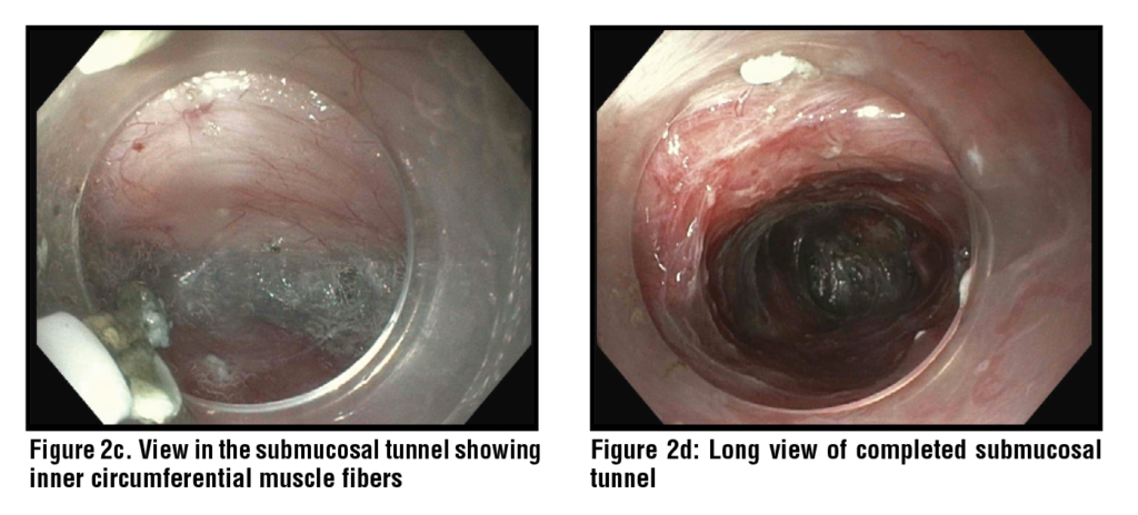
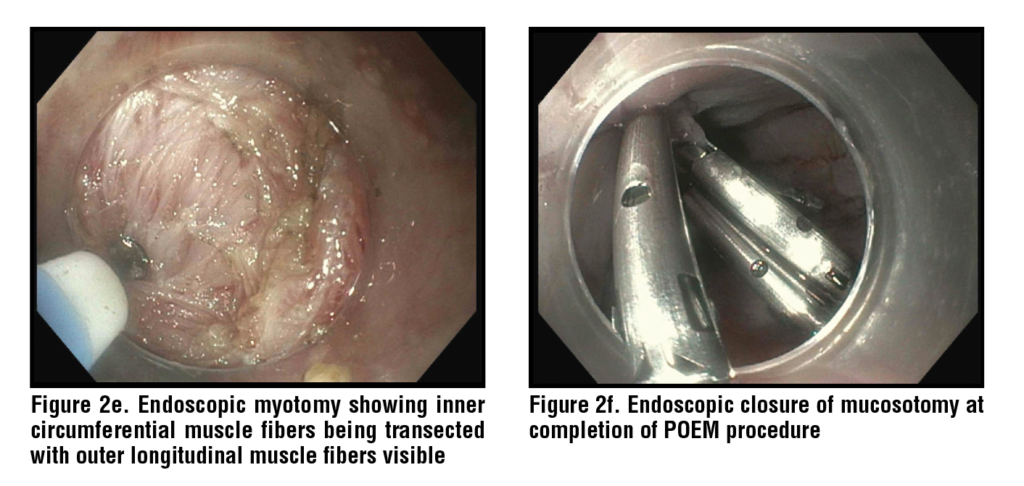
There are many variations to POEM, but in general, a mucosal entry site is made within the esophagus to expose the submucosal space. Once inside, a submucosal tunnel is created, extending a short distance past the GEJ. Then, circular LES muscle fibers are dissected until the endoscope can be easily passed through the GEJ to confirm successful myotomy. Finally, the mucosal entry site is closed. (Figures 2a-f)
Efficacy
Inoue et al. published the first prospective cohort study in 2010 which showed a significant reduction in symptom score (10 to 1.3; p=0.0003) and LES pressure (52.4mmHg to 19.9mmHg; p=0.0001).47 Since then, many meta-analyses have demonstrated high clinical success in 90-97% of patients.,,,, Vespa et al. performed one of the largest meta-analyses of 31 studies with 3,023 patients revealing a clinical success rate of 91% at a median follow-up of 29-months. Zhang et al. analyzed symptom resolution over time and reported clinical success rates of 91.3%, 90.4%, 89.8%, and 82.2% at 2, 3, 4, and 5 years, respectively. Post-POEM quality of life also significantly (p<0.05) improved according to a meta-analysis by Zhong et al.
Shou et al. identified risk factors for POEM failure in a meta-analysis of 27 studies with 9,371 patients. Worse outcomes occurred in patients with a sigmoid esophagus (a manifestation of advanced achalasia) (OR 1.90, 95% CI 1.45-2.47), type I achalasia (OR 1.30, 95% CI 1.04-1.63), type III achalasia (OR 1.26, 95% CI 0.89-1.78), prior Heller myotomy (OR 5.75, 95% CI 3.97-8.34), and prior PD (OR 1.18, 95% CI 1.07-1.29).56 Better outcomes occurred in patients with type II achalasia (OR 0.59, 95% CI 0.47-0.75).56 In contrast, there was no significant difference in symptom resolution between achalasia subtypes according to meta-analyses by Zhang et al. and Andolfi et al.54, POEM was examined in the elderly patient population by Hayat et al. who found no significant difference in success among patients ≥65-years-old (95.74% vs 95.31%) or ≥75-years-old (99.01% vs 97.97%) compared to their younger counterparts.
When initial POEM fails, repeat POEM may be attempted with good outcomes. Hayat et al. analyzed 6 studies with 123 patients who underwent repeat POEM 12.2-13.5 months on average after their initial failed POEM. 82.69% of patients achieved clinical success after repeat POEM.59
To optimize outcomes, many variations in POEM technique have been studied. Dhoop et al. compared full-thickness and circular myotomy and found no significant difference in clinical success at 1 or 3 years. However, full-thickness myotomy had an increased risk for post-POEM GERD (RR 3.03, 95% CI 1.53-6.02; p=0.002) and esophagitis (RR 1.79, 95% CI 1.09-2.76; p=0.02) compared to circular myotomy.61 Rodriguez de Santiago et al. and Mohan et al. performed meta-analyses comparing anterior and posterior wall tunneling, each finding no significant difference between myotomy approaches.52, Procedure time was shorter with posterior (62.1 minutes) compared to anterior (82.7 minutes) wall tunneling.62 In practice, individual operators are free to perform POEM as they see fit.
Optimal myotomy length has been extensively studied, comparing short and long myotomy lengths. Zhang et al., Weng et al., and Ghazaleh et al. performed meta-analyses which showed shorter myotomy resulted in shorter procedure times, as expected, with the same or less risk of reflux but no significant difference in clinical success.,,
POEM has also been successfully used as rescue therapy following failed improvement with other treatment modalities for achalasia. Tan et al. and Zhong et al. demonstrated clinical success in 90.8% and 91.0% of these patients, respectively.,
Adverse Events
Symptomatic reflux and reflux esophagitis are two of the most common adverse events seen following POEM with rates ranging from 22.0-26.2% and 5.9-19.0%, respectively.47,48,49,53,54,, Mota et al. performed a meta-analysis of 25 studies and found no statistical difference in post-POEM GERD rates when comparing full-thickness, partial-thickness and circular myotomy, anterior and posterior myotomy, or subtypes of achalasia. This differs from Dhoop et al.’s more recent meta-analysis which showed an increased risk of post-POEM GERD and esophagitis with full-thickness myotomy.61 POEM combined with fundoplication (POEM-F) has been trialed in an attempt to reduce post-POEM reflux. The rate of post-POEM-F reflux reported by Gopakumar et al.’s meta-analysis of 7 studies with 127 patients was 13.8% which is lower than most reflux rates after POEM alone but comes with far higher costs.
Serious adverse events include subcutaneous emphysema (21.8-31.6% of patients), pneumoperitoneum (16.2-30.6%), pneumothorax (8.7-11.0%), and in very rare instances, mediastinal emphysema/leak (0.3-4.9%), esophageal/gastric perforation (2.6%), bleeding (0.9-1.1%), and death (0.09%).48,69
Conclusion
Achalasia is a complex and debilitating esophageal motility disorder, but there are many efficacious endoscopic treatment options available, enabling individualized treatment approaches. Botulinum toxin injections have shown decent short-term clinical success with few adverse events and are best suited for older patients and/or those who are not candidates for more invasive endoscopic modalities. Pneumatic dilation has shown better short-term outcomes but is limited by high rates of symptom relapse and a small risk of serious adverse events, most notably esophageal perforation. Peroral endoscopic myotomy offers the highest long-term success rates but may be accompanied by post-procedural reflux symptoms and/or reflux esophagitis. Treatment should be tailored to patient preferences and institutional expertise.
References
1 Vaezi MF, Pandolfino JE, Yadlapati RH, Greer KB, Kavitt RT. ACG Clinical Guidelines: Diagnosis and Management of Achalasia. Am J Gastroenterol. 2020;115(9):1393-1411. doi:10.14309/ajg.0000000000000731
2 Gaber CE, Eluri S, Cotton CC, et al. Epidemiologic and Economic Burden of Achalasia in the United States. Clin Gastroenterol Hepatol. 2022;20(2):342-352.e5. doi:10.1016/j.cgh.2021.02.035
3 Savarino E, Bhatia S, Roman S, et al. Achalasia. Nat Rev Dis Primers. 2022;8(1):28. Published 2022 May 5. doi:10.1038/s41572-022-00356-8
4 Park W, Vaezi MF. Etiology and pathogenesis of achalasia: the current understanding. Am J Gastroenterol. 2005;100(6):1404-1414. doi:10.1111/j.1572- 0241.2005.41775.x
5 Francis DL, Katzka DA. Achalasia: update on the disease and its treatment. Gastroenterology. 2010;139(2):369-374. doi:10.1053/j.gastro.2010.06.024
6 Kahrilas PJ, Boeckxstaens G. The spectrum of achalasia: lessons from studies of pathophysiology and high-resolution manometry. Gastroenterology. 2013;145(5):954-965. doi:10.1053/j.gastro.2013.08.038
7 Panesso-Gómez S, Pavia P, Rodríguez-Mantilla IE, et al. Trypanosoma cruzi Detection in Colombian Patients with a Diagnosis of Esophageal Achalasia. Am J Trop Med Hyg. 2018;98(3):717-723. doi:10.4269/ajtmh.17-0417
8 Khashab MA, Vela MF, Thosani N, et al. ASGE guideline on the management of achalasia. Gastrointest Endosc. 2020;91(2):213-227.e6. doi:10.1016/j.gie.2019.04.231
9 Spechler SJ, Souza RF, Rosenberg SJ, Ruben RA, Goyal RK. Heartburn in patients with achalasia. Gut. 1995;37(3):305- 308. doi:10.1136/gut.37.3.305
10 Eckardt VF, Aignherr C, Bernhard G. Predictors of outcome in patients with achalasia treated by pneumatic dilation. Gastroenterology. 1992;103(6):1732-1738. doi:10.1016/0016-5085(92)91428-7
11 Pandolfino JE, Kwiatek MA, Nealis T, Bulsiewicz W, Post J, Kahrilas PJ. Achalasia: a new clinically relevant classification by high-resolution manometry. Gastroenterology. 2008;135(5):1526-1533. doi:10.1053/j.gastro.2008.07.022
12 Vespa E, Barchi A, Mandarino FV, et al. Standard length of peroral endoscopic myotomy (POEM) for achalasia: a systematic review and meta-analysis. Dis Esophagus. 2024;37(12):doae069. doi:10.1093/dote/doae069
13 Nassri A, Ramzan Z. Pharmacotherapy for the management of achalasia: Current status, challenges and future directions. World J Gastrointest Pharmacol Ther. 2015;6(4):145- 155. doi:10.4292/wjgpt.v6.i4.145
14 Storr M, Born P, Frimberger E, et al. Treatment of achalasia: the short-term response to botulinum toxin injection seems to be independent of any kind of pretreatment [published correction appears in BMC Gastroenterol. 2003 Nov 7;3(1):32]. BMC Gastroenterol. 2002;2:19. doi:10.1186/1471-230x-2-19
15 Pasricha PJ, Ravich WJ, Hendrix TR, Sostre S, Jones B, Kalloo AN. Intrasphincteric botulinum toxin for the treatment of achalasia [published correction appears in N Engl J Med 1995 Jul 6;333(1):75]. N Engl J Med. 1995;332(12):774- 778. doi:10.1056/NEJM199503233321203
16 D’Onofrio V, Annese V, Miletto P, et al. Long-term follow-up of achalasic patients treated with botulinum toxin. Dis Esophagus. 2000;13(2):96-103. doi:10.1046/j.1442- 2050.2000.00094.x
17 Campos GM, Vittinghoff E, Rabl C, et al. Endoscopic and surgical treatments for achalasia: a systematic review and meta-analysis. Ann Surg. 2009;249(1):45-57. doi:10.1097/ SLA.0b013e31818e43ab
18 Zhu Q, Liu J, Yang C. Clinical study on combined therapy of botulinum toxin injection and small balloon dilation in patients with esophageal achalasia. Dig Surg. 2009;26(6):493-498. doi:10.1159/000229784
19 Annese V, Bassotti G, Coccia G, et al. A multicentre randomised study of intrasphincteric botulinum toxin in patients with oesophageal achalasia. GISMAD Achalasia Study Group. Gut. 2000;46(5):597-600. doi:10.1136/gut.46.5.597
20 D’Onofrio V, Annese V, Miletto P, et al. Long-term follow-up of achalasic patients treated with botulinum toxin. Dis Esophagus. 2000;13(2):96-103. doi:10.1046/j.1442- 2050.2000.00094.x
21 Leyden JE, Moss AC, MacMathuna P. Endoscopic pneumatic dilation versus botulinum toxin injection in the management of primary achalasia. Cochrane Database Syst Rev. 2014;2014(12):CD005046. doi:10.1002/14651858. CD005046.pub3
22 Patti MG, Feo CV, Arcerito M, et al. Effects of previ
ous treatment on results of laparoscopic Heller myotomy for achalasia. Dig Dis Sci. 1999;44(11):2270-2276. doi:10.1023/a:1026660921776
23 Smith CD, Stival A, Howell DL, Swafford V. Endoscopic therapy for achalasia before Heller myotomy results in worse outcomes than heller myotomy alone. Ann Surg. 2006;243(5):579-586. doi:10.1097/01. sla.0000217524.75529.2d
24 Kadiyala J, Canakis A, Lee DU, Xue P, Fan GH, Kim RE. Comparing clinical success and procedural difficulty between treatment-naïve and treatment-refractory patients with esophageal motility disorders during peroral endoscopic myotomy. Gastrointest Endosc. 2023;98(1):19-27. e11. doi:10.1016/j.gie.2023.01.052
25 Khan SZ, Benson J, Boutros C, Lyons J, Hashimoto DA, Marks JM. Do preoperative endoscopic interventions predict symptom recurrence or reintervention after peroral endoscopic myotomy?. Surg Endosc. 2023;37(9):7153-7158. doi:10.1007/s00464-023-10196-6
26 Jones EL, Meara MP, Pittman MR, Hazey JW, Perry KA. Prior treatment does not influence the performance or early outcome of per-oral endoscopic myotomy for achalasia. Surg Endosc. 2016;30(4):1282-1286. doi:10.1007/s00464-015- 4339-y
27 Becker K, Biesenbach S, Erckenbrecht JF, Frieling T. Effect of balloon compliance on symptomatic success of pneumatic dilation in achalasia patients. Z Gastroenterol. 2001;39(10):831-836. doi:10.1055/s-2001-17868
28 Uppal DS, Wang AY. Update on the endoscopic treatments for achalasia. World J Gastroenterol. 2016;22(39):8670- 8683. doi:10.3748/wjg.v22.i39.8670
29 Khan AA, Shah SW, Alam A, Butt AK, Shafqat F, Castell DO. Pneumatic balloon dilation in achalasia: a prospective comparison of balloon distention time. Am J Gastroenterol. 1998;93(7):1064-1067. doi:10.1111/j.1572- 0241.1998.00330.x
30 Ghoshal UC, Chaudhuri S, Pal BB, Dhar K, Ray G, Banerjee PK. Randomized controlled trial of intrasphincteric botulinum toxin A injection versus balloon dilatation in treatment of achalasia cardia. Dis Esophagus. 2001;14(3-4):227- 231. doi:10.1046/j.1442-2050.2001.00189.x
31 Ponds FA, Fockens P, Lei A, et al. Effect of Peroral Endoscopic Myotomy vs Pneumatic Dilation on Symptom Severity and Treatment Outcomes Among Treatment-Naive Patients With Achalasia: A Randomized Clinical Trial. JAMA. 2019;322(2):134-144. doi:10.1001/jama.2019.8859
32 Bansal R, Nostrant TT, Scheiman JM, et al. Intrasphincteric botulinum toxin versus pneumatic balloon dilation for treatment of primary achalasia. J Clin Gastroenterol. 2003;36(3):209-214. doi:10.1097/00004836-200303000- 00005
33 Gideon RM, Castell DO, Yarze J. Prospective randomized comparison of pneumatic dilatation technique in patients with idiopathic achalasia. Dig Dis Sci. 1999;44(9):1853- 1857. doi:10.1023/a:1018898824135
34 Leyden JE, Moss AC, MacMathuna P. Endoscopic pneumatic dilation versus botulinum toxin injection in the management of primary achalasia. Cochrane Database Syst Rev. 2014;2014(12):CD005046. doi:10.1002/14651858. CD005046.pub3
35 van Hoeij FB, Prins LI, Smout AJPM, Bredenoord AJ. Efficacy and safety of pneumatic dilation in achalasia: A systematic review and meta-analysis. Neurogastroenterol Motil. 2019;31(7):e13548. doi:10.1111/nmo.13548
36 Farhoomand K, Connor JT, Richter JE, Achkar E, Vaezi MF. Predictors of outcome of pneumatic dilation in achalasia. Clin Gastroenterol Hepatol. 2004;2(5):389-394. doi:10.1016/s1542-3565(04)00123-5
37 Eckardt VF, Aignherr C, Bernhard G. Predictors of outcome in patients with achalasia treated by pneumatic dilation. Gastroenterology. 1992;103(6):1732-1738. doi:10.1016/0016-5085(92)91428-7
38 Baumann AJ, Carlson DA. EsoFLIP for esophageal dilation: proposed advantages. Curr Opin Gastroenterol. 2020;36(4):329-335. doi:10.1097/MOG.0000000000000639
39 Iqbal U, Yodice M, Ahmed Z, et al. Safety and efficacy of EsoFLIP dilation in patients with esophageal dysmotility: a systematic review. Dis Esophagus. 2024;37(8):doae036. doi:10.1093/dote/doae036
40 Schnurre L, Murray FR, Schindler V, et al. Short-term outcome after singular hydraulic EsoFLIP dilation in patients with achalasia: A feasibility study. Neurogastroenterol Motil. 2020;32(9):e13864. doi:10.1111/nmo.13864
41 Kappelle WF, Bogte A, Siersema PD. Hydraulic dilation with a shape-measuring balloon in idiopathic achalasia: a feasibility study. Endoscopy. 2015;47(11):1028-1034. doi:10.1055/s-0034-1392481
42 Chan B, Ding J, Bilal M et al. Safety and efficacy of EsoFLIP hydraulic dilation for achalasia. Gastrointest Endosc 2020; 91(6): AB136–7. https://doi.org/10.1016/j. gie.2020.03.1055
43 Carlson D A, Atluri S, Balla M, Listernick Z, Kahrilas P, Pandolfino J E. 871 hydraulic dilation for achalasia: a series utilizing a 30-mm functional lumen imaging probe dilator, the EsoFLIP. Gastrointest Endosc 2016; 83(5): AB173. https://doi.org/10.1016/j.gie.2020.03.1055
44 Tsai L, Kunkel D. Treatment outcomes from EsoFLIP dilaiton in patients with achalasia vary by subtype, Digestive Disease Week 2021, AGA Abstracts, Supplemental Su207, S-649. 2021.
45 Pasricha PJ, Hawari R, Ahmed I, et al. Submucosal endoscopic esophageal myotomy: a novel experimental approach for the treatment of achalasia. Endoscopy. 2007;39(9):761- 764. doi:10.1055/s-2007-966764
46 Inoue, H., Minami, H., Satodate, H., & Kudo, S. (2009). First Clinical Experience of Submucosal Endoscopic Esophageal Myotomy for Esophageal Achalasia with No Skin Incision. Gastrointestinal Endoscopy, 69. doi:10.1016/j. gie.2009.03.133
47 Inoue H, Minami H, Kobayashi Y, et al. Peroral endoscopic myotomy (POEM) for esophageal achalasia. Endoscopy. 2010;42(4):265-271. doi:10.1055/s-0029-1244080
48 Talukdar R, Inoue H, Nageshwar Reddy D. Efficacy of peroral endoscopic myotomy (POEM) in the treatment of achalasia: a systematic review and meta-analysis. Surg Endosc. 2015;29(11):3030-3046. doi:10.1007/s00464-014-4040-6
49 Barbieri LA, Hassan C, Rosati R, Romario UF, Correale L, Repici A. Systematic review and meta-analysis: Efficacy and safety of POEM for achalasia. United European Gastroenterol J. 2015;3(4):325-334. doi:10.1177/2050640615581732
50 Evensen H, Kristensen V, Larssen L, Sandstad O, Hauge T, Medhus AW. Outcome of peroral endoscopic myotomy (POEM) in treatment-naive patients. A systematic review. Scand J Gastroenterol. 2019;54(1):1-7. doi:10.1080 /00365521.2018.1549271
51 Li H, Peng W, Huang S, et al. The 2 years’ long-term
efficacy and safety of peroral endoscopic myotomy for the treatment of achalasia: a systematic review. J Cardiothorac Surg. 2019;14(1):1. Published 2019 Jan 3. doi:10.1186/ s13019-018-0811-9
52 Rodríguez de Santiago E, Mohammed N, Manolakis A, Shimamura Y, Onimaru M, Inoue H. Anterior versus posterior myotomy during poem for the treatment of achalasia: systematic review and meta-analysis of randomized clinical trials. J Gastrointestin Liver Dis. 2019;28(1):107-115. doi:10.15403/jgld.2014.1121.281.pom
53 Vespa E, Barchi A, Mandarino FV, et al. Standard length of peroral endoscopic myotomy (POEM) for achalasia: a systematic review and meta-analysis. Dis Esophagus. 2024;37(12):doae069. doi:10.1093/dote/doae069
54 Zhang H, Zeng X, Huang S, et al. Mid-Term and Long- Term Outcomes of Peroral Endoscopic Myotomy for the Treatment of Achalasia: A Systematic Review and Meta- Analysis. Dig Dis Sci. 2023;68(4):1386-1396. doi:10.1007/ s10620-022-07720-4
55 Zhong C, Tan S, Ren Y, et al. Quality of Life Following Peroral Endoscopic Myotomy for Esophageal Achalasia: A Systematic Review and Meta-Analysis. Ann Thorac Cardiovasc Surg. 2020;26(3):113-124. doi:10.5761/atcs. ra.19-00273
56 Shou Y, Wang X, Liu D. Factors associated with peroral endoscopic myotomy for achalasia outcomes: systematic review and meta-analysis. Surg Endosc. 2024;38(7):3503- 3519. doi:10.1007/s00464-024-10862-3
57 Andolfi C, Fisichella PM. Meta-analysis of clinical outcome after treatment for achalasia based on manometric subtypes. Br J Surg. 2019;106(4):332-341. doi:10.1002/ bjs.11049
58 Hayat U, Kamal F, Memon A, et al. Peroral Endoscopic Myotomy for Achalasia Among the Elderly Population: A Systematic Review and Meta-Analysis. J Clin Gastroenterol. Published online February 10, 2025. doi:10.1097/ MCG.0000000000002103
59 Hayat U, Kamal F, Rana UI, et al. Efficacy and Safety of Repeat Per-oral Endoscopic Myotomy After Failed POEM for Achalasia: A Systematic Review and meta-analysis. J Clin Gastroenterol. Published online December 5, 2024. doi:10.1097/MCG.0000000000002117
60 Mota RCL, de Moura EGH, de Moura DTH, et al. Risk factors for gastroesophageal reflux after POEM for achalasia: a systematic review and meta-analysis. Surg Endosc. 2021;35(1):383-397. doi:10.1007/s00464-020-07412-y
61 Dhoop S, Abu-Rumaileh M, Sayeh W, et al. Efficacy and safety of full-thickness versus circular peroral endoscopic myotomy for treatment of achalasia: a systematic review and meta-analysis. Ann Gastroenterol. 2025;38(2):143-155. doi:10.20524/aog.2025.0946
62 Mohan BP, Ofosu A, Chandan S, et al. Anterior versus posterior approach in peroral endoscopic myotomy (POEM): a systematic review and meta-analysis. Endoscopy. 2020;52(4):251-258. doi:10.1055/a-1090-0788
63 Zhang H, Zeng X, Huang S, et al. Safety and Efficacy of Peroral Endoscopic Shorter Myotomy versus Longer Myotomy for Patients with Achalasia: A Systematic Review and Meta-analysis. Gastroenterol Res Pract. 2022;2022:6770864. Published 2022 Mar 30. doi:10.1155/2022/6770864
64 Weng CY, He CH, Zhuang MY, Xu JL, Lyu B. Peroral endoscopic longer vs shorter esophageal myotomy for achalasia treatment: A systematic review and meta-analysis. World J Gastrointest Surg. 2022;14(3):247-259. doi:10.4240/wjgs. v14.i3.247
65 Ghazaleh S, Beran A, Khader Y, et al. Short versus standard peroral endoscopic myotomy for esophageal achalasia: a systematic review and meta-analysis. Ann Gastroenterol. 2021;34(5):634-642. doi:10.20524/aog.2021.0644
66 Tan S, Zhong C, Ren Y, et al. Efficacy and Safety of Peroral Endoscopic Myotomy in Achalasia Patients with Failed Previous Intervention: A Systematic Review and Meta- Analysis. Gut Liver. 2021;15(2):153-167. doi:10.5009/ gnl19234
67 Zhong C, Ni B, Liu S, et al. The Effect of Peroral Endoscopic Myotomy in Achalasia Patients with Prior Endoscopic Intervention: A Systematic Review and Meta-Analysis. Dig Surg. 2021;38(2):136-148. doi:10.1159/000512627
68 Vespa E, Pellegatta G, Chandrasekar VT, et al. Long-term outcomes of peroral endoscopic myotomy for achalasia: a systematic review and meta-analysis. Endoscopy. 2023;55(2):167-175. doi:10.1055/a-1894-0147
69 Patel K, Abbassi-Ghadi N, Markar S, Kumar S, Jethwa P, Zaninotto G. Peroral endoscopic myotomy for the treatment of esophageal achalasia: systematic review and pooled analysis. Dis Esophagus. 2016;29(7):807-819. doi:10.1111/ dote.12387
70 Mota RCL, de Moura EGH, de Moura DTH, et al. Risk factors for gastroesophageal reflux after POEM for achalasia: a systematic review and meta-analysis. Surg Endosc. 2021;35(1):383-397. doi:10.1007/s00464-020-07412-y
71 Gopakumar H, Annor E, Vohra I, et al. Peroral endoscopic myotomy with fundoplication (POEM-F) for achalasia: Systematic review and meta-analysis [published correction appears in Endosc Int Open. 2025 Mar 19;13:a25624787. doi: 10.1055/a-2562-4787.]. Endosc Int Open. 2025;13:a25368132. Published 2025 Mar 14. doi:10.1055/a-2536-8132