
 Douglas G. Adler
Introduction
Malignant gastric outlet obstruction has a variety of treatments. This case highlights a patient with malignant gastric outlet obstruction who was treated via endoscopic gastroenterostomy with the assistance of a newly available double-balloon catheter.
Case Report
A 73-year-old female with known cholangiocarcinoma presented with 5 days of progressive nausea with vomiting. The patient could swallow food but noted undigested gastric contents several hours after each meal. The patient had lost 10 pounds in just 5 days. On CT scan, the stomach was markedly distended with fluid with an abrupt change in caliber at the level of the pylorus and proximal duodenum, highly suggestive of an obstruction. The distal stomach and proximal small bowel were thickened. There was an adjacent soft tissue mass in the porta hepatis consistent with known cholangiocarcinoma, and the gastric outlet obstruction was felt to be due to direct tumor involvement of the gastric outlet. There was no ascites. A previously placed metal biliary stent was felt to be patent. A nasogastric tube was placed for gastric decompression.
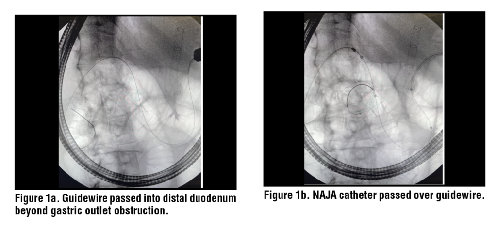
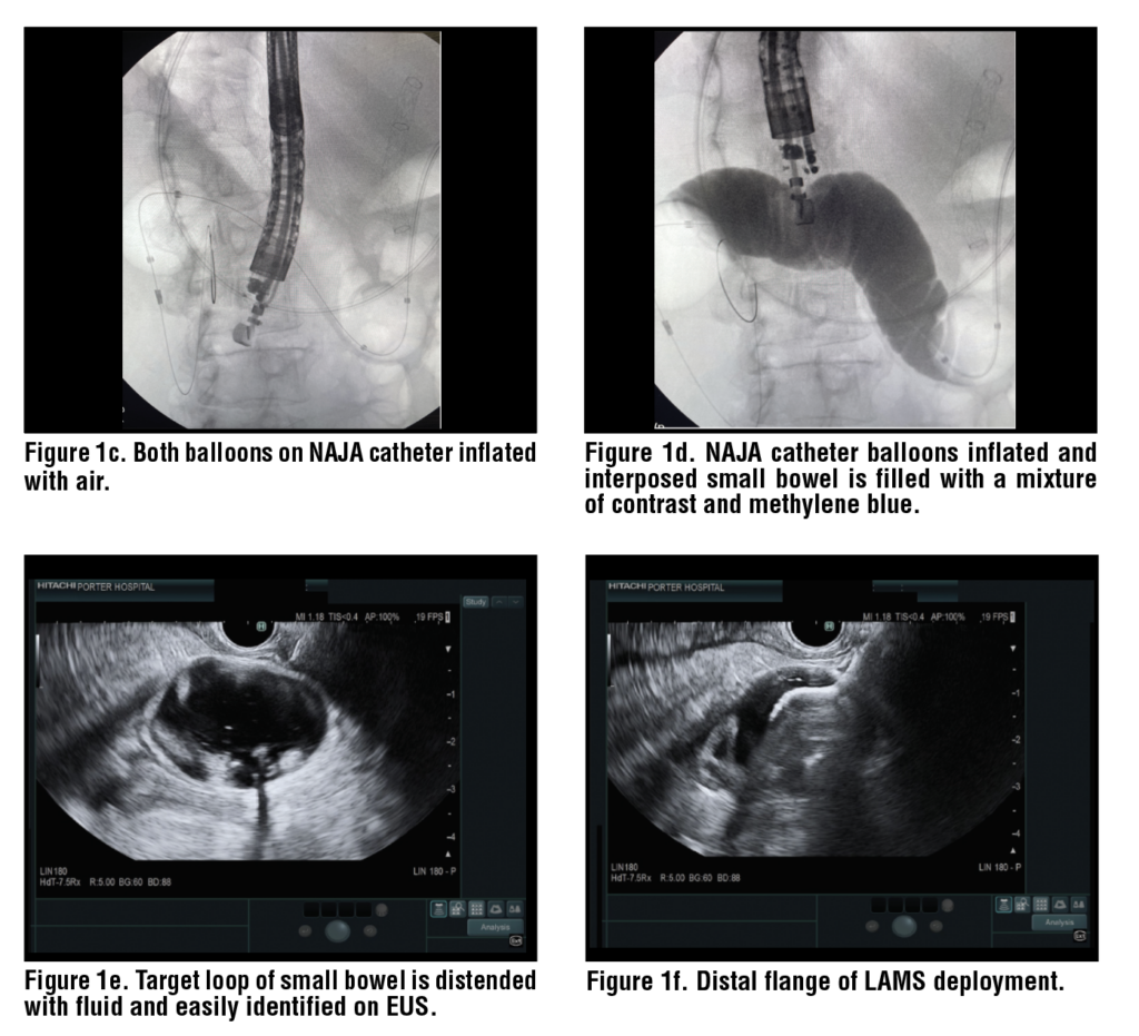
The patient was not felt to be a surgical candidate for resection of her primary tumor. Gastroenterology was consulted and the patient was given the options of pursuing a surgical gastrojejunostomy, a pyloric/duodenal stent, or an endoscopic ultrasound-guided gastroenterostomy (EUS-GE). The patient elected to undergo EUS-GJ. We elected to perform the EUS-GE using the NAJA catheter/balloon catheter device (Chess Medical, Gaithersburg, MD). The catheter consists of a dual balloon catheter that can be used to create a fluid-filled region of small bowel between two air-filled balloons to create a target for EUS-GE.
Procedure In Detail
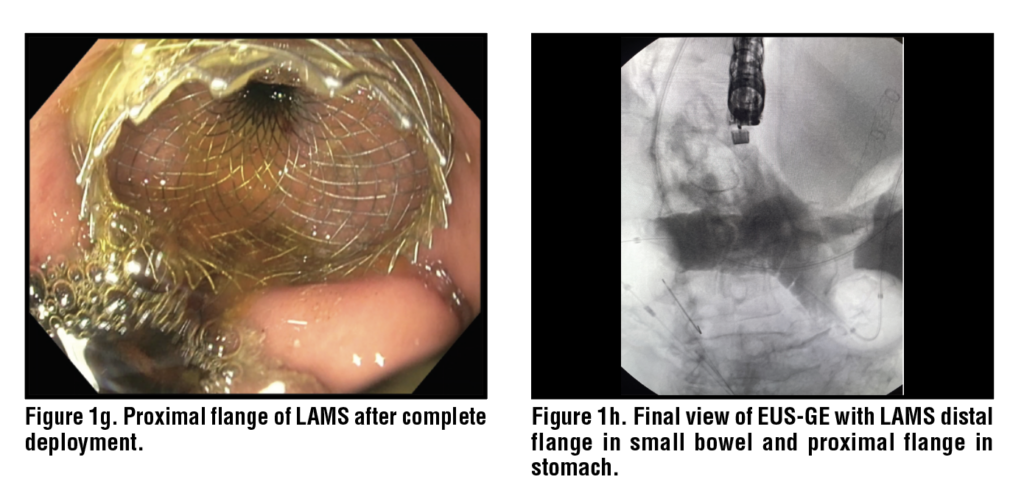
The nasogastric tube was removed. LA Grade D esophagitis with no bleeding was found in the entire esophagus and was felt to be secondary to her gastric outlet obstruction. A large amount of solid food was found in the stomach. A large fungating and ulcerated mass with no bleeding was found in the duodenal bulb. An acquired malignant-appearing, intrinsic severe stenosis was found in the duodenal bulb. Using a therapeutic channel upper endoscope, a 0.025-inch x 450 cm guidewire was advanced across the duodenal bulb stricture into the distal duodenum/proximal jejunum under fluoroscopic and endoscopic guidance. The endoscope was removed over the wire. The NAJA GI balloon catheter was advanced over the wire under fluoroscopic guidance across the malignant stricture into the distal duodenum. The proximal catheter balloon was inflated with 30 cc of air. The distal catheter balloon was subsequently inflated with 30 cc of air. Methylene blue mixed with contrast was injected between the balloons in the third/fourth portion of the duodenum. Then a linear echoendoscope was passed down to the stomach. The loop of dilated duodenum was noted in the third to fourth portion of the duodenum on EUS and fluoroscopy. The proximal and distal balloons were subsequently deflated and re-inflated with 40 cc of air, respectively, and the interposed segment was again filled with fluid. Once the location of the target small bowel between the two balloons was again located, an electrocautery enhanced 15 mm x 10 mm Axios Stent (Boston Scientific, Natick MA) was deployed across the gastroenterostomy. Upon deployment, there was immediate drainage of methylene blue and bile consistent with good attainment of enteral access. There was also clear endoscopic visualization of small bowel through the Axios stent, confirming successful deployment. The NAJA catheter balloons were subsequently deflated and removed from the patient along with the guidewire. At this point the procedure was complete. (Figure 1.)
Clinical Outcome
The patient tolerated the procedure with no adverse events. She was started on a clear liquid diet that day and slowly advanced. Her nausea and vomiting resolved, and on post-procedure day three she was discharged tolerating a soft diet with plans to continue dietary advancement as an outpatient.
Discussion
Malignant gastric outlet obstruction is commonly encountered among patients with pancreatic, biliary, gastric, and ampullary cancers. Patients often present with nausea and vomiting in the setting of weight loss. Treatments for malignant gastric outlet obstruction include surgical bypass/gastrojejunostomy, enteral stents, and EUS-GE. Feeding tubes, including direct jejunostomy tubes with a venting gastrostomy tube, or a percutaneous endoscopic gastrostomy with a jejunal feeding arm, can also be used. Total parenteral nutrition is also an option. Surgical gastrojejunostomy was historically the first-line treatment, but many patients with advanced malignancy are poor surgical candidates. These patients often have malignant ascites and/or are malnourished with hypoalbuminemia, giving surgeons significant pause in this situation. The procedure can be performed laparoscopically or via robotic surgery and can often be combined with a biliary bypass at the same time in patients with combined biliary obstruction. In many centers, surgical gastrojejunostomy is reserved for patients who are felt to have a potentially long lifespan, but this is relatively uncommon among patients with malignant gastric outlet obstruction.,
Duodenal/enteral stents have been in widespread use for over 20 years. Duodenal/enteral stents are placed under a combination of endoscopic and fluoroscopic guidance. These devices can be placed quickly and, in general, patients do not have to stop taking anticoagulants before implantation. In the United States, on-label duodenal/enteral stents are uncovered, so they are susceptible to tissue/tumor ingrowth and overgrowth, which can cause recurrent outlet obstruction. This is usually treated by tissue ablation within the stent or, more commonly, placement of a new stent inside the old one. LAMS can also be used to treat malignant gastric outlet obstruction if they are placed within the lumen in an off-label manner.,, LAMS are generally only placed for short segment luminal strictures given their limited saddle length.
EUS-GE represents the newest treatment for malignant gastric outlet obstruction. Using a LAMS in an off-label manner, the endoscopist is able to create a stable gastroenterostomy (often to the distal duodenum or proximal jejunum), allowing patients to resume oral nutrition, hydration, and medication delivery. The technique is still mostly performed by expert endoscopists at high-volume centers but is slowly becoming more widely adopted. In the past, EUS-GE was often performed with the aid of a catheter passed across the stricture to “flood” the small bowel distal to the obstruction with fluid/saline to create a better target for stent placement. In some cases, a loop of suspected small bowel distal to the obstruction was simply accessed via a “freehand” approach without distension with fluid.
A large systematic review and meta-analysis reported on outcomes and adverse events of EUS-GE. The authors included 36 studies (n = 1846) in the meta-analysis. The pooled technical success rate was 96.9% and the clinical success rate was 90.6%. The incidence of adverse events was 13.0%. The incidence of serious adverse events was 1.2% and procedure-related mortality 0.3%. A separate systematic review and meta-analysis of EUS-GE found an overall adverse event rate of 17%. It seems likely that adverse events with EUS-GE may be under-reported in the literature given the complexity and relatively novel nature of the procedure. No definitive standard technique for EUS-GE exists, but efforts have been made towards identifying best practices to reduce adverse events and improve outcomes.,
Balloon-catheter assisted EUS-GE with the NAJA device aims to reduce the rate of adverse events by creating a stable target for LAMS deployment and by increasing the diameter of the target loop of small bowel via distension with fluid. The device reduces the risk of accidentally accessing an undesired loop of small bowel or interposed colon and significantly enhances visualization during LAMS targeting and deployment. In practice, it obviates the need for “freehand” techniques that may be more dangerous for the patient and increase the risk of adverse events. Other balloon-assisted versions of EUS-GE have been reported as well with similar good outcomes. , The EPASS system (Create Medic Co., Ltd, Yokohama, Japan) has been available outside of the USA for some time and is similar to the NAJA device. Studies have shown high technical and clinical success with acceptable rates of adverse events with EPASS assisted EUSGE. EPASS is not available in the United States.
Devices like NAJA and EPASS do increase cost of EUS-GE, but likely with the benefit of reduced risk of certain adverse events, most notably perforation and stent misdeployment/maldeployment. NAJA has only very recently been introduced in the USA and is not widely available. Still, it seems likely that balloon catheter-assisted techniques will likely become more common over time given the potential to reduce adverse events when compared with freehand EUS-GE.
References
1. Acharya T, Spathis A, Godfrey E, Corbett G, Kuhn I, Etkind SN. Palliative management of malignant gastric outlet obstruction: A practice review. Palliat Med. 2026 Mar;40(3):333-343. doi: 10.1177/02692163251403430. Epub 2025 Dec 27. PMID: 41454660; PMCID: PMC12936159.
2. Acharya T, Spathis A, Godfrey E, Corbett G, Kuhn I, Etkind SN. Palliative management of malignant gastric outlet obstruction: A practice review. Palliat Med. 2026 Mar;40(3):333-343. doi: 10.1177/02692163251403430. Epub 2025 Dec 27. PMID: 41454660; PMCID: PMC12936159.
3. Jang S, Stevens T, Lopez R, Bhatt A, Vargo JJ. Superiority of Gastrojejunostomy Over Endoscopic Stenting for Palliation of Malignant Gastric Outlet Obstruction. Clin Gastroenterol Hepatol. 2019 Jun;17(7):1295-1302.e1. doi: 10.1016/j. cgh.2018.10.042. Epub 2018 Oct 31. PMID: 30391433.
4. Adler DG. Should Patients With Malignant Gastric Outlet Obstruction Receive Stents or Surgery? Clin Gastroenterol Hepatol. 2019 Jun;17(7):1242-1244. doi: 10.1016/j. cgh.2018.11.049. Epub 2018 Nov 29. PMID: 30503965.
5. Adler DG, Baron TH. Endoscopic palliation of malignant gastric outlet obstruction using self-expanding metal stents: experience in 36 patients. Am J Gastroenterol. 2002 Jan;97(1):72-8. doi: 10.1111/j.1572-0241.2002.05423.x. PMID: 11808972.
6. Mizrahi M, Fahmawi Y, Merritt L, Kumar M, Tharian B, Khan SA, Inamdar S, Sharma N, Uppal D, Shami VM, Kashif MS, Gabr M, Pleskow D, Berzin TM, James TW, Croglio M, Baron TH, Adler DG. Luminal-apposing stents for benign intraluminal strictures: a large United States multicenter study of clinical outcomes. Ann Gastroenterol. 2021;34(1):33-38. doi: 10.20524/aog.2020.0538. Epub 2020 Oct 2. PMID: 33414619; PMCID: PMC7774669.
7. Mohan BP, Chandan S, Garg R, Mohamed S, Shakhatreh M, Dugyala S, Mashiana HS, Ponnada S, Asokkumar R, Adler DG. Lumen-apposing Metal Stents, Fully Covered Self-expanding Metal Stents, and Biodegradable Stents in the Management of Benign of GI Strictures: A Systematic Review and Meta-Analysis. J Clin Gastroenterol. 2019 Sep;53(8):560-573. doi: 10.1097/MCG.0000000000001228. PMID: 31149932.
8. Larson B, Adler DG. Lumen-apposing metal stents for gastrointestinal luminal strictures: current use and future directions. Ann Gastroenterol. 2019 Mar-Apr;32(2):141-146. doi: 10.20524/aog.2018.0337. Epub 2018 Dec 14. PMID: 30837786; PMCID: PMC6394263.
9. Giri S, Harindranath S, Mohan BP, Jearth V, Varghese J, Kozyk M, Kale A, Sundaram S. Adverse events with endoscopic ultrasound-guided gastroenterostomy for gastric outlet obstruction-A systematic review and meta-analysis. United European Gastroenterol J. 2024 Sep;12(7):879- 890. doi: 10.1002/ueg2.12576. Epub 2024 May 15. PMID: 38747173; PMCID: PMC11497769.
10. Li JS, Lin K, Tang J, Liu F, Fang J. EUS-guided gastroenterostomy for gastric outlet obstruction: a comprehensive meta-analysis. Minim Invasive Ther Allied Technol. 2023 Dec;32(6):285-299. doi: 10.1080/13645706.2023.2221336. Epub 2023 Jun 13. PMID: 37310282.
11. Vanella G, Frigo F, Bronswijk M, van Wanrooij RLJ, Chen YI, Binmoeller KF, Perez-Miranda M, Leone R, Chahal P, Chan SM, Jovani M, Tyberg A, Pérez-Cuadrado-Robles E, Sharaiha R, Barthet M, Deprez P, Baron T, Kahaleh M, Adler DG, Khashab M, Teoh AYB, Itoi T, Lakhtakia S, Kunda R, Van der Merwe S, Arcidiacono PG. Standardizing Success and Troubleshooting in EUS-Guided Gastroenterostomy: An International Technical Review (With Videos). J Clin Gastroenterol. 2026 Mar 1;60(3):197-215. doi: 10.1097/ MCG.0000000000002242. PMID: 40966230.
12. Vanella G, Frigo F, Bronswijk M, van Wanrooij RLJ, Chen YI, Binmoeller KF, Perez-Miranda M, Leone R, Chahal P, Chan SM, Jovani M, Tyberg A, Pérez-Cuadrado-Robles E, Sharaiha R, Barthet M, Deprez P, Baron T, Kahaleh M, Adler DG, Khashab M, Teoh AYB, Itoi T, Lakhtakia S, Kunda R, Van der Merwe S, Arcidiacono PG. Standardizing Success and Troubleshooting in EUS-Guided Gastroenterostomy: An International Technical Review (With Videos). J Clin Gastroenterol. 2026 Mar 1;60(3):197-215. doi: 10.1097/ MCG.0000000000002242. PMID: 40966230.
13. Chen YI, Menard C, Khashab M, May G, Miller C, Forbes N, White S, Bessissow A. EUS-guided gastroenterostomy using a novel through-the-scope exchangeable dual-balloon enteroclysis catheter: a potentially secure and scalable approach. VideoGIE. 2023 Sep 12;8(12):500-502. doi: 10.1016/j.vgie.2023.07.014. PMID: 38155826; PMCID: PMC10751377.
14. Ngamruengphong S, Kumbhari V, Tieu AH, Haito-Chavez Y, Bukhari M, Hajiyeva G, Ismail A, Aguila G, Chen YI, Khashab MA. A novel “balloon/snare apparatus” technique to facilitate easy creation of fistula tract during EUS-guided gastroenterostomy. Gastrointest Endosc. 2016 Sep;84(3):527. doi: 10.1016/j.gie.2016.03.1493. Epub 2016 Apr 2. PMID: 27048972.
15. Tarantino I, Ligresti D, Barresi L, Curcio G, Granata A, Traina M. One-step, exchange-free, single-balloon-assisted endoscopic ultrasound-guided gastroenterostomy with lumen-apposing metal stent in malignant gastric outlet obstruction. Endoscopy. 2017 Feb;49(S 01):E92-E94. doi: 10.1055/s-0043-101509. Epub 2017 Feb 13. PMID: 28192804.