
 Lakshmipriya Subbaraj
 Linda Huang
 Priya Kathpalia
Eosinophilic esophagitis (EoE) is a chronic, food-allergen-induced, immune-mediated inflammatory disorder of the esophagus with increasing incidence and prevalence in both pediatric and adult populations. Once viewed as a manifestation of gastroesophageal reflux disease, EoE is now recognized as a distinct condition driven by food antigens and type 2 inflammation. Over the past three decades, advances in diagnostic criteria, disease monitoring, dietary strategies, and pharmacologic therapy have significantly altered the landscape of EoE detection and management. This review summarizes recent advances in the understanding, diagnosis, monitoring, and treatment of EoE with practical quick reference summaries to primary care physicians to aid in the longitudinal care and coordination of these patients.
Introduction
Developing an understanding of eosinophilic esophagitis (EoE) begins with a review of its evolution as a disease entity. EoE was first described as a distinct disease in 1993. EoE is a chronic, allergen-induced, type 2 immune-mediated inflammatory condition of the esophagus characterized by symptoms of esophageal dysfunction, which can include nausea, vomiting, regurgitation, dysphagia, and weight loss. If left untreated, development of fibrosis and strictures leads to food impactions, aspiration events, and emergency room visits. Diagnosis is made with esophageal biopsy demonstrating 15 or more eosinophils per high-power field and exclusion of other causes of esophageal eosinophilia.1
Over the 30 years since recognition of EoE as a disease entity, the incidence and prevalence rates in the United States have rapidly increased. The increases in incidence and prevalence have outpaced endoscopy and biopsy rates, suggesting a true rise in disease occurrence rather than just increased recognition of the disease. A global meta-analysis showed that pooled prevalence gradually increased from 8.18 cases per 100,000 inhabitant-years in 1976 to 2001 to 74.42 cases per 100,000 inhabitant-years in 2017 to 2022.2 An updated review of data in the United States from 2009 to 2022 estimated a prevalence rate of 1 in 700 people with an estimated healthcare cost of $1.32 billion.3 The disease continues to affect males more than females with a bimodal age distribution with peak incidence from 1-5 years of age followed by a peak in 40-50 years of age. Geographic variation exists with higher incidence in higher-income countries in North America as opposed to Europe and Asia.
Advances in Understanding EOE Pathophysiology
The pathophysiology of EoE arises from a complex interplay of genetic predisposition, environmental exposures, and a dysregulated Th2 immune response, leading to chronic inflammation and tissue remodeling in the esophagus.
Individuals with EoE develop sensitization to certain food antigens and, in some cases, environmental aeroallergens. Antigen exposure triggers activation of Th2 immune cells, which play a key role in driving the inflammatory response through Th2-mediated release of cytokines including interleukins (IL)-4, IL-5, and IL-13.4 This response promotes recruitment and activation of eosinophils, mast cells and basophils within the esophagus, leading to further release of cytotoxic proteins and profibrotic factors such as TGF-β.5 The epithelial barrier is also compromised in EoE, allowing for increased penetration of allergens and immune cells into esophageal tissue.6 Chronic inflammation and tissue damage leads to progressive esophageal remodeling, resulting in dense subepithelial fibrosis, mucosal hypertrophy, and reduced esophageal compliance.7 Over time, the disease can progress from an inflammatory to a fibrostenotic phenotype, characterized by structural changes of the esophagus such as strictures that result in symptoms of esophageal dysfunction.8
Genetic factors, early life exposures, and an atopic state all contribute to increased disease susceptibility in EoE. Genetic predisposition plays a significant role in the pathogenesis of EoE, as first-degree relatives of EoE patients have a 10 to 65 times higher risk of developing the condition compared to the general population.9 Twin studies reported an EoE frequency of 41% in monozygotic twins and 24% in dizygotic twins, while the incidence among siblings was 2.4%.9 Moreover, genome wide association studies have identified various susceptibility loci associated with EoE, including 5q22 (TSLP) and 2p23 (CAPN14), which are involved in epithelial barrier function and immune signaling.10 Environmental factors, particularly early-life exposures such as antibiotics during infancy, caesarean delivery, and formula feeding, have also been associated with increased risk of EoE.11
Advances in Diagnosis
Initially, esophageal eosinophilia was thought to be a manifestation solely of gastroesophageal reflux disease (GERD). However, in the mid 1990s, the first major paradigm shift occurred when clinicians discovered esophageal eosinophilia in patients with symptoms that did not respond to acid suppression or anti-reflux surgery but did respond to elemental diets, suggesting a condition distinct from GERD. In the first three clinical guidelines in 2007, 2011, and 2013, an initial 8-week trial of proton pump inhibitors (PPIs) was mandated prior to achieving a diagnosis of EoE. The rationale for this was to exclude a diagnosis of GERD and an entity termed PPI-responsive esophageal eosinophilia (PPI-REE). A second fundamental shift in the diagnostic algorithm for EoE came during the 2018 AGREE (A Working Group on PPI-REE) Consensus Meeting in which the PPI trial mandate prior to diagnosis of EoE was removed.12 This change was driven by an increasing body of research that showed EoE and GERD are not necessarily mutually exclusive – EoE and PPI-REE are endoscopically, microscopically, and immunologically indistinguishable. PPI-REE patients also responded to classic EoE treatments of dietary interventions and topical steroids. We also now know that PPIs have anti-inflammatory properties to explain their mechanism of action in a non-acid mediating disease.13 These initial diagnostic paradigm shifts have propelled recent advances in our understanding of the pathophysiology of EoE and management strategies, which we explore further in this review.
Advances in Disease Monitoring
EoE is a chronic condition without a cure and therefore requires long-term management. Cessation of treatment leads to disease recurrence with risk of esophageal remodeling and development of fibrostenotic strictures leading to recurrent food impactions and hospital visits. The hypothesis of various endotypes of EoE may be an explanation for varying disease courses.14 Disease monitoring has historically focused on clinical symptoms to guide assessment of disease management. However, studies have shown only a moderate correlation between symptoms and histological eosinophilic inflammation, which signaled a need to identify other disease aspects that can serve as surrogate markers for fibrosis risk and improvement in quality of life.15
At present, there are no evidence-based recommendations on the clinical monitoring of patients with EoE and thus current surveillance strategies have been developed through international, multi-disciplinary groups.16 A treat-to-target algorithmic approach to EOE disease monitoring has been proposed, which was adapted from the management of inflammatory bowel disease (IBD) given considerable overlaps of chronic inflammatory conditions of the GI tract.17 Analogous to the treat-to-target approach for IBD, in EoE, the therapeutic targets identified include symptomatic response and remission, normalized quality of life, endoscopic healing, and histologic healing.
In practice, the dysphagia symptom questionnaire (DSQ) is a validated patient reporting outcome for monitoring of clinical symptoms. Clinical response is defined as a 30% symptom decrease using the DSQ.18 The Endoscopic Reference Score (EREFs) provides a simple standardized reporting system for endoscopic response assessments. Histologic response is defined as less than 15 eosinophils per high power field.19 Endoscopic assessment occurs 6 to 12 months after treatment change with biopsies to confirm histologic healing followed by surveillance endoscopy every 2 years to confirm ongoing clinicopathologic remission, which can guide tapering of therapy to lowest effective dose.
Recent noninvasive testing developments may decrease the need for endoscopic procedures moving forward. The Esophageal String Test is deployed via a capsule containing a string. Upon swallowing, the capsule dissolves. The string is withdrawn after at least an hour, which allows time for inflammatory mediators to be absorbed by the string. Subsequent staining of the string can reveal eosinophil-derived protein markers that can help distinguish active EoE from EoE in remission.20 The Cytosponge is an ingestible gelatin capsule filled with a compressed sponge attached to a string. Once the capsule is swallowed, the gelatin dissolves, releasing the sponge which is then collected and the surface epithelial and inflammatory cells are analyzed.21 Both tests have shown correlation of noninvasively collected levels of eosinophil-derived proteins from string and sponge samples with peak eosinophil counts in esophageal biopsies. Other alternatives being explored include blood tests to monitor serum markers of Type 2 inflammation and breath tests measuring exhaled nitric oxide and metabolites of Th2 inflammation with high-resolution mass spectrometry. A summary of these surveillance tests is displayed in Table 1.
Table 1. Monitoring Tools for Eosinophilic Esophagitis
| Tool | What It Tells You | How to Use It | Take-Home Point |
| Dysphagia Symptom Questionnaire (DSQ) | Change in dysphagia severity over time | Clinical symptom response monitoring | Greater than 30% improvement suggests clinical response |
| Endoscopy (EREFS + biopsy) | Direct structural and inflammatory evaluation | Current gold standard surveillance method | <15 eos/hpf suggests remission |
| Esophageal String Test | Immunologic activity based on eosinophil-derived inflammatory proteins | Capsule with string retrieval | Studied but not standard in clinical practice |
| Cytosponge | Immunologic activity based on surface epithelial and eosinophil sampling | Gelatin capsule with sponge retrieval | Studied but not standard in clinical practice |
| Serum blood test | Levels of serum biomarkers (e.g. eotaxin-3, IL-4,5,6,9, 13; transforming growth factors alpha and beta, thymic stromal lymphopoetin, proteoglycan 2, pro eosinophil major basic protein, ribonuclease A family member 2) | Not ready for clinical use | Promising method in development |
| Exhaled breath test | Measurement of fractional nitric oxide and metabolites of Th2 inflammation | Not ready for clinical use | Promising method in development |
Advances in Dietary Management
Dietary therapy offers an effective management option that focuses on elimination of food allergens. The elemental diet, which consists of only an amino acid-based liquid formulation, is more effective than that of allergy testing-based food elimination (91% vs 46%).22 However, it is an extremely difficult diet to follow long-term and therefore rarely recommended to patients except in refractory cases. An alternative six-food elimination diet, excluding cow’s milk, wheat, egg, soy, tree nuts, and shellfish, has shown great clinical efficacy in regard to symptomatic and histologic remission. Most recently, studies have found less restrictive diets—including the four-food elimination diet, the two-food elimination diet (excluding dairy and wheat), and even the one-food elimination diet (excluding dairy)—to show no significant difference in efficacy compared to the six-food elimination diet.23 This suggests cow’s dairy as the biggest culprit food allergen and has changed the dietary management strategy to step-up therapy instead of step-down therapy.
Advances in Medical Management
Swallowed topical corticosteroids were among the first medical therapies used for EOE along with dietary modifications. The conceptual approach was to coat the esophagus with anti-inflammatory steroids analogous to applying a steroid cream to the skin in atopic dermatitis. Placebo-controlled trials found patients using topical corticosteroids achieved histologic remission in 65% of patients compared to 13% of patients for placebo.24 Initially, asthma inhalers containing budesonide or fluticasone were administered off label by swallowing rather than inhaling, primarily targeting symptom relief.
Proton pump inhibitors (PPIs) inadvertently became the first line medical therapy for management of EoE by virtue of the evolution of diagnostic criteria, particularly after 2018 when the requirement of a trial of PPI at least daily for 8 weeks to rule out GERD or PPI-REE prior to a diagnosis of EoE was removed. Prior to this change to diagnostic criteria, patients with EoE on PPI were seen to have some clinical response, and yet this was not entirely explained by acid mediated disease. The clinical response was explained by the anti-inflammatory properties of PPIs targeting eotaxin-3 expression that are separate from its acid suppression mechanism.25
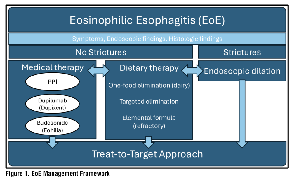
With the shift to a treat-to-target approach with specific target endpoints of symptomatic as well as endoscopic and histologic remission came the development of targeted, esophagus-specific formulations in earnest (Figure 1). Dupilumab (Dupixent) became the first United States Food and Drug Administration (FDA)-approved biologic treatment for eosinophilic esophagitis in 2022, paving the way for the new management landscape for EoE. Dupilumab is a human monoclonal antibody that blocks the receptor component for interleukin-4 and interleukin-13, which are key drivers of type 2 inflammation characteristic of EOE. It is approved for both the adult and pediatric (age >1) patient population. The phase 3 LIBERTY EE TREET trial showed histologic remission in 60% of patients compared to 5% receiving placebo at 24 weeks with 300 mg weekly subcutaneous injection dosing.26,27 Eohilia, a budesonide oral suspension, became the first oral FDA-approved medication for EoE based on two separate multicenter randomized controlled studies showing Eohilia 2 mg twice daily achieved histologic remission over placebo at 12 weeks.28 Current limitations on the use of Eohilia include lack of clinical data supporting the use for maintenance therapy.
Many other emerging biologics are now in the investigation pipeline for EoE, including monoclonal antibodies to interleukin-5 (benralizumab) and interleukin-13 (cendakimab).29 Other immune system targets being investigated include anti-tumor necrosis factor, anti-IgE, anti-Siglec 8, sphingosine-1-phosphate, and thymic stromal lymphopoietin. The emergence of these studies is promising for the future landscape of EoE therapies.
Endoscopic Management of Fibrostenotic Disease
Endoscopic dilation plays a key role in the management of fibrostenotic disease in patients with dysphagia and esophageal strictures. Endoscopic dilation alone, however, does not address the underlying inflammatory process or prevent disease progression. While dilation can be highly effective in relieving symptoms of dysphagia, up to 50% of EoE patients will have recurrent dysphagia at 15 months after dilation if not treated with maintenance anti-inflammatory therapy.30 The American Society of Gastrointestinal Endoscopy and the American College of Gastroenterology therefore recommend that endoscopic dilation be performed as an adjunct to medical treatment.31,32 Achieving histologic remission (<15 eosinophils/hpf) and being on long-term maintenance therapy have been associated with reduced need for subsequent dilations to maintain the same esophageal caliber.33,34
Despite early concerns regarding safety of dilation in EoE, endoscopic dilation can be performed even in the presence of active inflammation and in those not on treatment. Meta-analyses of large population-based studies of EoE patients undergoing esophageal dilation found a low rate (<1%) of serious complications, such as perforation and hemorrhage, with an adverse event rate similar to that of endoscopic dilation of benign strictures, and clinical improvement occurring in up to 85% of patients.35,36 A goal luminal diameter of at least 16-18 mm can relieve symptoms of dysphagia and reduce the risk of food impactions.37,38 Repeated endoscopic procedures with serial dilations may be needed to gradually achieve this target endpoint while optimizing medical therapy, depending on the initial luminal caliber and effect of each dilation on the esophageal mucosa. In patients with persistent dysphagia despite achieving histologic remission, an empiric dilation can be performed even if a stricture is not visualized endoscopically.
Specialized Populations and Clinical Scenarios: IBD Overlap and Pregnancy
EoE is frequently associated with other immune-mediated and atopic conditions, such as asthma, allergic rhinitis, and atopic dermatitis. Emerging evidence has also supported an association between coexisting EoE and inflammatory bowel disease (IBD), including Crohn’s disease and ulcerative colitis. Multiple large population-based studies have demonstrated an apparent bidirectional increased risk of EoE and IBD.39-40 This overlap may reflect shared genetic susceptibility and immune dysregulation, particularly through Th2-mediated pathways, although the precise mechanisms remain under investigation. Patients with EoE and IBD have been shown to experience a higher rate of IBD-related complications, including increased need for glucocorticoids, biologic therapy, and abdominal surgery, while conversely having a lower risk of EoE-related complications such as food impaction.41-42 Dysphagia symptoms in IBD patients should prompt further workup and consideration of a concurrent EoE diagnosis. Current treatment modalities for IBD are not effective for EoE, and patients with overlap of EoE and IBD who have active eosinophilic inflammation and symptoms of esophageal dysfunction should receive EoE-specific therapy in a treat-to-target approach.
During pregnancy, EoE management requires special considerations to ensure effective disease control and maternal-fetal safety. Despite EoE often affecting young patients of reproductive age, there is a paucity of data on pregnancy outcomes in EoE. A nationwide cohort study in Sweden identifying 23 births to 19 patients with EoE over a nearly 25-year period found that patients with EoE were overall not at increased risk of preterm birth or adverse pregnancy outcomes compared to controls.43 A retrospective survey-based study of 20 patients with EoE representing 34 pregnancies found that 56% of respondents reported improvement of dysphagia during pregnancy and 20% experienced worsening symptoms, with dysphagia returning to pre-pregnancy levels in majority of cases after delivery.44 Case reports have also highlighted several concerns that can arise when treating EoE patients who desire pregnancy, including safety of therapies during pregnancy and potential nutritional risks with dietary elimination.45 Patients should be advised that abrupt discontinuation of treatment can result in exacerbation of EoE symptoms and risk of disease progression. Dysphagia due to untreated disease and overly restrictive elimination diets can lead to nutritional risks. Therefore close monitoring and consultation with a dietician should be considered in EoE patients during pregnancy to ensure adequate nutrition.
Conclusion
Since the recognition of eosinophilic esophagitis as a disease entity, multiple key paradigm shifts have altered the course of our understanding and management of EoE. First, the recognition of dietary triggers in the mid-1990s established food-based antigens as dominant mediators for EoE and created a unique entity separate from GERD. Second, the elimination of the PPI trial requirement and removal of PPI-REE in 2018 fundamentally changed the diagnostic approach to EoE. The dissociation between symptom persistence despite histologic normalization after medical therapy or symptom resolution after dilation led to a treat-to-target approach in 2020 which underlined the importance of reaching multiple therapeutic targets with induction and maintenance therapy. Recognition of one-food elimination diet being just as effective as the six-food elimination diet has been instrumental in offering patients a more sustainable dietary intervention for management of their disease. Finally, the emergence of dupilumab as the first FDA-approved biologic therapy has completely changed the landscape for EoE management and has encouraged investigation for additional immune targets for this chronic inflammatory condition. Collectively, these paradigm shifts have transformed EoE into a manageable chronic disease with expanding therapeutic and monitoring options.
References
- Dellon ES, Muir AB, Katzka DA, et al. ACG Clinical Guideline: Diagnosis and Management of Eosinophilic Esophagitis. Am J Gastroenterol. 2025;120(1):31-59.
- Hahn JW, Lee K, Shin JI, et al. Global Incidence and Prevalence of Eosinophilic Esophagitis, 1976-2022: A Systematic Review and Met-analysis. Clin Gastroenterol Hepatol. 2023;21(13):3270-3284.e77.
- Thel HL, Anderson C, Xue AZ et al. Prevalence and Costs of Eosinophilic Esophagitis in the United States. Clin Gastroenterol Hepatol. 2025;23(7):272-280.e8.
- Massironi S, Mulinacci G, Gallo C, Elvevi A, Danese S, Invernizzi P, Vespa E. Mechanistic Insights into Eosinophilic Esophagitis: Therapies Targeting Pathophysiological Mechanisms. Cells. 2023 Oct 18;12(20):2473.
- O’Shea KM, Aceves SS, Dellon ES, Gupta SK, Spergel JM, Furuta GT, Rothenberg ME. Pathophysiology of Eosinophilic Esophagitis. Gastroenterology. 2018 Jan;154(2):333-345.
- Furuta GT, Katzka DA. Eosinophilic Esophagitis. N Engl J Med. 2015 Oct 22;373(17):1640-8.
- Underwood B, Troutman TD, Schwartz JT. Breaking down the complex pathophysiology of eosinophilic esophagitis. Ann Allergy Asthma Immunol. 2023 Jan;130(1):28-39.
- Dellon ES, Kim HP, Sperry SL, Rybnicek DA, Woosley JT, Shaheen NJ. A phenotypic analysis shows that eosinophilic esophagitis is a progressive fibrostenotic disease. Gastrointest Endosc. 2014 Apr;79(4):577-85.e4.
- Alexander ES, Martin LJ, Collins MH, Kottyan LC, Sucharew H, He H, Mukkada VA, Succop PA, Abonia JP, Foote H, Eby MD, Grotjan TM, Greenler AJ, Dellon ES, Demain JG, Furuta GT, Gurian LE, Harley JB, Hopp RJ, Kagalwalla A, Kaul A, Nadeau KC, Noel RJ, Putnam PE, von Tiehl KF, Rothenberg ME. Twin and family studies reveal strong environmental and weaker genetic cues explaining heritability of eosinophilic esophagitis. J Allergy Clin Immunol. 2014 Nov;134(5):1084-1092.e1.
- Kottyan LC, Davis BP, Sherrill JD, Liu K, Rochman M, Kaufman K, Weirauch MT, Vaughn S, Lazaro S, Rupert AM, Kohram M, Stucke EM, Kemme KA, Magnusen A, He H, Dexheimer P, Chehade M, Wood RA, Pesek RD, Vickery BP, Fleischer DM, Lindbad R, Sampson HA, Mukkada VA, Putnam PE, Abonia JP, Martin LJ, Harley JB, Rothenberg ME. Genome-wide association analysis of eosinophilic esophagitis provides insight into the tissue specificity of this allergic disease. Nat Genet. 2014 Aug;46(8):895-900.
- Jensen ET, Dellon ES. Environmental factors and eosinophilic esophagitis. J Allergy Clin Immunol. 2018 Jul;142(1):32-40.
- Dellon ES, Liacrouas CA, Molina-Infante J, et al. Updated International Consensus Diagnostic Criteria for Eosinophilic Esophagitis: Proceedings of the AGREE Conference. Gastroenterology. 2018;155(4):1022-1033.e10.
- Kim HP, Dellon ES. An Evolving Approach to the Diagnosis for Eosinophilic Esophagitis. Gastroenterol Hepatol (N Y). 2018;14(6):358-366.
- Shoda T, Wen T, Aceves SS, et al. Eosinophilic oesophagitis endotype classification by molecular, clinical, and histopathological analyses: a cross-sectional study. Lancet Gastroenterol Hepatol. 2018;3(7):477-488.
- Safroneeva E, Straumann A, Coslovksy M, et al. Symptoms Have Modest Accuracy in Detecting Endoscopic and Histologic Remission in Adults with Eosinophilic Esophagitis. Gastroenterology. 2016; 150(3):581-580.e4.
- Arnim UV, Biedermann L, Aceves SS, et al. Monitoring Patients With Eosinophilic Esophagitis in Routine Clinical Practice – International Expert Recommendations. Clin Gastroenterol Hepatol. 2023;21(10):2526-2533.
- Greuter T, Straumann A. Rationale for an Eosinophilic Esophagitis Treat-to-Target Concept. Gastroenterol Hepatol (N Y). 2024;20(10):583-590.
- Dellon ES, Irani AM, Hill MR, Hirano I. Development and field testing of a novel patient-reported outcome measure of dysphagia in patients with eosinophilic esophagitis. Aliment Pharmacol Ther. 2013;38(6):634-642.
- Cotton CC, Woosley JT, Moist SE, et al. Determination of a treatment response threshold for the Eosinophilic Esophagitis Endoscopic Reference Score. Endoscopy. 2022;54(7):635-643.
- Furuta GT, Kagalwalla AF, Lee JJ, et al. THe oesophageal string test: a novel, minimally invasive method measures mucosal inflammation in eosinophilic esophagitis. Gut. 2013;62(10):1395-405.
- Katzka DA, Geno DM, Ravi A, et al. Accuracy, safety, and tolerability of tissue collection by Cytosponge vs endoscopy for evaluation of eosinophilic esophagitis. Clin Gastroenterol Hepatol. 2015;13(1):77-83.e2.
- Arias A, Gonzalez-Cervera J, Tenias JM, et al. Efficacy of dietary interventions for inducing histologic remission in patients with eosinophilic esophagitis: a systematic review and meta-analysis. Gastroenterology. 2014;14697):1639-48.
- Kliewer KL, Gonsalves N, Dellon ES, et al. One-food versus six-food elimination diet therapy for the treatment of eosinophilic oesophagitis: a multicentre, randomized, open-label trail. Lancet Gastroenterol Hepatol. 2023;8(5):408-421.
- Muir A, Falk GW. Eosinophilic Esophagitis: A Review. 2021;3236(13):1310-1318.
- Cheng E, Zhang X, Huo X, et al. Omeprazole blocks eotaxin-3 expression by oesophageal squamous cells from patients with eosinophilic oesophagitis and gastro-oesophageal reflux disease. Gut. 2013;62(6):824-832. doi:10.1136/ gutjnl-2012-302250.
- Dellon ES, Rothenberg ME, Collins MH, et al. Dupilumab in Adults and Adolescents with Eosinophilic Esophagitis. NEJM. 2022;387(25):2317-2330.
- Rothenberg ME, Dellon ES, Collins MH, et al. Efficacy and safety of dupilumab up to 52 weeks in adults and adolescents with eosinophilic oesophagitis (LIBERTY EoE TREET study): a multicentre, double-blind, randomised, placebo-controlled, phase 3 trial. Lancet Gastroenterol Hepatol. 2023;8(11):990-1004.
- Dellon ES, Katzka DA, Collins MH, et al. Safety and Efficacy of Budesonide Oral Suspension Maintenance Therapy in Patients with Eosinophilic Esophagitis. Clin Gastroenterol Hepatol. 2019;17(4):666-673.e8.
- Musburger BG, Echeandia MG, Suskin EL, et al. Current and Emerging Therapies for Eosinophilic Esophagitis (EoE): A Comprehensive Review. Pharmaceutics. 2025;17(6):753.
- Schoepfer AM, Gonsalves N, Bussmann C, Conus S, Simon HU, Straumann A, Hirano I. Esophageal dilation in eosinophilic esophagitis: effectiveness, safety, and impact on the underlying inflammation. Am J Gastroenterol. 2010 May;105(5):1062-70.
- DelAceves SS, Alexander JA, Baron TH, Bredenoord AJ, Day L, Dellon ES, Falk GW, Furuta GT, Gonsalves N, Hirano I, Konda VJA, Lucendo AJ, Moawad F, Peterson KA, Putnam PE, Richter J, Schoepfer AM, Straumann A, McBride DL, Sharma P, Katzka DA. Endoscopic approach to eosinophilic esophagitis: American Society for Gastrointestinal Endoscopy Consensus Conference. Gastrointest Endosc. 2022 Oct;96(4):576-592.e1.
- Dellon ES, Muir AB, Katzka DA, Shah SC, Sauer BG, Aceves SS, Furuta GT, Gonsalves N, Hirano I. ACG Clinical Guideline: Diagnosis and Management of Eosinophilic Esophagitis. Am J Gastroenterol. 2025 Jan 1;120(1):31-59.
- Runge TM, Eluri S, Woosley JT, Shaheen NJ, Dellon ES. Control of inflammation decreases the need for subsequent esophageal dilation in patients with eosinophilic esophagitis. Dis Esophagus. 2017 Jul 1;30(7):1-7.
- Schupack DA, Ravi K, Geno DM, Pierce K, Mara K, Katzka DA, Alexander JA. Effect of Maintenance Therapy for Eosinophilic Esophagitis on Need for Recurrent Dilation. Dig Dis Sci. 2021 Feb;66(2):503-510.
- Moawad FJ, Cheatham JG, DeZee KJ. Meta-analysis: the safety and efficacy of dilation in eosinophilic oesophagitis. Aliment Pharmacol Ther. 2013 Oct;38(7):713-20.
- Moole H, Jacob K, Duvvuri A, Moole V, Dharmapuri S, Boddireddy R, Uppu A, Puli SR. Role of endoscopic esophageal dilation in managing eosinophilic esophagitis: A systematic review and meta-analysis. Medicine (Baltimore). 2017 Apr;96(14):e5877.
- Schoepfer AM, Gonsalves N, Bussmann C, Conus S, Simon HU, Straumann A, Hirano I. Esophageal dilation in eosinophilic esophagitis: effectiveness, safety, and impact on the underlying inflammation. Am J Gastroenterol. 2010 May;105(5):1062-70.
- Nicodème F, Hirano I, Chen J, Robinson K, Lin Z, Xiao Y, Gonsalves N, Kwasny MJ, Kahrilas PJ, Pandolfino JE. Esophageal distensibility as a measure of disease severity in patients with eosinophilic esophagitis. Clin Gastroenterol Hepatol. 2013 Sep;11(9):1101-1107.e1.
- Yanofsky R, Jogendran R, Hoxha T, Pathak A, Orchanian- Cheff A, Chhibba T, Sasson AN, Tandon P. The Association of Inflammatory Bowel Disease and Eosinophilic Esophagitis: A Systematic Review and Meta-analysis. Inflamm Bowel Dis. 2025 Oct 1;31(10):2895-2906.
- Sonnenberg A, Turner KO, Genta RM. Comorbid Occurrence of Eosinophilic Esophagitis and Inflammatory Bowel Disease. Clin Gastroenterol Hepatol. 2021 Mar;19(3):613- 615.e1.
- Malik A, Liu BD, Zhu L, Kaelber D, Song G. A Comprehensive Global Population-Based Analysis on the Coexistence of Eosinophilic Esophagitis and Inflammatory Bowel Disease. Dig Dis Sci. 2024 Mar;69(3):892-900.
- Limketkai BN, Shah SC, Hirano I, Bellaguarda E, Colombel JF. Epidemiology and implications of concurrent diagnosis of eosinophilic oesophagitis and IBD based on a prospective population-based analysis. Gut. 2019 Dec;68(12):2152- 2160.
- Röjler L, Uchida AM, Garber JJ, Stephansson O, Söderling J, Roelstraete B, Ludvigsson JF. Pregnancy Outcomes in Females with Eosinophilic Esophagitis: A Nationwide Population-Based Study. Inflamm Intest Dis. 2023 Oct 5;8(4):143-152.
- Röjler L, Uchida AM, Garber JJ, Stephansson O, Söderling J, Roelstraete B, Ludvigsson JF. Pregnancy Outcomes in Females with Eosinophilic Esophagitis: A Nationwide Population-Based Study. Inflamm Intest Dis. 2023 Oct 5;8(4):143-152.
- Burk CM, Long MD, Dellon ES. Management of Eosinophilic Esophagitis During Pregnancy. Dig Dis Sci. 2016 Jul;61(7):1819-25.